

Isolated calcific deposition within the subacromial bursa is a rare presentation of calcific tendinopathy that can mimic other pathologies; arthroscopic management allows accurate diagnosis and complete removal, leading to excellent clinical outcomes.
Dr Vasileios Kosmas, Orthopaedic Clinic OSTEON, Athens Medical Center, Athens, Greece. E-mail: v.d.kosmas@hotmail.com
Abstract
Introduction: Calcific tendinopathy is a common disorder of the shoulder characterized by the deposition of hydroxyapatite crystals within the rotator cuff tendons. It is part of the spectrum of basic calcium phosphate crystal deposition disease. However, isolated deposition within the subacromial–subdeltoid bursa is rare and may present diagnostic challenges.
Case Report: A 40-year-old female presented with persistent right shoulder pain and limited range of motion. Imaging revealed calcific deposits within the subacromial region. After failure of conservative management, arthroscopic removal was performed. Histopathological examination confirmed dystrophic calcification without malignancy. At 6-month follow-up, the patient demonstrated complete symptom resolution.
Conclusion: Subacromial bursal calcific deposition is an uncommon manifestation of calcific tendinopathy. Arthroscopic management is both diagnostic and therapeutic, with excellent clinical outcomes.
Keywords: Calcific tendinopathy, subacromial bursa, hydroxyapatite, arthroscopy, shoulder.

Calcific tendinopathy of the shoulder is a frequently encountered condition in orthopaedic practice and represents one of the most common causes of non-traumatic shoulder pain [1,2,3]. It is characterised by the deposition of hydroxyapatite crystals within the rotator cuff tendons, most commonly affecting the supraspinatus tendon [2,3]. This entity is considered part of the broader category of basic calcium phosphate crystal deposition disease, which includes a spectrum of disorders such as calcific tendinitis and bursitis [4,5]. The clinical presentation varies widely, ranging from asymptomatic calcifications to acute inflammatory episodes with severe pain and functional limitation [5]. Long-term studies have demonstrated variable clinical courses of calcific tendinitis, ranging from spontaneous resolution to persistent symptoms requiring intervention [6]. The natural history of calcific tendinopathy has been described as a dynamic, cell-mediated process involving three distinct phases: pre-calcific, calcific (formative and resting), and post-calcific (resorptive) [5,7]. The resorptive phase is typically associated with acute symptoms due to an intense inflammatory response triggered by crystal fragmentation and phagocytosis [7]. Although calcific deposits are usually confined within the tendon substance, extension or migration into adjacent anatomical structures has been described [8]. Involvement of the subacromial–subdeltoid bursa is considered a rare complication and may result in atypical clinical and imaging findings, often leading to diagnostic uncertainty [8]. The pathogenesis of this migration is not fully understood, but proposed mechanisms include increased intratendinous pressure during the resorptive phase, rupture of the tendon fibres, and extrusion of calcific material into surrounding tissues [7,8]. In this report, we present a rare case of isolated calcific deposition within the subacromial bursa, confirmed histopathologically and successfully treated with arthroscopic removal.
A 40-year-old female patient presented to our clinic with a history of progressive right shoulder pain over a period of several months. The pain was exacerbated by overhead activities and was associated with significant functional limitation. The patient had previously undergone conservative treatment, including oral non-steroidal anti-inflammatory drugs, physiotherapy, and activity modification, without significant improvement. On physical examination, the patient demonstrated:
- Painful arc during abduction between 60° and 120°
- Restricted active range of motion, particularly in abduction and forward flexion
- Localized tenderness over the anterolateral aspect of the shoulder
- No signs of instability or neurological deficit.
Plain radiographs revealed dense calcific deposits in the subacromial region. Magnetic resonance imaging confirmed the presence of calcific material adjacent to the rotator cuff, with associated subacromial bursitis (Fig. 1).
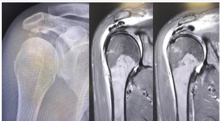
Figure 1: Imaging evaluation with X-rays and magnetic resonance imaging demonstrating calcific deposits within the subacromial space.
Given the persistence of symptoms and failure of conservative therapy, surgical intervention was recommended. The procedure was performed under general anaesthesia with the patient in the beach-chair position. Standard posterior and lateral arthroscopic portals were established. Diagnostic arthroscopy of the glenohumeral joint revealed no intra-articular pathology. Attention was then directed to the subacromial space. On bursectomy, a collection of calcific material was identified within the subacromial bursa (Fig. 2).
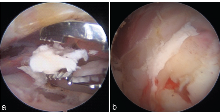
Figure 2: Presence of calcific material (a) in the subacromial region, (b) in the subacromial bursa.
The deposits were carefully evacuated using a combination of shaver and irrigation, ensuring complete removal. No significant rotator cuff tear or additional pathology was identified. Adequate decompression and lavage were performed before closure. Macroscopic examination revealed multiple whitish, chalky tissue fragments measuring approximately 1.6 × 0.7 × 0.2 cm (Fig. 3).
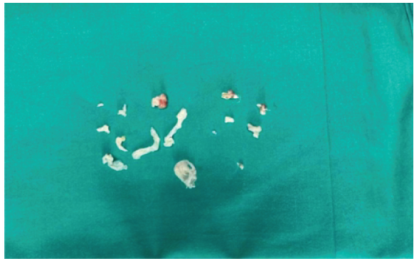
Figure 3: Macroscopic appearance of multiple tissue fragments removed arthroscopically from the subacromial space.
Microscopic analysis demonstrated synovial tissue with basophilic granular calcium deposits consistent with hydroxyapatite crystals. The surrounding stroma exhibited mild fibrosis and a mild inflammatory infiltrate. No evidence of malignancy, cellular atypia, or granulomatous reaction was identified. These findings confirmed the diagnosis of dystrophic calcification within the subacromial bursa, consistent with crystal-induced pathology [4,5]. At 6-month follow-up, the patient reported complete resolution of symptoms. Clinical examination demonstrated a full, pain-free range of motion with restoration of shoulder function. No recurrence of calcification was observed on follow-up imaging.
Calcific tendinopathy is a well-documented condition; however, isolated involvement of the subacromial bursa is rare [8]. Most calcific deposits are located within the rotator cuff tendons, and migration beyond the tendon is considered an uncommon phenomenon. The migration of calcific material into the subacromial bursa is thought to occur during the resorptive phase, when increased intratendinous pressure and enzymatic activity may lead to rupture of tendon fibres and extrusion of calcium deposits [7,8]. Clinically, such cases may present with more severe pain and acute inflammatory symptoms compared to typical calcific tendinopathy. Imaging findings may also be misleading, as bursal calcifications can mimic infection, tumours, or other inflammatory conditions. High-resolution ultrasonography has been shown to facilitate the detection, characterisation, and management of calcific deposits in the rotator cuff [9]. Dystrophic calcification, as observed in this case, typically occurs in degenerated or injured tissues and is not associated with systemic metabolic abnormalities [10]. Differential diagnosis includes synovial chondromatosis, which is characterised by the presence of cartilaginous nodules within the synovium [11]. Histopathological examination is essential to distinguish between these entities. Arthroscopic management has been shown to be highly effective in treating calcific tendinopathy, particularly in cases resistant to conservative therapy [12,13,14]. It allows direct visualisation, accurate localisation, and complete removal of deposits, resulting in excellent clinical outcomes. The present case highlights the importance of recognising atypical presentations of calcific tendinopathy and supports the role of arthroscopy as both a diagnostic and therapeutic modality.
Isolated calcific deposition within the subacromial bursa is a rare manifestation of calcific tendinopathy. Arthroscopic removal provides effective treatment, leading to complete symptom resolution and excellent functional recovery.
Atypical localization of calcific deposits should be considered in patients with persistent shoulder pain. Arthroscopy is a valuable tool for both diagnosis and management in such cases.
[refernces_numbered]1. Bosworth BM. Calcium deposits in the shoulder and subacromial bursitis: A survey of 12,122 shoulders. JAMA 1941;116:2477-82.
2. Uhthoff HK, Loehr JW. Calcific tendinopathy of the rotator cuff: Pathogenesis, diagnosis, and management. J Am Acad Orthop Surg 1997;5:183-91.
3. Speed CA, Hazleman BL. Calcific tendinitis of the shoulder. N Engl J Med 1999;340:1582-4.
4. McCarty DJ. Crystal-induced inflammation of the joints. Annu Rev Med 1970;21:357-66.
5. Oliva F, Via AG, Maffulli N. Calcific tendinopathy of the rotator cuff tendons. Sports Med Arthrosc Rev 2011;19:237-43.
6. DePalma AF, Kruper JS. Long-term study of shoulder joints afflicted with and treated for calcific tendinitis. Clin Orthop Relat Res 1961;20:61-72.
7. Uhthoff HK, Sarkar K, Maynard JA. Calcifying tendinitis: A new concept of its pathogenesis. Clin Orthop Relat Res 1976;118:164-8.
8. Della Valle V, Bassi EM, Calliada F. Migration of calcium deposits into subacromial-subdeltoid bursa and into humeral head as a rare complication of calcifying tendinitis: Sonography and imaging. J Ultrasound 2015;18:259-63.
9. Chiou HJ, Chou YH, Wu JJ, Huang TF, Ma HL, Hsu CC, et al. The role of high-resolution ultrasonography in management of calcific tendonitis of the rotator cuff. Ultrasound Med Biol 2001;27:735-43.
10. Resnick D, Niwayama G. Diagnosis of Bone and Joint Disorders. 4th ed. Australia: Saunders; 2002.
11. Chen A, Wong LY, Sheu CY, Chen BF. Synovial chondromatosis differentiation. Skeletal Radiol 2002;31:119-21.
12. Louwerens JK, Sierevelt IN, Van Hove RP, van den Bekerom MPJ, van Noort A, Eygendaal D et al. Comparing surgical and conservative treatment of calcific tendinitis. Arthroscopy 2014;30:1019-27.
13. Ark JW, Flock TJ, Flatow EL, Bigliani LU. Arthroscopic treatment of calcific tendinitis of the shoulder. Arthroscopy 1992;8:183-8.
14. Mole D, Kempf JF, Gleyze P, Rio B, Bonnomet F, Walch G. Results of endoscopic treatment of non-broken tendinopathies of the rotator cuff. 2. Calcifications of the rotator cuff. Rev Chir Orthop Reparatrice Appar Mot 1993;79:532-41.[/refernces_numbered]




Related Articles in Journal of Orthopaedic Case Reports
 December 10, 2023 Osteonecrosis of the Humeral Head Following Arthroscopic Repair of Rotator Cuff Tears: A Case Report
 October 1, 2026 Myositis Ossificans Affecting the Rotator Cuff Muscles and Proximal Humerus: A Rare Clinical Entity
 August 1, 2026 Intra-articular Ganglionic Cysts of Knee: A Cause for Anterior Knee Pain
 July 1, 2026 Post-traumatic Hemosiderotic Synovitis Mimicking Pigmented Villonodular Synovitis in a Non-hemophilic Child: First Reported Case from the United Arab Emirates