

Deformities of the oblique plane are difficult to identify, let alone treat. This article provides a description of the management of pediatric tibial deformity.
Dr. Rohit Kumar Yadav, Department of Orthopaedics, All India Institute of Medical Sciences, Deoghar, Jharkhand, India. E-mail: rk169816@gmail.com
Abstract
Introduction: Tibial shaft fractures are among the most common fractures encountered in the pediatric population. They require appropriate diagnosis and treatment to minimize complications and optimize outcomes. Treatment is individualized based on patient age, concomitant injuries, fracture pattern, associated soft tissue and neurovascular injury, and surgeon experience. Although there is potential for remodeling, this may not be adequate with more significant deformities, thus requiring re-manipulation or, rarely, operative intervention.
Case report: We have performed deformity correction in a 12-year-old male patient, who presented with a malunited fracture of the shaft of both bones in the right leg, with gait abnormality and cosmetic concerns. The aim of our study was to determine the feasibility of using the Ilizarov ring fixator to correct long-bone deformities in pediatric tibial malunited shaft fractures presenting with oblique plane deformities.
Conclusion: The deformity was analyzed, and the center of rotation of angulation was identified. Our case belonged to variant “3” in the classification system of Dror Paley.
Keywords: Oblique plane deformity, graph method, ilizarov, oblique plane, tibia, trigonometry.

A malunion is broadly defined as a fracture that has healed in a clinically unacceptable position, often resulting in deformity or dysfunction. Malunions of the tibia have the potential to have significant short- and long-term effects on the individual’s biomechanics, cosmesis, and ultimately quality of life. Proximal tibial physeal injuries are rare, comprising only 0.6% of long-bone fractures in children [1]. The proximal tibial epiphysis is well protected by the contralateral knee and surrounding soft tissues, including the fibular head ligaments, patellar tendon, semitendinosus insertion, and medial collateral ligament. In a series of 214 surgically treated tibial shaft fractures, 43 involved the distal third, with 32.6% showing an associated distal tibial physeal injury [2]. We present a case of a malunited tibial diaphyseal fracture associated with proximal lateral physeal growth arrest, resulting in ipsilateral limb shortening, valgus malalignment, and a biplanar deformity.
This is a case of a 12-year-old male patient who came to the orthopedic department in December 2024 with ipsilateral limb shortening, valgus malalignment, and biplanar deformity of the tibia. After a thorough review of the patient’s history and available documentation, it was established that the injury had occurred at 9 years of age and had been treated conservatively with a plaster application for 3 months before the patient presented to our department. He presented to us as if he had a leg deformity and limb shortening, which led to gait difficulties and significant cosmetic concerns for his parents. On examination, the patient was found to have a valgus deformity of the knee and a procurvatum deformity of the mid-diaphyseal tibia, accompanied by a limb-length discrepancy of 3 cm compared with the contralateral limb (Fig. 1).
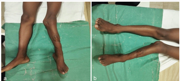
Figure 1: (a) Valgus deformity in frontal, (b) procurvatum deformity in sagittal.
The patient was walking without support with a limp. No neurovascular deficit was encountered. There was no flexion deformity in the knee, but movement restriction at the ankle joint was present. Radiological findings revealed a malunited diaphyseal fracture and high tibial slope, a non-union fibular diaphyseal fracture in the lateral view, and a varus deformity in the anteroposterior (AP) view. (Fig. 2) On measurement, a varus deformity of 7 degrees & procurvatum of 36 degrees was found. (Fig. 3a & 4)
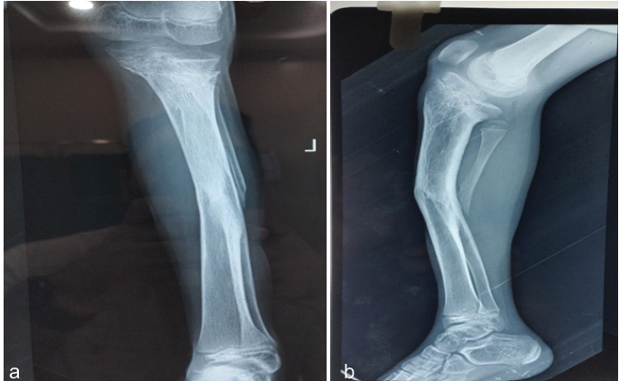
Figure 2: (a) Anteroposterior view shows malunion tibia and non-union fibula, (b) lateral view shows malunion tibia.
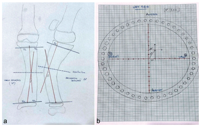
Figure 3: (a) Varus deformity of 7° and procurvatum of 36°, (b) Graph method for hinge placement.
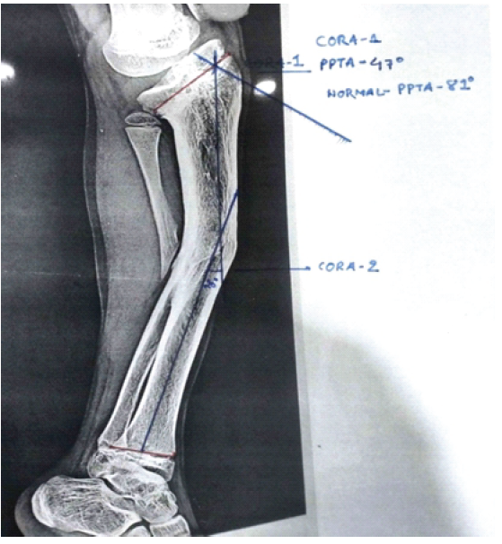
Figure 4: X-ray lateral view shows procurvatum of 20° in diaphysis and another center of rotation of angulation at proximal tibia with abnormal posterior proximal tibial angle of correction 10°.
Management:
Preoperative planning regarding the following parameters was done: preoperative templating, frame design, position of hinges, level of corticotomy, latent period, and distraction protocol. Preoperative templating was done to identify (a) the mechanical axis deviation plane, magnitude, and level of deformity; (b) the anatomical and mechanical axis and center of rotation of angulation (CORA). The level of placement of hinges in the ring was identified using trigonometry and the graph method; the hinge on the medial side was anteromedial, and the hinge on the lateral side was posterolateral. (Fig. 3b). The “Rule of Similar Triangles” was used to identify the rate of distraction in the rod. The distraction rate at the level of the distraction rod (to achieve a distraction of 1 mm/day at the osteotomy site) was calculated to be 7 mm, which is 2 full turns 4 times a day (Fig. 5).
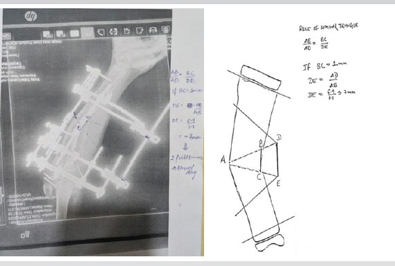
Figure 5: Rule of similar triangles.
After templating the bone, a construct was made with hinges anteromedially and posterolateral and a distraction rod posteromedial. After fixing the ring to the bone with wires, there was no need for a fibular osteotomy, as the fibular diaphyseal fracture was not united. In our case, two CORAs were identified: one situated proximally in the metaphyseal region and the other at the malunited diaphyseal shaft of the tibia. The proximal deformity was corrected acutely through osteotomy, aided by the use of multiple Kirschner wires intraoperatively. The diaphyseal recurvatum and varus deformity were subsequently managed with an Ilizarov external fixator (Figs. 6 & 7).
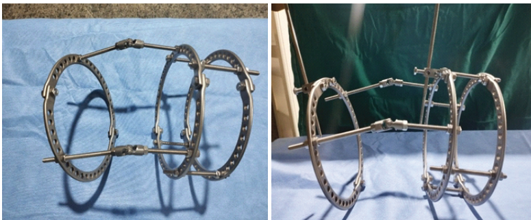
Figure 6: Ilizarov construct.
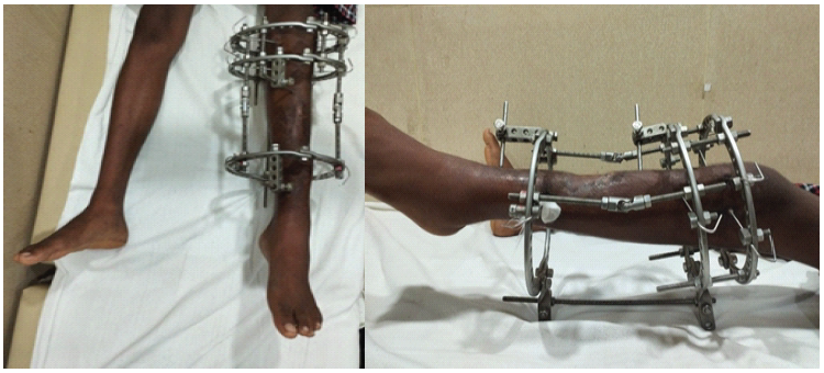
Figure 7: Ilizarov construct application.
Corticotomy (open wedge) was performed at the level of CORA identified in the lateral radiograph (which is at the level of the malunion site). The immediate post-operative radiograph shows hinges to be placed at the level of CORA in the lateral view. Distraction was initiated on the 7th day. 7 mm/day of distraction resulted in the opening of the osteotomized site at 1 mm/day. Full correction was achieved after 40 days. After complete correction of deformity, the fixator was replaced with a patellar tendon-bearing cast. Consolidation was achieved in 4 months. The tracing of the X-ray image and the relationship of the anatomical axis show that there is good alignment in the lateral view.
Follow-up:
Follow-up was done once in 2 weeks during the course of distraction and once a month after deformity correction. Follow-up after 9 months shows good consolidation of regeneration and correction of angular deformity. The patient has good functional and cosmetic satisfaction (Figs. 8 & 9).
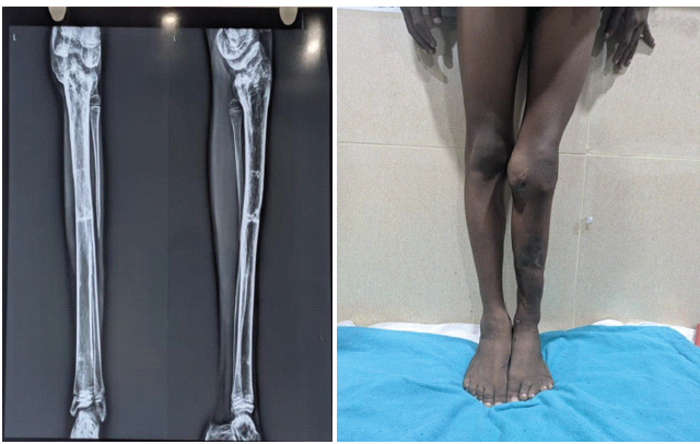
Figure 8: X-ray and clinical picture after correction.
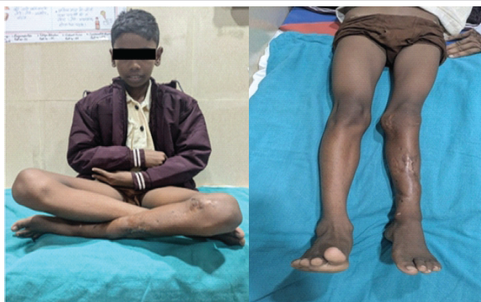
Figure 9: Normal function with no residual shortening.
With angular malunion of the tibia, which is particularly apparent with external fixation, there may be remodeling over a period of 2–3 years according to the age and sex of the patient. Significant remodeling of the tibial shaft occurs in girls <8 years and boys <10 years of age, compared to 50% of correction for girls 9–12 years and boys 11–12 years [3]. Once >13 years, <25% of correction occurs from remodeling, with complex, apex posterior, and varus deformities the least forgiving. Mild tibial growth inhibition can occur after tibial shaft fractures, especially in those over 8 years. Physeal injury to the proximal tibia may occur from a concomitant occult tibial tubercle or physeal crush (e.g., Salter-Harris I or V) injury, or indeed pin placement from external fixation [4]. Altered load transmission across joints can lead to premature degenerative changes secondary to deformity [5,6,7]. The goals of deformity-correction surgery are to alleviate any existing symptoms and to protect adjacent joints from the development of osteoarthrosis [7,8]. Various treatment modalities are available, ranging from splintage and corrective plaster casting to plating, intramedullary interlocking nailing, monolateral external fixation, Ilizarov ring fixation, and six-axis correction devices such as the Ortho-SUV and the Taylor Spatial Frame. However, we utilized the Ilizarov ring fixator for deformity correction. The technique involved frame design and application, subperiosteal corticotomy, and pin and wire insertion [9]. The Ilizarov frame offers a highly versatile fixation system that ensures stability, soft-tissue preservation, adjustability, and functionality—all of which are essential for bone to achieve its full osteogenic potential. Bone stability, a critical factor in osteogenesis, depends largely on the stability of the external frame [10]. Frame stability is significantly influenced by ring characteristics, with larger-diameter rings providing less stability compared to smaller ones [11].
Oblique plane deformity:
Angular deformity may occur in any plane. The standard reference planes are the two anatomic planes, coronal and sagittal. The standard reference radiographs that correspond to the planes are the AP and lateral radiographs, respectively. Uni-apical angular deformities for which angulation is seen on both AP and lateral radiographs are often incorrectly referred to as biplanar angular deformities. These biplanar angular deformities are actually uniplanar angular deformities in an oblique plane. Oblique plane deformity is not a biplanar deformity. Here, the true plane of angulation lies between the frontal and sagittal planes. In such cases, trigonometry is useful to find out the plane of deformity (while using regular AP and lateral views) [8].
CORA:
By definition, CORA is the apex of the deformity and is identified as the point of intersection of the axis drawn through the proximal and distal fragments [10,11].
Angulation correction axis (ACA):
The axis around which the deformity has to be corrected. When the CORA falls below the level of the apex of the deformity, Dror Paley has named this translation as compensatory translation, and the CORA as angulation–translation CORA. In our case, the osteotomy and ACA were placed at the same level of CORA in the AP view, and this has contributed to angular correction without translation (i.e., osteotomy rule 1) [8].
Order of correction:
There is a slight difference between the order of correction of deformities during fracture fixation and gradual deformity correction according to Lambotte’s principle [12,13] and Paley’s principle [8]. In both, the angulation is corrected first and the translation last. In our case, rule 2 happened in procurvatum deformity, and rule 1 happened in varus deformity in the frontal plane, so there was a secondary translation seen in the lateral view. In our case, we achieved comparable limb length without any residual limb-length discrepancy. Complete correction of the diaphyseal procurvatum deformity from the malunited tibial shaft fracture, as well as correction of the valgus deformity; however, some amount of residual deformity secondary to physeal growth arrest was present with a correction angle of 10°. These outcomes were confirmed through post-operative and follow-up radiographs and clinical evaluations.
The Ilizarov ring fixator can be used to correct deformities in multiple planes [4], but the correction has to be done in multiple stages in the order mentioned previously. Alternatively, six-axis correction devices like Ortho-SUV can be used to correct all deformities in a single stage. Although several options are available regarding deformity correction, the technique that should be instituted depends on deformity pattern, soft tissue status, and level of deformity. Proper planning is needed preoperatively, intraoperatively, and postoperatively. Revision of planning to be done at any point of time during management. Proper counseling of the parents should be done to gain compliance in case revision surgery is needed.
Ilizarov ring fixation can be a useful modality in complex deformities of the tibia, even in the pediatric population, with considerable gain in length.
References
- 1. Vrettakos AN, Evaggelidis DC, Kyrkos MJ, Tsatsos AV, Nenopoulos A, Beslikas T. Lower limb deformity following proximal tibia physeal injury: Long-term follow-up. J Orthop Traumatol 2012;13:7-11. [Google Scholar] [PubMed]
- 2. McGraw-Heinrich JA, Ezeokoli EU, Mitchell P, Silverstein RS, Smith BG. Pediatric distal third tibial shaft fractures: A comparison of surgical fixation methods and incidence of concomitant physeal fractures. J Pediatr Orthop Part B 2023;32:393-400. [Google Scholar] [PubMed]
- 3. Egol KA. Handbook of fractures. In: Pediatric Fracture and Dislocations. 7ed., Part. V. Berlin: Springer; 2025. [Google Scholar] [PubMed]
- 4. Patel NK, Horstman J, Kuester V, Sambandam S, Mounasamy V. Pediatric tibial shaft fractures. Indian J Orthop 2018;52:522-8. [Google Scholar] [PubMed]
- 5. Zhang L, Liu G, Han B, Wang Z, Yan Y, Ma J, et al. Knee joint biomechanics in physiological conditions and how pathologies can affect it: A systematic review. Appl Bionics Biomech 2020;2020:7451683. [Google Scholar] [PubMed]
- 6. Zhang L, Liu G, Yan Y, Han B, Li H, Ma J, et al. A subject-specific musculoskeletal model to predict the tibiofemoral contact forces during daily living activities. Comput Methods Biomech Biomed Engin 2023;26:972-85. [Google Scholar] [PubMed]
- 7. Sv B. Oblique Plane Deformity Correction with Ilizarov Technique in Case of Malunited Tibia – Case Report. Germany: dSpace Home; 2012. [Google Scholar] [PubMed]
- 8. Paley D, Tetsworth K. Deformity ’Correction by the Ilizarov Technique. Berlin: Springer; 2009. [Google Scholar] [PubMed]
- 9. Pathania VP, Sharma AK, Joshi GR, John JT. Correction of lower limb deformities using Ilizarov’s technique. Med J Armed Forces India 2005;61:322-5. [Google Scholar] [PubMed]
- 10. Spiegelberg B, Parratt T, Dheerendra SK, Khan WS, Jennings R, Marsh DR. Ilizarov principles of deformity correction. Ann R Coll Surg Engl 2010;92:101-5. [Google Scholar] [PubMed]
- 11. Cross AR, Lewis DD, Murphy ST, Rigaud S, Madison JB, Kehoe MM, et al. Effects of ring diameter and wire tension on the axial biomechanics of four-ring circular external skeletal fixator constructs. Am J Vet Res 2001;62:1025-30. [Google Scholar] [PubMed]
- 12. Campbell’s operative orthopaedics. In: Fractures and Dislocation in Children. 14th ed. New York: Thieme; 2013. [Google Scholar] [PubMed]
- 13. Garg M, Kumar S, Agrawal HK, Jaiman A. Proximal femoral stress reaction in a military recruit – a treatment prospect. J Orthop Case Rep 2014;4:25-8. [Google Scholar] [PubMed]






Related Articles in Journal of Orthopaedic Case Reports
 September 1, 2026 Limb Salvage for Entire Tibial Ewing Sarcoma in a 4-year-Old Child Using Extracorporeal Irradiation and Free Flap Reconstruction: A Case Report and Surgical Technique
 August 1, 2026 Plastic Bowing of the Pediatric Tibia is Extraordinarily Rare and Not Represented in the Literature, the Unicorn of Pediatric Fractures: A Case Report
 August 1, 2026 Primary Tuberculous Osteomyelitis of the Proximal Tibia Presenting as Chronic Osteomyelitis: A Diagnostic Challenge in Musculoskeletal Tuberculosis
 August 1, 2026 Extensive Giant Cell Tumor Involving Entire Tibia with Talar Extension and Pulmonary Metastases: Successful Limb Preservation with Denosumab and Angioembolization