

In high-energy ankle trauma, clinicians should consider concomitant Achilles tendon rupture, as careful examination and advanced imaging can prevent missed diagnosis and guide timely operative management.
Samuel T. Hovland, University of Central Florida College of Medicine, Orlando, Florida, United States. E-mail: sam_hovland@ucf.edu
Abstract
Introduction: Ankle fractures and Achilles tendon ruptures most commonly occur as isolated injuries. The simultaneous occurrence of an ankle fracture with an Achilles tendon rupture is rare. It may be overlooked during initial evaluation, particularly in elderly patients or in the presence of distracting injuries. Previous reports have described Achilles tendon rupture in association with medial malleolar, bimalleolar, trimalleolar, and distal tibial fractures. To our knowledge, this is the first reported case of an Achilles tendon rupture occurring in conjunction with a pilon fracture. Reporting this case is important to raise awareness of this uncommon injury pattern and to emphasize the need for comprehensive clinical assessment in ankle trauma.
Case Report: A 78-year-old male with a history of Parkinson’s disease sustained a right ankle injury after falling from a ladder. He initially presented to the emergency department with ankle pain and superficial lacerations. Radiographs were obtained, but no fracture was diagnosed. Upon follow-up with an orthopedic surgeon, advanced imaging and physical examination revealed a fracture of the tibial plafond with an ipsilateral Achilles tendon rupture. The patient subsequently underwent open reduction and internal fixation of the pilon fracture and surgical repair of the Achilles tendon.
Conclusion: This case highlights an exceptionally rare injury combination that can be easily missed during initial evaluation. It underscores the importance of maintaining a high index of suspicion and performing thorough physical examination and appropriate imaging when assessing ankle trauma, particularly in elderly patients. This report contributes a novel injury pattern to the orthopedic literature and demonstrates that concurrent operative management of both injuries can be safely performed in selected patients. Increased awareness of this association may help prevent delayed diagnosis, reduce complications, and improve functional outcomes in future cases.
Keywords: Pilon fracture, Achilles rupture, ankle trauma.

Tibial pilon fractures and Achilles tendon ruptures, in isolation, are common and potentially devastating injuries [1]. The simultaneous occurrence of an ankle fracture with an ipsilateral Achilles tendon rupture is exceedingly rare and may be overlooked during initial evaluation [2,3]. Failure to recognize this combined injury pattern can significantly alter surgical planning, increase technical complexity, and potentially worsen outcomes [4,5,6,7]. Although combined osseous and tendon injuries about the ankle have been described, prior reports have largely involved fracture patterns around the malleoli or distal tibial metaphysis [8,9]. Cases in the literature include Achilles tendon rupture associated with medial malleolar, lateral malleolar, bimalleolar, and trimalleolar fractures, as well as open distal tibia and fibula injuries [8,9,10,11,12,13,14,15,16,17,18,19,20,21]. In addition, malleolar fractures with associated superior peroneal retinaculum (SPR) disruption and peroneal tendon instability have been reported [22,23,24]. The mechanisms of injury and patient demographics from these cases are summarized in Table 1.
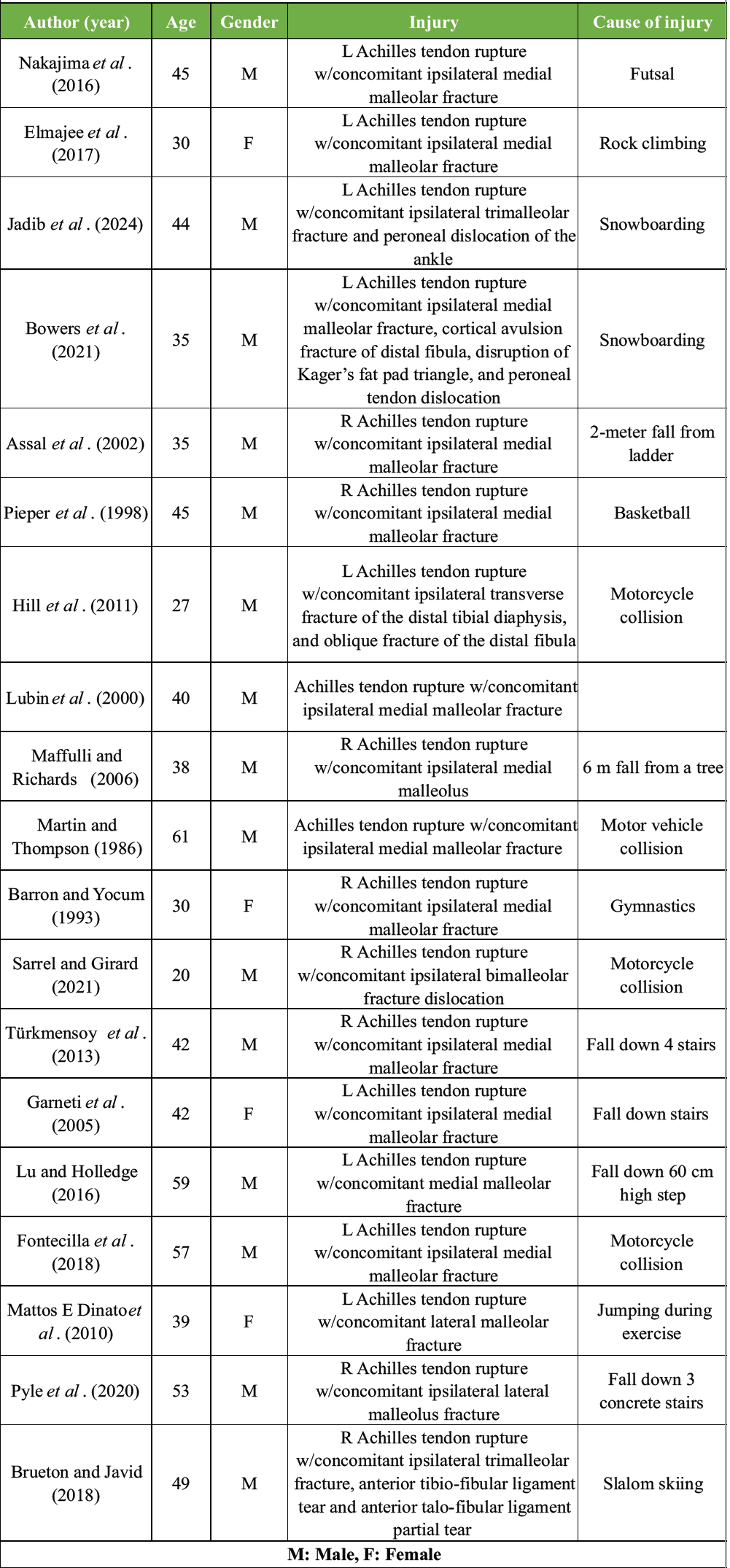
Table 1: Patient demographics: Patient demographics, injury, and mechanism of injury of the reported case reports of similar injuries found upon literature search
We present a ladder-related fall resulting in a displaced tibial pilon fracture with a simultaneous acute Achilles tendon rupture. This case underscores the importance of maintaining a high index of suspicion for associated tendon injury in high-energy ankle trauma. It highlights the need for careful clinical examination and imaging review to avoid missed diagnoses and optimize operative management.
A 78-year-old male with a history of Parkinson’s disease (PD) presented to the emergency department after a fall from a ladder, reporting right ankle pain and superficial lacerations. Radiographs were obtained, and no fracture was diagnosed (Fig. 1).
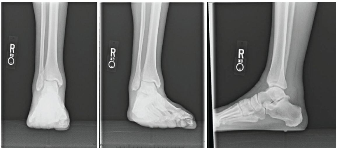
Figure 1: Pre-operative radiographs.
He was discharged with crutches for a suspected ankle sprain. On presentation to our clinic 2 weeks after initial injury, physical examination demonstrated moderate swelling of the right ankle and scattered healing scabs along the anterior tibia without signs of infection. There was tenderness to palpation of the medial malleolus, lateral malleolus, and anterior ankle. He had moderate swelling throughout the right ankle, and the right Achilles tendon had a palpable defect. The Thompson test was positive. Active and passive ankle range of motion was preserved but limited secondary to pain. Given the high-energy mechanism of injury and concern for soft-tissue injury, magnetic resonance imaging (MRI) of the ankle was performed. The MRI demonstrated a combination of an acute non-insertional Achilles tendon rupture (Fig. 2) and an acute tibial plafond fracture (Fig. 3).
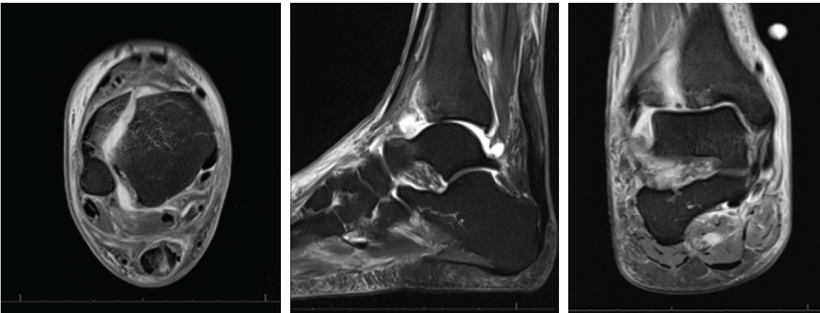
Figure 2: Pre-operative magnetic resonance imaging (MRI): Pre-operative MRI demonstrating recent non-insertional Achilles tendon rupture as well as syndesmotic and deltoid ligament ruptures with associated malalignment.
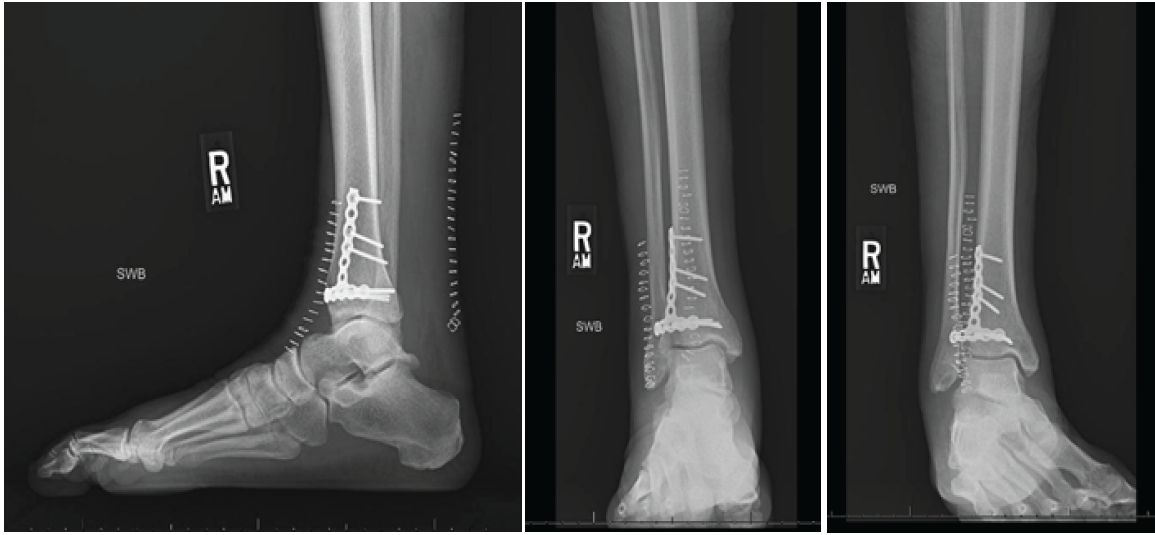
Figure 3: 2 Week post-operative radiographs: 2 week post-operative X-rays showing a healing fracture and intact hardware.
Given the displaced intra-articular fracture pattern and malreduction of the fibula, operative fixation of the tibial plafond was recommended. Non-operative and operative management were discussed at length with the patient regarding his Achilles tendon rupture. After shared decision-making, the patient elected to proceed with operative management of both injuries. A popliteal and saphenous nerve block was administered for post-operative analgesia, and a thigh tourniquet was applied. The patient was positioned supine for fixation of the pilon fracture. An anterolateral approach to the distal tibia was performed. The superficial peroneal nerve was identified and protected. After division of the retinaculum, dissection was carried to the distal anterior tibia. Fracture hematoma and fibrous tissue were evacuated. There was focal comminution of the articular surface, including a 4 × 6 mm segment of non-viable cartilage, which was excised. The joint was irrigated, and the fracture was reduced under fluoroscopic guidance. A mini-fragment T-plate (Stryker) was selected and applied using an antiglide technique. The plate was secured proximally and distally with non-locking screws following contouring. Final fluoroscopic imaging confirmed anatomic reduction and stable fixation. The retinaculum was repaired, and the skin was closed in layered fashion. The patient was then repositioned prone for Achilles tendon repair. A posterior midline incision was made approximately 8–9 cm proximal to the calcaneal insertion, centered over the palpable defect. The tendon ends demonstrated fibrous tissue and early scar formation without complete healing. The scar tissue was debrided, and the tendon ends were mobilized to allow direct repair. The tendon was then repaired using a standard Krackow suture technique proximally and distally, supplemented with a modified Kessler suture anteriorly. With the ankle held in plantarflexion, the sutures were sequentially tied, achieving satisfactory tension and restoration of tendon continuity. The plantaris tendon was harvested proximally and woven through the repair site for augmentation. The paratenon was closed, followed by layered skin closure. A well-padded posterior splint was applied with the ankle in plantar flexion, and the patient was maintained non-weight-bearing. At 2 weeks postoperatively, staples were removed, and the patient was transitioned to a controlled ankle motion boot with stacked heel lifts. Partial weight-bearing was initiated. At 4 weeks, there was concern that Parkinsonian rigidity may affect tendon relaxation and repair tension; therefore, dorsiflexion beyond neutral was restricted. Physical therapy was initiated, focusing on gait training, a gentle range of motion, and progressive strengthening. At 6 weeks, one heel wedge was removed. He then transitioned to flat-foot ambulation in the boot the following week and gradually out of the boot thereafter. At 10 weeks, the patient was ambulating in regular footwear with mild swelling and residual plantarflexion weakness. Radiographs demonstrated maintained alignment without hardware failure. The Achilles repair demonstrated active gastrocnemius contraction, though weakness persisted. Strengthening was intensified, and dorsiflexion beyond neutral remained restricted for an additional 2 weeks. At 4 months postoperatively, radiographs confirmed fracture union (Fig. 4). The patient had returned to baseline daily activities, was ambulating independently without pain, and reported overall satisfaction with his outcome.
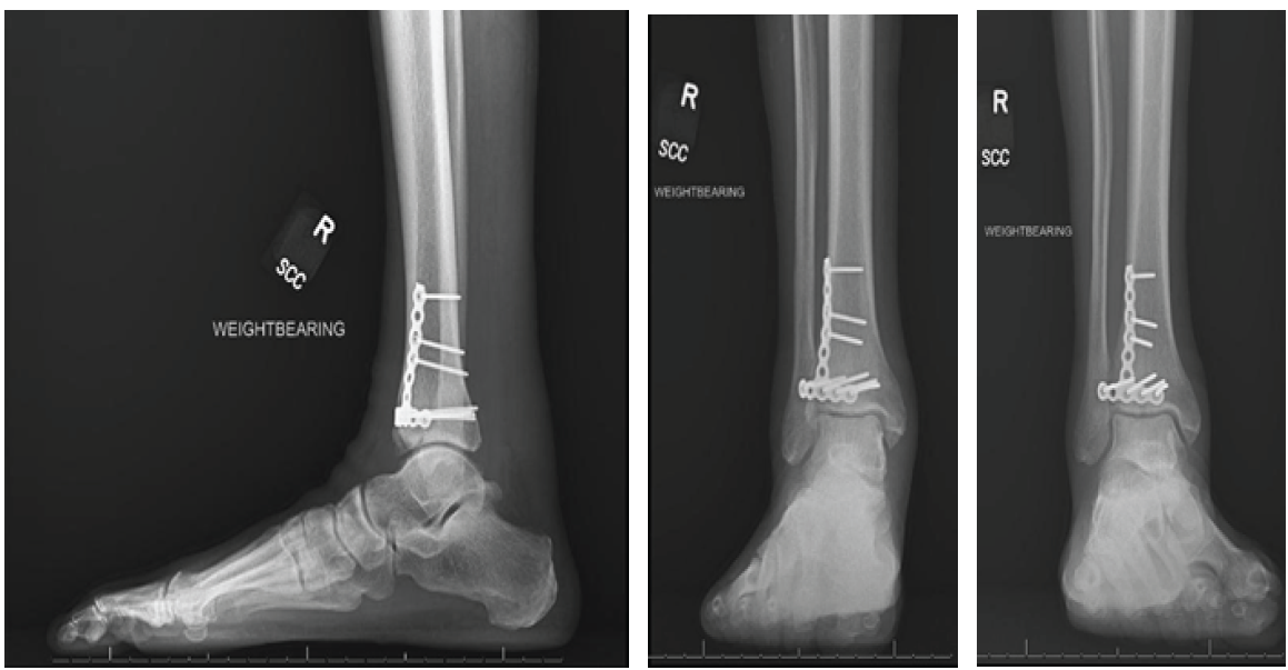
Figure 4: Four month post-operative radiographs: 4-month post-operative X-rays demonstrating a well-healing fracture and intact hardware.
Achilles tendon rupture and fracture are generally seen in isolation due to a traumatic event or a sports-related injury. However, in rare circumstances, these injuries can occur in combination. To date, there have been 13 case reports of Achilles tendon rupture with associated medial malleolar fracture, 2 case reports of Achilles rupture with concomitant lateral malleolar fracture, 1 case report of Achilles rupture with associated bimalleolar fracture, 2 cases of Achilles rupture with associated trimalleolar fracture, and 1 case report of Achilles tendon rupture with concomitant open distal tibia and fibula fracture. There have also been three reports of malleolar fractures associated with apparent SPR interruption and dislocation or subluxation of the peroneus tendons. To the best of the authors’ knowledge, our presented case is the first to describe an injury containing the combination of an Achilles tendon rupture with an associated pilon fracture of the tibia. The injuries resulting in isolated Achilles rupture or ankle fracture are often the result of a low-energy fall or rotational injury. However, the cases that have reported a combination of these injuries illustrate that the mechanism of injury is often due to higher-energy trauma such as a fall from height, motor vehicle collision, or snowboarding accident. These injuries could potentially be explained by an abrupt dorsiflexion force applied to the foot that is followed by axial loading of the hindfoot, with or without a rotational movement. The mechanism of action of the injury presented in this case is in alignment with previously reported mechanisms in the literature. Recovery from Achilles tendon repair can be significantly influenced by comorbid conditions such as PD, which may complicate both post-operative management and rehabilitation. PD is a progressive neurodegenerative disorder characterized by bradykinesia, rigidity, postural instability, and gait disturbances. These features can impair the patient’s ability to comply with weight-bearing restrictions, participate effectively in physical therapy, and maintain appropriate joint positioning during healing. Furthermore, muscle stiffness and abnormal resting tone may increase mechanical stress on the tendon repair site, potentially elevating the risk of rerupture or delayed healing. Cognitive challenges associated with PD can also hinder adherence to rehabilitation protocols, ultimately compromising functional outcomes. These factors were a concern during the post-operative course of our patient and contributed to a more cautious rehabilitation strategy. A missed or delayed diagnosis is common in the combination of the injuries described. Of the cases reported in the literature, eight Achilles ruptures were initially missed [14,15,16,18,21,25,26], and three fractures were initially missed [8,13,27]. In the report, we present that the fracture and the Achilles rupture were not diagnosed at the initial visit, and the correct diagnosis was not made until 2 weeks after the injury. This delay in diagnosis may have further complicated the recovery of the patient and contributed to the choice of a more cautious rehabilitation strategy. Moonen et al. attributed diagnostic errors in emergency departments to a lack of history taking and inadequate medical examinations [28,29,30], as was seen in our case. A delayed diagnosis can result in significant patient morbidity and may cause an increase in long-term consequences [4,5,6,7]. Given these adverse events caused by a delayed diagnosis, physicians should have an increased suspicion for the combination of the injuries described after a high-energy traumatic event to the ankle. If the mechanism of injury and physical examination are concerning, physicians should consider advanced imaging to ensure the proper diagnosis is made and prevent the delayed treatment of injuries.
This case highlights a rare but clinically significant combination of injuries—Achilles tendon rupture with an associated pilon fracture—that has not been previously described in the literature. The initial missed diagnosis underscores the diagnostic challenges posed by complex lower extremity trauma, particularly in elderly patients with comorbidities such as PD. A high index of suspicion is essential when evaluating patients with high-energy ankle injuries, especially when clinical findings suggest both soft tissue and osseous involvement. Timely imaging and thorough physical examination are critical to avoid delays in diagnosis and management, which can increase morbidity and complicate rehabilitation. By sharing this unique case, we aim to raise awareness of this injury pattern and reinforce the importance of a comprehensive diagnostic approach in similar clinical scenarios.
In patients presenting after high-energy ankle trauma, clinicians should maintain a high index of suspicion for concomitant Achilles tendon rupture even when a fracture is identified or initial radiographs appear negative. Early recognition through careful physical examination and advanced imaging can prevent missed diagnoses, optimize surgical planning, and improve functional outcomes.
[refernces_numbered]1. Lemme NJ, Li NY, DeFroda SF, Kleiner J, Owens BD. Epidemiology of Achilles tendon ruptures in the United States: Athletic and nonathletic injuries from 2012 to 2016. Orthop J Sports Med 2018;6(11):2325967118808238. doi:10.1177/2325967118808238.
2. Lyons JG, Berkay FB, Minhas A. Epidemiology of sports-related tendon ruptures presenting to emergency departments in the United States. Am J Sports Med 2024;52:3396-403.
3. Robertson GA, Wood AM, Aitken SA, Court Brown C. Epidemiology, management, and outcome of sport-related ankle fractures in a standard UK population. Foot Ankle Int 2014;35:1143-52.
4. He SK, Liao JP, Huang FG. Higher rate of postoperative complications in delayed Achilles tendon repair compared to early Achilles tendon repair: A meta-analysis. J Invest Surg 2022;35:157-63.
5. Maffulli N, Ziello S, Maisto G, Migliorini F, Oliva F. Local tendon transfers for chronic ruptures of the Achilles tendon: A systematic review. J Clin Med 2023;12:707.
6. Zelle BA, Johnson TR, Ryan JC, Martin CW, Cabot JH, Griffin LP, et al. Fate of the uninsured ankle fracture: Significant delays in treatment result in an increased risk of surgical site infection. J Orthop Trauma 2021;35:154-9.
7. Pilskog K, Gote TB, Odland HE, Fjeldsgaard KA, Dale H, Inderhaug E, et al. Association of delayed surgery for ankle fractures and patient-reported outcomes. Foot Ankle Int 2022;43:762-71.
8. Nakajima K, Taketomi S, Inui H, Nakamura K, Sanada T, Tanaka S. Missed medial malleolar fracture associated with Achilles tendon rupture: A case report and literature review. J Foot Ankle Surg 2016;55:169-72.
9. Elmajee M, Rafee A, Williams T. Ankle fracture associated with rupture of the Achilles tendon: Case report and review of the literature. J Foot Ankle Surg 2017;56:1320-2.
10. Assal M, Stern R, Peter R. Fracture of the ankle associated with rupture of the Achilles tendon: Case report and review of the literature. J Orthop Trauma 2002;16:358-61.
11. Lubin JW, Miller RA, Robinson BJ, Blevins FT. Achilles tendon rupture associated with ankle fracture. Am J Orthop (Belle Mead NJ) 2000;29:707-8.
12. Bowers M, Hunt KJ, Metzl J. High-energy Achilles tendon rupture with associated medial malleolus fracture and traumatic peroneal dislocation: A case report. Foot Ankle Spec 2021;14:438-44.
13. Pieper HG, Radas CB, Quack G, Krahl H. Mediomalleolar fracture combined with Achilles tendon rupture–a rare simultaneous injury of the ankle. Int J Sports Med 1998;19:68-70.
14. Martin JW, Thompson GH. Achilles tendon rupture. Occurrence with a closed ankle fracture. Clin Orthop Relat Res 1986;210:216-8.
15. Barron JL, Yocum LA. Unrecognized Achilles tendon rupture associated with ipsilateral medial malleolar fracture. Am J Sports Med 1993;21:629-31.
16. Lu J, Maruo Holledge M. Medial malleolus fracture of the ankle combined with rupture of the Achilles tendon. J Surg Case Rep 2016;2016:rjw062.
17. Pyle LH, Al-Gharib RW, Kissel EC. Lateral malleolar fracture with concurrent Achilles tendon rupture: A case report and literature review. Case Rep Emerg Med 2020;2020:6479140.
18. Sarrel K, Girard P. Gustilo IIIA open bimalleolar fracture dislocation of ankle with ipsilateral Achilles tendon rupture: A case report and review of literature. JBJS Case Connect 2021;11(2):e20.00435. doi:10.2106/JBJS.CC.20.00435.
19. Brueton RN, Javid KS. Trimalleolar fracture and associated Achilles tendon rupture: Ten year follow up of an unusual water-skiing injury. Trauma Case Rep 2018;17:33-8.
20. Jadib I, Veyrat G, Ionescu S, Belvisi B, Chirpaz-Cerbat JM. Trimalleolar fracture associated with subcutaneous rupture of the achilles tendon and traumatic peroneal dislocation: A case report and review of the literature. JBJS Case Connect 2024;14:e23.00564.
21. Hill GN, Barksfield R, Ramesh P. Achilles tendon rupture with open distal tibia and fibula fracture. Foot Ankle Spec 2011;4:171-4.
22. Kopp F. Traumatic peroneal dislocation with medial malleolus fracture: A case report. Foot Ankle Int 2008;29:756-8.
23. Malik AK, Mehta S, Solan M. Significance of flake fracture with medial malleolar fracture. Foot Ankle Int 2013;34:749-52.
24. Vosoughi AR, Erfani MA. Concomitant traumatic peroneal tendon dislocation and medial malleolus fracture: A case report. Malays Orthop J 2018;12:57-9.
25. Garneti N, Muralikuttan K, Shenolikar A. Ankle fracture associated with an Achilles tendon rupture: A rare injury. Foot Ankle Surg 2005;11:219-22.
26. Mattos E Dinato MC, De Farias Freitas M, D’Elia CO, Bitar AC, Rodrigues Gonçalves FM. Acute calcaneus tendon rupture associated with ipsilateral malleolar fracture: Case report and literature review. J Foot Ankle Surg 2010;49:565.e1-4.
27. Fontecilla N, Hube M, Bastías G, Fuentes P, Arancibia M. Acute Achilles tendon rupture associated with medial malleolar fracture: Is it still a rare injury? Case report and review of the literature. Int J Foot Ankle 2018;2:017.
28. Moonen PJ, Mercelina L, Boer W, Fret T. Diagnostic error in the emergency department: Follow up of patients with minor trauma in the outpatient clinic. Scand J Trauma Resusc Emerg Med 2017;25:13.
29. Maffulli N, Richards PJ. Subcutaneous rupture of the Achilles tendon and ipsilateral fracture of the medial malleolus. BMC Musculoskelet Disord. 2006;7:59. doi:10.1186/1471-2474-7-59.
30. Türkmensoy F, Türkmen I, Turhan Y, Özkan K, Akçal MA. Achilles Tendon Rupture with Isolated Medial Malleolar Fracture in Ipsilateral Ankle: A Case Report. Open Journal of Orthopedics. 2013;3:224-226. doi:10.4236/ojo.2013.35042.[/refernces_numbered]





Related Articles in Journal of Orthopaedic Case Reports
 June 1, 2025 Management of Ipsilateral Pilon with Calcaneal Fractures: A Rare Case Report
 February 1, 2025 Non-union of a Tibial Plafond Fracture in a COVID-Positive Patient: A Case Report
 November 23, 2017 Rotatory Subluxation and Facet Deformity in the Atlanto-occipital Joint in Patients with Chronic Atlantoaxial Rotatory Fixation: Two Case Reports
 November 1, 2025 Superior Capsular Reconstruction Using Biceps Tendon Autograft with Novel Biceps Tenodesis Arthroscopic Fixation: A Case Report