

Primary tuberculous osteomyelitis of the proximal tibia can closely mimic Brodie's abscess or chronic pyogenic osteomyelitis, making early biopsy with CBNAAT and histopathology essential for accurate diagnosis and timely treatment, even in immunocompetent patients.
Dr. Anirudh Dwajan, Department of Orthopaedics, All India Institute of Medical Sciences, Bilaspur, Himachal Pradesh, India. E-mail: anirudhdwajan@gmail.com
Abstract
Introduction: Tuberculous involvement of long bones is an uncommon form of extrapulmonary tuberculosis (TB) and frequently poses a diagnostic dilemma because of its indolent presentation and non-specific imaging features. Tibial tuberculous osteomyelitis is particularly rare and may closely mimic subacute pyogenic osteomyelitis or Brodie’s abscess, especially in immunocompetent individuals, leading to delayed diagnosis.
Case Report: A 70-year-old immunocompetent male presented with a short duration of pain and swelling over the proximal tibia without constitutional symptoms. Radiographs showed a lytic metaphyseal lesion with cortical thinning, while magnetic resonance imaging revealed an intramedullary abscess cavity with surrounding marrow edema and minimal soft-tissue extension, suggestive of subacute osteomyelitis. Routine bacterial cultures were negative. Intraoperative aspirate tested positive for Mycobacterium TB on cartridge-based nucleic acid amplification test, confirming tuberculous osteomyelitis. The patient underwent intramedullary debridement, abscess drainage, and dead-space management with antibiotic-loaded calcium sulfate beads, followed by initiation of standard multidrug antitubercular therapy. Post-operative radiographs demonstrated adequate lesion clearance and maintained alignment, with progressive clinical improvement on follow-up.
Conclusion: Tibial tuberculous osteomyelitis should be considered in the differential diagnosis of metaphyseal lytic lesions resembling Brodie’s abscess, even in immunocompetent patients with short-duration symptoms. Early biopsy and molecular diagnostics such as cartridge-based nucleic acid amplification tests are crucial for timely diagnosis. Combined surgical debridement and antitubercular therapy resulted in a favorable outcome in this patient. Further studies are required to determine the reproducibility of this approach.
Keywords: Tuberculous osteomyelitis, tibia, Brodie’s abscess, cartridge-based nucleic acid amplification tests, intramedullary abscess, extrapulmonary tuberculosis.

Tuberculosis (TB) continues to impose a substantial global health burden, with musculoskeletal involvement representing a small but clinically significant subset of cases. Although recent national and regional reports indicate improved detection of extrapulmonary TB, osteoarticular involvement still accounts for only 1–3% of all TB cases and remains a diagnostic challenge due to its slow, indolent evolution, and non-specific early symptoms [1,2]. Contemporary data from India and other endemic regions show an increasing proportion of long-bone TB among musculoskeletal presentations, though isolated tibial involvement without pulmonary disease remains distinctly uncommon [3].
Diagnosing tuberculous osteomyelitis is often difficult because clinical findings such as localized pain and swelling may be indistinguishable from low-grade pyogenic osteomyelitis or subacute presentations like Brodie’s abscess. This overlap frequently contributes to delayed recognition, especially in immunocompetent adults who lack constitutional symptoms and exhibit sterile bacterial cultures despite progressive bone destruction [4]. Recent imaging reviews and institutional case series emphasize that cortical erosions, marrow edema, and periosseous abscesses are common but non-specific features that mimic a spectrum of subacute infective or neoplastic bone lesions [1,3].
The advent of rapid molecular diagnostics has significantly improved the accuracy and timeliness of identifying skeletal TB. Cartridge-based nucleic acid amplification tests (CBNAAT) have shown high sensitivity and are now recommended as frontline tests to identify tuberculous osteomyelitis [5,6]. Despite these advances, atypical presentations involving the long bones continue to create diagnostic uncertainty, particularly in older adults.
Here, we describe a rare case of isolated proximal tibial tuberculous osteomyelitis in an immunocompetent elderly patient whose clinical and radiological features closely resembled subacute pyogenic osteomyelitis.
A 70-year-old man from a rural background presented with a 3-week history of gradually increasing swelling over the anteromedial aspect of his left upper leg, accompanied by deep, localized pain for 2 weeks that limited walking and routine activities. He denied trauma, prior surgeries, chronic systemic illness, anti-tuberculosis treatment intake, or TB exposure. His only significant history was cataract surgery.
On admission, he was afebrile and hemodynamically stable. Local examination revealed a firm, tender 5 × 6 cm swelling over the anteromedial proximal tibia, warm to touch and non-fluctuant, without any sinus or overlying skin changes. Knee and ankle movements were preserved, and neurovascular status was intact.
Plain radiographs of the left knee and upper tibia showed subtle cortical irregularity and mild periosteal reaction along the anteromedial cortex of the proximal tibia. Although the findings were subtle, a faint lucent area corresponding to the clinically tender zone suggested an underlying subacute infective process. The joint spaces were maintained, and the fibula appeared normal (Fig. 1).
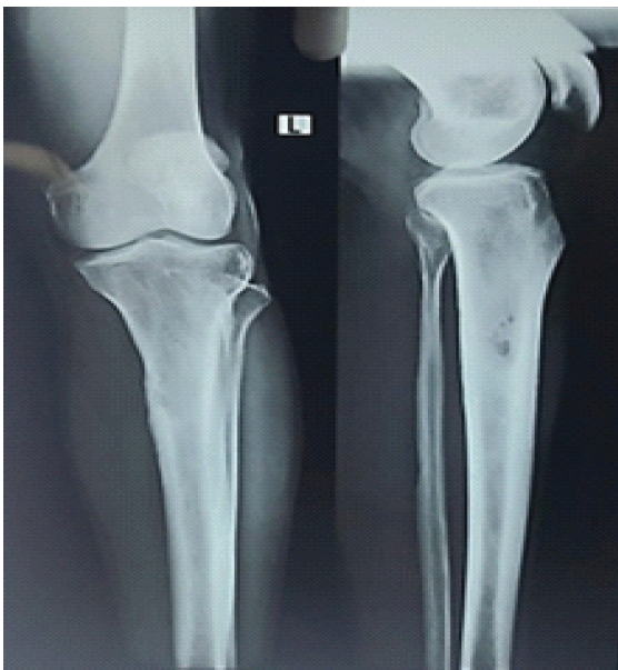
Figure 1: Preoperative anteroposterior and lateral radiographs of the left tibia showing a well-defined lytic lesion in the proximal tibial metaphysis with cortical thinning and early breach, suggestive of a subacute osteomyelitic process.
Laboratory evaluation revealed markedly elevated inflammatory markers (erythrocyte sedimentation rate [ESR] 54 mm/h; C-reactive protein [CRP] 151 mg/L) with normal total leukocyte count and preserved renal and hepatic profiles.
A contrast-enhanced computed tomography scan demonstrated a focal cortical erosion on the anteromedial aspect of the proximal tibia with surrounding periosteal reaction, extending into the adjacent subcutaneous soft tissues. There was associated soft-tissue edema tracking along the proximal one-third of the leg. The overall appearance was consistent with evolving osteomyelitis (Figs. 2 and 3).
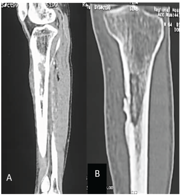
Figure 2: Preoperative computed tomography scan of the left tibia. (a)
Saggital reconstruction showing a lytic intramedullary cavity in the proximal
tibial metaphysis with cortical thinning and early breach. (b) Coronal
reconstruction demonstrating longitudinal extension of the lesion with
surrounding sclerosis and subperiosteal reaction. Findings were suggestive of a
chronic osteomyelitic focus.
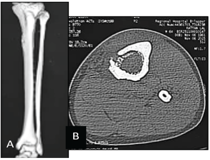
Figure 3: Preoperative computed tomography (CT) assessment of the left tibia. (a) Three-dimensional CT reconstruction demonstrating a well-defined defect in the proximal tibial metaphysis with cortical thinning. (b) Axial CT image revealing an intramedullary cavity with cortical erosion and adjacent periosteal reaction, consistent with a chronic osteomyelitic lesion.
Further characterization using magnetic resonance imaging (MRI) revealed diffuse marrow edema in the proximal tibial diaphysis with a well-defined intramedullary abscess cavity measuring approximately 2.1 × 1.4 cm. The intramedullary component exhibited T1 hypointensity and T2/short tau inversion recovery hyperintensity and was associated with a periosseous abscess on the anteromedial surface, extending into the subcutaneous plane. There was associated edema in the tibialis anterior and extensor hallucis longus muscles, though the joint surfaces and neurovascular bundles were preserved (Fig. 4).
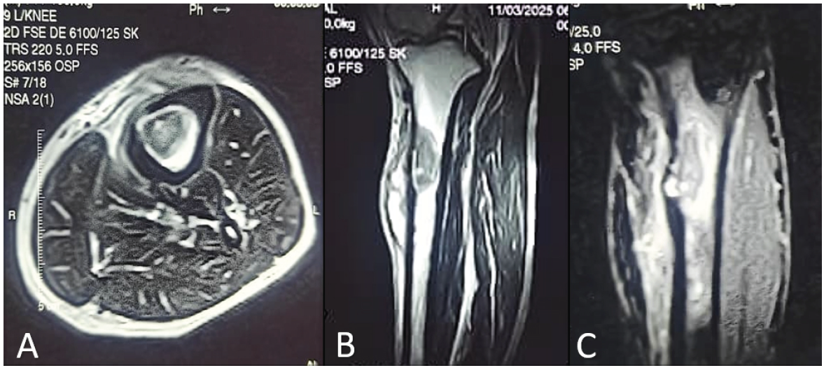
Figure 4: Preoperative magnetic resonance imaging of the left tibia. (a) Axial T2-weighted image showing a hyperintense intramedullary abscess in the proximal tibia with surrounding marrow edema. (b) Coronal sequence demonstrating cortical thinning and an anteromedial cortical breach. (c) Sagittal sequence revealing longitudinal intramedullary extension with associated soft-tissue inflammatory changes. The overall appearance initially suggested subacute osteomyelitis or Brodie’s abscess.
The findings favored a subacute infective etiology, with the differential including chronic pyogenic osteomyelitis, atypical mycobacterial infection, or an indolent abscess such as Brodie’s.
Given the progression and imaging characteristics, surgical exploration was undertaken. Intramedullary abscess drainage and thorough debridement were performed with placement of antibiotic-impregnated Stimulan beads.
Intraoperatively, thick purulent material was noted tracking through the cortical breach into adjacent soft tissues, matching the imaging predictions.
Bacterial and fungal cultures showed no growth. However, CBNAAT of the pus confirmed Mycobacterium tuberculosis (M. TB). establishing a diagnosis of tuberculous osteomyelitis of the proximal tibia – a rare, extrapulmonary manifestation often mistaken for chronic pyogenic infection in early stages. Histopathological examination of the curetted tissue showed granulomatous inflammation with epithelioid cells and Langhans-type giant cells, a pattern consistent with tuberculous osteomyelitis and supportive of the CBNAAT-positive finding.
The absence of systemic symptoms, lack of a draining sinus, and relatively short symptom duration made this an atypical and diagnostically challenging presentation.
The patient had an uneventful recovery, with a healthy postoperative wound and drain removal by day 4. He was commenced on standard antitubercular therapy and discharged with instructions for non-weight-bearing ambulation, limb elevation, nutritional supplementation, and structured follow-up. The patient was followed up for 6 months. Serial inflammatory markers showed progressive improvement, with ESR decreasing from 54 mm/h preoperatively to 22 mm/h at 3 months and 10 mm/h at 6 months and CRP reducing from 151 mg/L to 10 mg/L at 3 months and 2 mg/L at final follow-up. At 6 months, radiographs showed sclerosis and reduction of the intramedullary cavity with no new cortical destruction (Fig. 5). Clinically, the wound had healed well without discharge, and the patient was able to ambulate without support (Fig. 6).
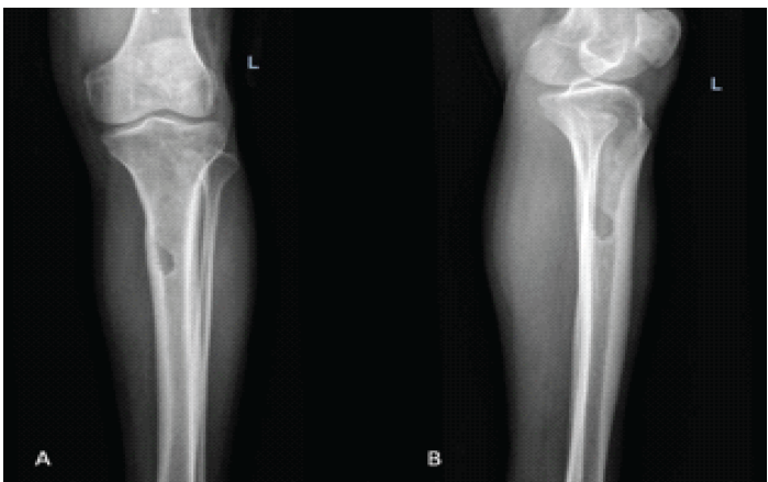
Figure 5: Follow-up radiographs (AP and lateral views) at 6 months showing interval sclerosis and reduction in the intramedullary cavity, with no evidence of progression or cortical destruction.
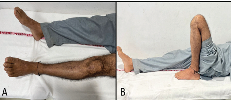
Figure 6: Clinical photographs at 6 months follow-up. (a) Healed surgical site over the proximal tibia without swelling or sinus formation. (b) Functional outcome demonstrating preserved knee range of motion.
Tuberculous involvement of long bones is uncommon and often presents with non-specific symptoms, leading to diagnostic delay. In immunocompetent adults, the absence of constitutional features and sterile cultures may further obscure the diagnosis [1,2,3]. Our patient’s short symptom duration, preserved mobility, and unremarkable constitutional profile exemplify this typical yet misleading presentation.
Radiologically, the proximal tibia presents a diagnostic challenge in subacute infections. Early tuberculous osteomyelitis may exhibit imaging features that overlap significantly with Brodie’s abscess and low-grade pyogenic osteomyelitis, including intramedullary cavities, cortical breaches, and surrounding marrow edema. Fine needle aspiration cytology-based analyses and small clinical series have repeatedly highlighted this overlap, stressing that radiology alone is insufficient to differentiate the entities in early stages [4]. The resemblance between these pathologies explains why tuberculous involvement is frequently mistaken for indolent pyogenic disease or even benign bone tumors.
Coinfection involving M. TB and pyogenic bacteria in bone is an even rarer phenomenon. While isolated pulmonary and soft-tissue coinfections have been recorded, true mixed infection within bone has only been described in highly unusual cases, such as coexistent Streptococcus agalactiae with tibial TB osteomyelitis [7]. These rare reports demonstrate how secondary bacterial seeding of a partially necrotic, tuberculous focus can accelerate symptoms and misguide clinical decision-making. Although our case did not yield bacterial growth, the radiological suspicion and clinical evolution warranted careful exclusion of this possibility.
Accurate diagnosis hinges on obtaining tissue and integrating multiple diagnostic modalities. Even in experienced hands, conventional smear microscopy and culture may fail to detect M. TB in skeletal lesions due to the paucibacillary milieu [4]. With the advent of CBNAAT, clinicians now possess a rapid and reliable tool that enhances diagnostic accuracy in extrapulmonary TB [5]. Several recent investigations – including large-scale evaluations of bone and joint TB – have reaffirmed CBNAAT’s advantage in detecting mycobacterial DNA when smear and culture are inconclusive [6]. The decisive CBNAAT positivity in our patient aligns with these findings, demonstrating the indispensable role of molecular diagnostics in ambiguous skeletal infections.
The diagnostic landscape is further complicated by case reports highlighting the protean nature of tibial TB. Sari et al. [8] and Kwan et al. [9] described presentations nearly indistinguishable from subacute osteomyelitis, with some cases even progressing to pathological fractures before TB was suspected. Mayo et al. [10] similarly reported distal tibial involvement initially mistaken for neoplastic pathology. The clinical spectrum of
osteoarticular TB is broad and often deceptive, with presentations ranging from indolent metaphyseal lesions to unusual multifocal or bilateral involvement. Ranjan et al. reported a case of bilateral tubercular dactylitis in an adult, highlighting how even common forms of TB may present in unexpected locations and mimic other chronic inflammatory or
infective conditions [11]. Such reports underscore the variability of musculoskeletal TB and reinforce the need to consider TB in any atypical or non-resolving bone lesion in endemic regions.
Management of tuberculous osteomyelitis traditionally focuses on prolonged antitubercular therapy, yet surgery retains an important role when abscesses, necrotic bone, or structural compromise is present. Surgical debridement optimizes drug penetration, reduces bacterial load, and prevents deformity or joint extension. The addition of biodegradable calcium sulfate carriers, such as Stimulan®, offers a modern adjunct to traditional debridement by providing dead-space management, delivering high local antibiotic concentrations, and supporting early granulation. Although its application is well established in pyogenic osteomyelitis, emerging case experiences – including ours – suggest that these carriers may also be beneficial in tuberculous lesions by improving local milieu and reducing the risk of recurrence.
In summary, this case underscores the multifaceted challenge of diagnosing tibial TB in immunocompetent adults. Subtle symptoms and nonspecific imaging often delay diagnosis. CBNAAT, histopathology, and early biopsy remain indispensable tools. As illustrated by the expanding clinical literature, timely recognition and combined surgical-medical management continue to offer excellent outcomes, even in anatomically complex or diagnostically deceptive presentations.
Limitations
This report has several limitations. As a single-patient case report, the findings cannot be generalized to all patients with skeletal TB. Follow-up was limited to six months and therefore does not permit assessment of long-term recurrence, bone remodeling, or late complications. Histopathology demonstrated granulomatous inflammation consistent with TB, but detailed characterization of caseous necrosis and bacillary burden was beyond the scope of this report. Disease resolution was evaluated clinically and radiographically without serial MRI. Standardized patient-reported and functional outcome measures were out of the scope of this report. Because surgical debridement, local calcium sulfate bead implantation, and antitubercular therapy were administered concurrently, the independent contribution of each intervention to clinical recovery cannot be determined. Finally, this report represents a rare clinical presentation managed at a single institution and cannot establish the superiority of combined surgical and medical treatment over medical therapy alone. Larger prospective studies are required to validate these findings.
Tuberculous osteomyelitis of the tibia remains an uncommon and diagnostically challenging entity, especially in immunocompetent adults presenting with short-duration symptoms and nonspecific imaging findings. As illustrated in this case, the close radiological resemblance to subacute pyogenic osteomyelitis or Brodie’s abscess can easily mislead early clinical evaluation. Timely diagnosis requires a high index of suspicion, prompt biopsy, and incorporation of molecular tools such as CBNAAT, particularly when routine cultures are negative. Surgical debridement combined with standard antitubercular therapy continues to offer excellent clinical recovery. Although this case demonstrates a favorable outcome with early surgical debridement and antitubercular therapy, these findings should be interpreted cautiously because they are based on a single patient. Larger studies with longer follow-up are required to determine the optimal management strategy for isolated tibial tuberculous osteomyelitis.
Tuberculous osteomyelitis should be considered in the differential diagnosis of metaphyseal lytic lesions resembling Brodie’s abscess, particularly in tuberculosis-endemic regions. Early tissue biopsy with CBNAAT and histopathology enables prompt diagnosis and timely initiation of appropriate treatment.
References
- 1. Kumar Si V, Hosthota A, D V, Hd B, S Pai H. Skeletal Tuberculosis: Clinical and Radiological Profile, Management, and Outcomes in a Retrospective Study. Cureus. 2025;17(11):e97948. Published 2025 Nov 27. doi:10.7759/cureus.97948</p> [Google Scholar] [PubMed] [CrossRef]
- 2. <p>2. Gopalaswamy R, Dusthackeer VNA, Kannayan S, Subbian S. Extrapulmonary tuberculosis—an update on the diagnosis, treatment and drug resistance. J Resp. 2021;1(2):141–164. doi:10.3390/jor1020015 [Google Scholar] [PubMed] [CrossRef]
- 3. Hazra D, Shaji V R, Dhall A, Rao AP, Bhat AK, Chawla K. A ten-year experience of musculoskeletal tuberculosis at a tertiary care hospital in South India. J Orthop. 2024;56:92-97. Published 2024 May 3. doi:10.1016/j.jor.2024.05.001 [Google Scholar] [PubMed] [CrossRef]
- 4. Dey B, Nigam JS, Bharti JN, Singh A, Nair V. Osteoarticular tuberculosis: A series of six cases diagnosed on fine-needle aspiration cytology. Cytojournal. 2022 Mar 4;19:11. doi: 10.25259/Cytojournal_22_2021. PMID: 35510112; PMCID: PMC9063557. [Google Scholar] [PubMed] [CrossRef]
- 5. Kashyap B, Sarkar K, et al. Evaluating diagnostic performance of extrapulmonary tuberculosis using CBNAAT: a retrospective cross-sectional study. EMJ Microbiol Infect Dis. 2024;9(4):89–96. doi:10.33590/emj/KBFU2758 [Google Scholar] [PubMed] [CrossRef]
- 6. Yuan Y, Li P, Qiu X, et al. Bone and joint tuberculosis: clinical manifestation, diagnostic techniques and drug resistance analysis. Front Cell Infect Microbiol. 2025;15:1687906. Published 2025 Nov 21. doi:10.3389/fcimb.2025.1687906 [Google Scholar] [PubMed] [CrossRef]
- 7. Ajit A, Jacob B, Warrier A. Coinfection of Streptococcus agalactiae and Tuberculous Osteomyelitis of Tibia Mimicking Brodie’s Abscess in an Immunocompetent Adult: A Case Report. J Orthop Case Rep. 2023;13(7):95-98. doi:10.13107/jocr.2023.v13.i07.3768 [Google Scholar] [PubMed] [CrossRef]
- 8. Sari A, Dinçel YM, Erdogdu IH, Sayıner HS, Agir I, Çetin MÜ. Tuberculosis osteomyelitis of the tibia mimicking Brodie abscess: A case report and review of the literature. SAGE Open Med Case Rep. 2019;7:2050313X19869455. Published 2019 Aug 8. doi:10.1177/2050313X19869455 [Google Scholar] [PubMed] [CrossRef]
- 9. Kwan CK, Tso SWS, Liu WH, et al. Primary diaphyseal tuberculosis of the tibia, presenting as a pathological fracture – A case report. Trauma Case Rep. 2025;57:101161. Published 2025 Apr 15. doi:10.1016/j.tcr.2025.101161 [Google Scholar] [PubMed] [CrossRef]
- 10. Mayo M, Osman OH, Alsakkar MB, Barrieh R, Abdulkareem A, Saheel M. Tuberculous osteomyelitis of the distal tibia: a rare case report from Syria. Int J Surg Case Rep. 2025;132:111475. doi:10.1016/j.ijscr.2025.111475 [Google Scholar] [PubMed] [CrossRef]
- 11. Ranjan R, Goel L, Sud A, Sinha A, Tulika, Kumar R. Bilateral Tubercular Dactylitis: Unusual presentation of an usual disease. Indian J Tuberc 2019;66:346-52. [Google Scholar] [PubMed]






Related Articles in Journal of Orthopaedic Case Reports
 February 1, 2026 When MRSA Misleads: Tuberculous Osteomyelitis of the Anterior Pelvic Bones
 October 1, 2024 Brodie’s Abscess of the Ankle Presenting as a Tumor: A Summary of Five Cases
 September 1, 2026 Limb Salvage for Entire Tibial Ewing Sarcoma in a 4-year-Old Child Using Extracorporeal Irradiation and Free Flap Reconstruction: A Case Report and Surgical Technique
 August 1, 2026 Plastic Bowing of the Pediatric Tibia is Extraordinarily Rare and Not Represented in the Literature, the Unicorn of Pediatric Fractures: A Case Report