

Metallosis although an uncommon occurrence after total hip arthroplasty, must be considered when patients present with pain, eccentric femoral head migration, osteolysis or suspected implant failure during management to aid in proper planning, especially in revision scenarios.
Dr. Anudeep Manne, Department of Orthopaedics, ESIC Medical College Hospital, Hyderabad - 500038, Telangana, India. E-mail: dr.anudeepmanne@gmail.com
Abstract
Introduction: Total hip arthroplasty (THA) is a common procedure for treating hip osteoarthritis but carries potential complications, including implant loosening and wear. Metallosis is a rare complication caused by metal debris from prosthetic components, particularly in metal-on-metal implants, leading to adverse local tissue reactions such as osteolysis, soft-tissue necrosis, and pseudotumor formation.
Case Report: A 45-year-old male presented with progressive right hip pain, difficulty walking, and shortening of the affected limb following trauma. He had undergone right total hip arthroplasty 10 years previously. On evaluation of the patient, the systemic parameters were under normal limits. The radiographs showed proximal and medial migration of the metal femoral head, giving an impression of subluxation of the metal femoral head. The patient was planned for a revision THA suspecting an osteolysis. Intraoperatively, blackish pigmentation of the surrounding soft tissue was noted along with hypertrophy of the capsule. The metal femoral head was intact and eroded into the polyethylene liner and acetabular cup. This showed that extensive Metallosis secondary to catastrophic polyethylene liner wear and acetabular shell failure could have caused the failure of the previous surgery, contrary to the earlier expected osteolysis as the causative phenomenon. This diagnosis was confirmed by the histopathological examination of the tissues slides obtained intraoperatively via biopsy of the stained tissues. Post-surgery, the patient regained his function, and remained pain-free with stable implants at the follow-up.
Conclusion: Metallosis must be a diagnosis kept in mind in cases where the patient presents with problems post-THA particularly when radiographs demonstrate eccentric femoral head migration, polyethylene wear, osteolysis, or component failure. Although initially asymptomatic, the presence of metal ions in the tissue and blood can cause soft-tissue damage locally and systemically as well. Thus, early diagnosis and planning can help in the proper management and are crucial to limit progressive soft-tissue destruction, bone loss, and potential deleterious effects caused by metal ions in the body.
Keywords: Metallosis, total hip arthroplasty, revision total hip arthroplasty.

Total hip arthroplasty (THA) is a very common procedure to treat hip osteoarthritis, with over one million patients undergoing the procedure every year [1]. Total hip replacement, like any surgery, has potential complications. These include infection, blood loss, nerve or blood vessel damage, loosening of the implant, dislocation, fractures, and wear. Metallosis is one of the rare complications of THA caused due to debris released from arthroplasty components secondary to implant wear [2,3,4]. There has been an escalating acknowledgment of both local and systemic adverse events linked to the discharge of metal ions and nanoparticles from arthroplasty components [5]. This increased vigilance originally arose after the implementation of large-diameter metal-on-metal (MoM) THA and hip resurfacing devices encompassing a metal-on-metal articulation. The majority of these failures are attributed to the production of metal debris resulting in adverse local tissue reactions which includes extensive tissue necrosis, osteolysis, and large sterile hip effusions, as well as periprosthetic solid and cystic masses known as pseudotumor [1,6,7,8,9]. We present a case report in which a patient who underwent THA a decade ago subsequently presented with hip pain accompanied by signs of loosening around the acetabular component. there were also features indicative of Metallosis observed during the intraoperative evaluation. The condition was addressed through a meticulous approach involving debridement, biopsy, and revision THA.
A 45-year-old male underwent primary right THA for osteonecrosis of the femoral head in the year 2014. Six months prior to the presentation, the patient had sustained trauma to the operated hip following a road traffic accident. Following the injury, the patient developed progressively worsening pain and difficulty in walking. At presentation, he had significant pain and a severe limp. Examination revealed a painful range of motion and limb length discrepancy of 2 cm. A 10 cm scar was seen on the posterolateral aspect of the right gluteal region, suggesting a posterior approach for the primary surgery. Laboratory investigations including inflammatory markers were within normal limits, making infection less likely. Hip radiographs suggested proximal migration of the metallic femoral head in the acetabulum with osteolysis around the acetabular component (Fig. 1). Considering a possibility of aseptic implant failure, the patient was planned for a revision hip arthroplasty procedure.
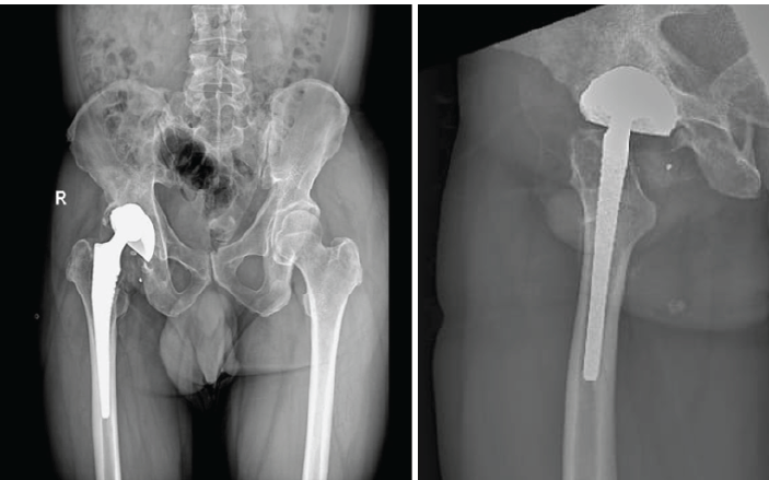
Figure 1: Anteroposterior radiograph of the pelvis with both hips and lateral radiograph of the right hip demonstrating eccentric positioning with proximal and medial migration of the femoral head, together with osteolysis around the acetabular component.
The patient underwent revision THA in the lateral decubitus position through the previous incision to approach the hip joint. Meticulous dissection of the deeper planes unveiled a notable blackish discoloration of the soft tissues, encompassing the gluteus maximus and external rotators, accompanied by gross hypertrophy of the capsule (Fig. 2a and b).
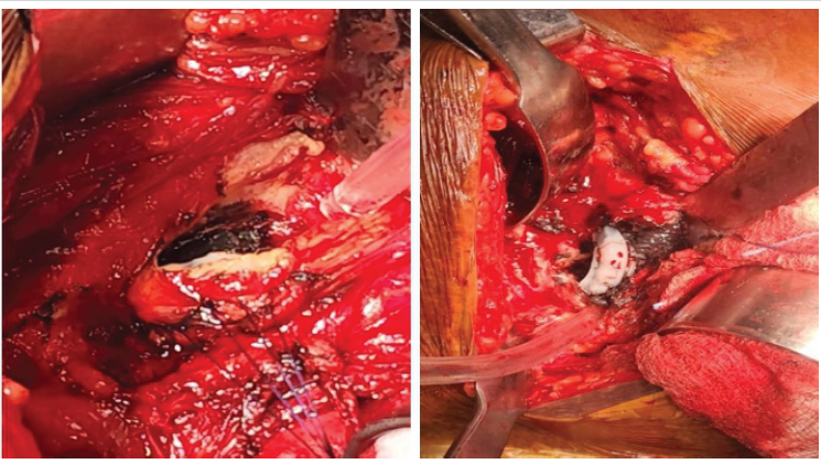
Figure 2: (a and b) Intraoperative images demonstrating diffuse black pigmentation of the hip capsule and surrounding soft tissues.
There was no gross damage to the abductors noted. The pigmented and abnormal tissues were meticulously debrided a sample was sent for histopathological evaluation and culture sensitivity testing. Following the dislocation of the femoral component, the acetabular component was visualized and it revealed significant polyethylene liner wear over the superolateral aspect. After removal of the polyethylene liner, the acetabular cup revealed circumferential aseptic loosening with catastrophic erosion over the superolateral aspect.(Fig. 3a, b, c). The intact metallic head had penetrated the polyethylene liner and protruded beyond the confines of the acetabular cup. Head explantation was completed, and the trunnion was carefully examined; no signs of wear were observed. The femoral stem was well fixed and appropriately positioned; therefore, it was retained.
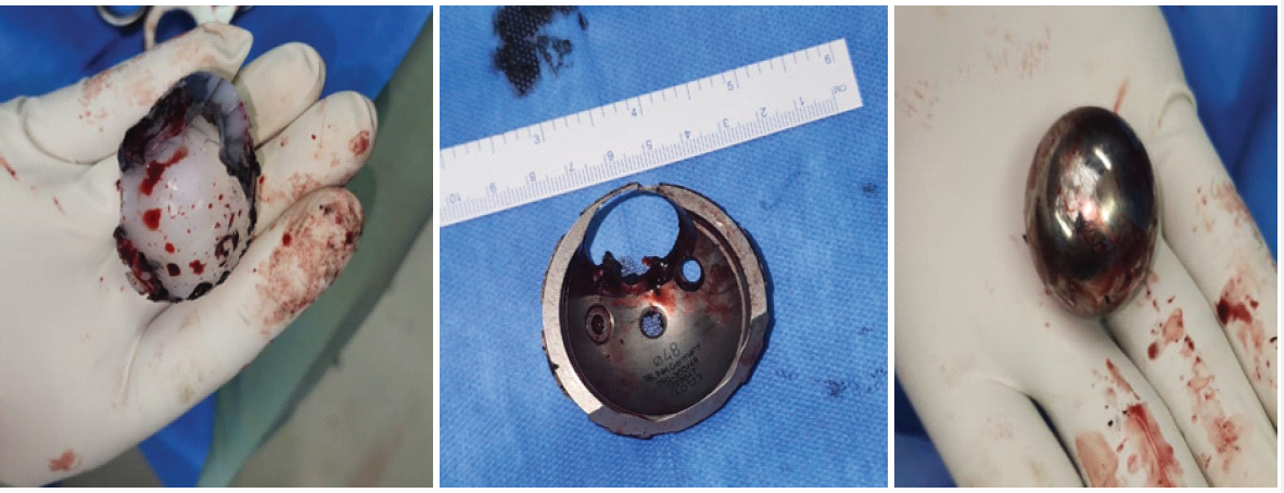
Figure 3: (a, b, and c) Intraoperative images demonstrating catastrophic polyethylene liner wear, acetabular shell erosion, and intact cobalt- chromium metallic femoral head after explantation.
A 52-mm size acetabular cup was implanted and fixed with two 6.5-mm titanium screws to increase fixation stability. Liner of size 52/36 and a 36-mm cobalt-chromium femoral head were used (Fig. 4).
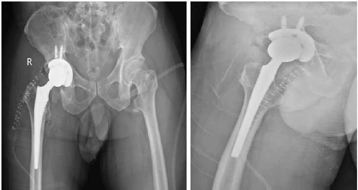
Figure 4: Post-operative anteroposterior and lateral radiographs demonstrating the revised acetabular component and total hip arthroplasty construct.
The histopathology report revealed coarse grey-black pigment within the synovial lining, histiocytes, and foreign-body giant cells, suggestive of metallosis (Fig. 5 and 6). Tissue cultures revealed no growth. At the 12-month follow-up, the patient was pain-free, ambulatory without support, and demonstrated stable implant positioning without radiographic evidence of loosening.
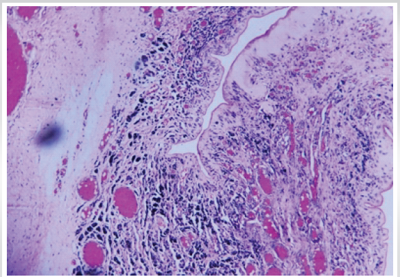
Figure 5: Hematoxylin and Eosin-stained histology image demonstrating coarse grey-black pigment in synovial lining and foreign-body giant cells.
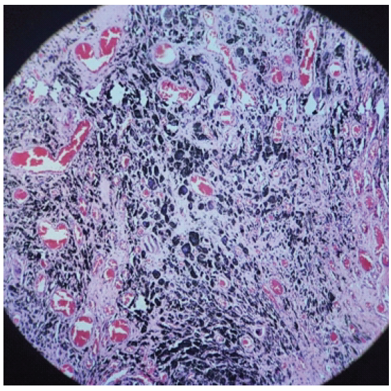
Figure 6: Hematoxylin and Eosin-stained histology image demonstrating the presence of grey-black pigment-laden histiocytes and foreign-body giant cells, consistent with metallosis.
Metallosis may occur when catastrophic polyethylene liner wear, fracture, or dissociation permits direct articulation between the metallic femoral head and the acetabular shell. This abnormal contact generates metallic and polyethylene debris, which may initiate macrophage activation, foreign-body giant-cell reaction, synovial hypertrophy, osteolysis, soft-tissue necrosis, and progressive aseptic loosening [1,5,10,13]. Burger and Nowakowski reported an massive eccentric acetabular wear following complete polyethylene liner abrasion and emphasized the potential for extensive component destruction and increased technical difficulty during revision surgery [13].
Metallosis is most commonly associated with metal-on-metal bearings. It has also been reported in metal-on-polyethylene and ceramic-bearing constructs when abnormal contact occurs between metallic components [5,10,12-15]. Previous comparative studies of different bearing surfaces show the effect of bearing materials and polyethylene wear properties on implant longevity [2–4]. Hjorth et al. reported a higher prevalence of mixed or solid pseudotumors in metal-on-polyethylene THA than in metal-on-metal THA and hip-resurfacing arthroplasty, indicating that adverse local tissue reactions are not confined to metal-on-metal articulations [12]. Malizos et al. described complete polyethylene liner and metallic shell destruction with protrusion of an intact ceramic head through the acetabular shell [14], whereas Needham et al. reported catastrophic failure of ceramic-on-polyethylene THA secondary to severe polyethylene wear [15]. Potential contributing factors include polyethylene thickness, failure of the locking mechanism, component malposition, impingement, edge loading, third-body wear, backside wear at the liner-shell interface, and increased acetabular inclination leading to accelerated wear [14,15,17,18].
Although metallosis gained greater recognition following the widespread use of metal-on-metal hip arthroplasty, it had previously been described in association with metallic fracture fixation devices, demonstrating that prolonged exposure to metallic wear debris can produce characteristic inflammatory tissue reactions [11]. Long-term survival of hard-on-hard bearing surfaces depends on bearing material, implant design, manufacturing technique, and component positioning rather than bearing surface alone [19]. Furthermore, adverse local tissue reactions and pseudotumor formation have been reported in ceramic-on-metal arthroplasties, which suggests that these complications are not unique to conventional metal-on-metal implants [20]. Walter et al. pointed out that impingement, edge loading, third-body particles, and loss of lubrication can also contribute to implant failure, all leading to abnormal wear patterns [21].
Chang et al defined three grades of metallosis: Grade 1, mild black spotting; Grade 2, geographically distributed black staining of the soft tissues; Grade 3, extensive black staining involving soft tissues and bone [16]. The diffuse but geographically distributed pigmentation observed intraoperatively in the present case was consistent with Grade 2 metallosis.
The clinical presentation of metallosis is variable and may include pain, limp, restricted movement, limb shortening, instability, or progressive loss of function. Reported radiographic findings include eccentric positioning or migration of the femoral head, polyethylene wear, periprosthetic osteolysis, component loosening, the cloud sign, and the bubble sign [10,24,25]. In the present case, proximal and medial migration of the femoral head with acetabular osteolysis strongly suggested catastrophic liner and acetabular component failure before surgery.
Macrophage activation due to metal debris stimulates the release of inflammatory cytokines that induce histiocytosis, fibrosis, osteolysis, soft-tissue necrosis and aseptic loosening [1,22]. In the current case report the patient did not have any systemic manifestations, but high levels of circulating metal ions have been associated with neurological, cardiovascular, endocrine and other systemic complications. Pesce et al. reported a case of vanadium metallosis following ceramic-on-ceramic THA, highlighting the importance of considering systemic toxicity in patients with prolonged exposure to metallic debris [23].
In this case, at revision the acetabular cup was eroded with the femoral head having penetrated the polyethylene liner, leading to direct metal-on-metal contact and extensive deposition of metallic debris. The femoral stem and trunnion were well fixed and there was no macroscopic damage, allowing stem retention. The management included extensive excision of metallosis affected tissue, removal of the failed acetabular components, microbiological and histopathological evaluation and acetabular revision. Histopathological analysis showed pigment-laden histiocytes and foreign-body giant cells, while tissue cultures showed no growth, confirming the diagnosis of metallosis.
This case also highlights that metallosis can occur in a standard metal-on-polyethylene THA after catastrophic failure of the polyethylene liner and acetabular shell. Suspicion for advanced bearing-surface wear and possible metallosis should be raised by eccentric migration of the femoral head and progressive osteolysis. The critical steps for preventing progressive destruction of soft tissue, bone loss, and technically difficult reconstruction are early recognition, exclusion of infection, timely revision surgery, and thorough debridement.
In the preoperative evaluation of a painful total hip arthroplasty, metallosis is often a diagnosis of exclusion, after more common causes such as periprosthetic joint infection, instability, and aseptic loosening have been ruled out. Early recognition, thorough debridement, and timely revision are essential to limit progressive soft-tissue destruction and bone loss.
Metallosis must be kept in mind when dealing with cases which present with pain and limitation after total hip arthroplasty, negligence of which can be detrimental for the patient.
References
- 1. Eltit F, Wang Q, Wang R. Mechanisms of adverse local tissue reactions to hip implants. Front Bioeng Biotechnol 2019;7:176. [Google Scholar] [PubMed]
- 2. Zichner LP, Willert HG. Comparison of alumina-polyethylene and metal-polyethylene in clinical trials. Clin Orthop 1992;282:86-94. [Google Scholar] [PubMed]
- 3. Sugano N, Nishii T, Nakata K, Masuhara K, Takaoka K. Polyethylene sockets and alumina ceramic heads in cemented total hip arthroplasty. A ten-year study. J Bone Joint Surg Br 1995;77:548-56. [Google Scholar] [PubMed]
- 4. Weightman B, Light D. The effect of the surface finish of alumina and stainless steel on the wear rate of UHMW polyethylene. Biomaterials 1986;7:20-4. [Google Scholar] [PubMed]
- 5. Mastel M, Boisvert A, Moore R, Sutherland F, Powell J. Metallosis following hip arthroplasty: Two case reports. J Med Case Rep 2022;16:115. [Google Scholar] [PubMed]
- 6. Bolognesi MP, Ledford C. Metal-on-metal total hip arthroplasty: Patient evaluation and treatment. J Am Acad Orthop Surg 2015;23:724-31. [Google Scholar] [PubMed]
- 7. Bozic KJ, Browne J, Dangles CJ, Manner PA, Yates AJ Jr., Weber KL, et al. Modern metal-on-metal hip implants. J Am Acad Orthop Surg 2012;20:402-6. [Google Scholar] [PubMed]
- 8. Cottino U, Dettoni F, Risitano S, Marmotti A, Rossi R. Two-stage treatment of a large pelvic cystic pseudotumor in a metal-on-metal total hip arthroplasty. Joints 2017;5:121-4. [Google Scholar] [PubMed]
- 9. Grote CW, Cowan PC, Anderson DW, Templeton KJ. Pseudotumor from metal on metal total hip arthroplasty causing unilateral leg edema: Case presentation and literature review. BioRes Open Access 2018;7:33-8. [Google Scholar] [PubMed]
- 10. Oliveira CA, Candelária IS, Oliveira PB, Figueiredo A, Caseiro-Alves F. Metallosis: A diagnosis not only in patients with metal-on-metal prostheses. Eur J Radiol Open 2014;2:3-6. [Google Scholar] [PubMed]
- 11. Von Lüdinghausen M, Meister P, Probst J. Metallosis after osteosynthesis. Pathol Eur 1970;5:307-14. [Google Scholar] [PubMed]
- 12. Hjorth MH, Mechlenburg I, Soballe K, Roemer L, Jakobsen SS, Stilling M. Higher prevalence of mixed or solid pseudotumors in metal-on-polyethylene total hip arthroplasty compared with metal-on-metal total hip arthroplasty and resurfacing hip arthroplasty. J Arthroplasty 2018;33:2279-86. [Google Scholar] [PubMed]
- 13. Burger D, Nowakowski AM. Massive eccentric wear of the acetabular cup after polyethylene liner abrasion in hip arthroplasty may lead to a high risk of injury during revision surgery. J Orthop Case Rep 2021;11:75-8. [Google Scholar] [PubMed]
- 14. Malizos K, Roidis NT, Poultsides L, Basdekis G, Moraitis T, Xenakis T. Protrusio of a ceramic femoral head through the acetabular metallic shell, extensive metallosis and ‘Bubble Sign’. Orthopedics 2009;32:128. [Google Scholar] [PubMed]
- 15. Needham J, Burns T, Gerlinger T. Catastrophic failure of ceramic-polyethylene bearing total hip arthroplasty. J Arthroplasty 2008;23:627-30. [Google Scholar] [PubMed]
- 16. Chang JD, Lee SS, Hur M, Seo EM, Chung YK, Lee CJ. Revision total hip arthroplasty in hip joints with metallosis: A single-center experience with 31 cases. J Arthroplasty 2005;20:568-73. [Google Scholar] [PubMed]
- 17. Hothi HS, Ilo K, Whittaker RK, Eskelinen A, Skinner JA, Hart AJ. Corrosion of metal modular cup liners. J Arthroplasty 2015;30:1652-6. [Google Scholar] [PubMed]
- 18. Hart AJ, Ilo K, Underwood R, Cann P, Henckel J, Lewis A, et al. The relationship between the angle of version and rate of wear of retrieved metal-on-metal resurfacings: A prospective, CT-based study. J Bone Joint Surg Br 2011;93:315-20. [Google Scholar] [PubMed]
- 19. Zywiel MG, Sayeed SA, Johnson AJ, Schmalzried TP, Mont MA. Survival of hard-on-hard bearings in total hip arthroplasty: A systematic review. Clin Orthop Relat Res 2011;469:1536-46. [Google Scholar] [PubMed]
- 20. Blau YM, Meyers AJ, Giordani M, Meehan JP. Pseudotumor in ceramic-on-metal total hip arthroplasty. Arthroplast Today 2017;3:220-4. [Google Scholar] [PubMed]
- 21. Walter WL, Yeung E, Esposito C. A review of squeaking hips. J Am Acad Orthop Surg 2010;18:319-26. [Google Scholar] [PubMed]
- 22. Gutman G, Hershkovich O, Amit Y, Israeli A. Catastrophic failure due to massive osteolysis of both acetabular and femoral components in a metal-on-metal hip arthroplasty: A demonstrative case report. Eur J Orthop Surg Traumatol 2013;23 Suppl 2:S225-8. [Google Scholar] [PubMed]
- 23. Pesce V, Maccagnano G, Vicenti G, Notarnicola A, Lovreglio P, Soleo L, et al. First case report of vanadium metallosis after ceramic-on-ceramic total hip arthroplasty. J Biol Regul Homeost Agents 2013;27:1063-8. [Google Scholar] [PubMed]
- 24. Su EP, Callander PW, Salvati EA. The bubble sign: A new radiographic sign in total hip arthroplasty. J Arthroplasty 2003;18:110-2. [Google Scholar] [PubMed]
- 25. Birkett N, El-Daly I, Ibraheim H, Mbubaegbu C. Metallosis following full thickness wear in total hip arthroplasty. J Surg Case Rep 2015;2015:rjv122. [Google Scholar] [PubMed]






Related Articles in Journal of Orthopaedic Case Reports
 August 1, 2026 Outcome of Large-Diameter Jumbo Cups or Acetabular Mesh with Impaction Bone Grafting for Acetabular Defects in Primary and Revision Total Hip Arthroplasty: A Case Series
 August 1, 2025 Progressive Loosening in Metal-on-Metal Total Hip Arthroplasty after Fifteen Years of Effective Function: Two Case Reports
 December 1, 2024 Diagnostic Dilemma: Unusual Post-replacement Hip Pain Following Trauma Leading to Metallosis – A Case Report
 September 1, 2024 Metallosis Following Non-Metal-on-Metal Hip Arthroplasty: A Case Report and Review