

Achilles tendon–calcaneal allograft with press-fit bone-block fixation and suture anchor augmentation is a reproducible reconstructive option for chronic pectoralis major tendon ruptures where primary repair is not feasible.
Dr. Kemal Gokkus, Department of Orthopaedics and Traumatology, Baskent University School of Medicine Alanya Research and Practice Center, Alanya/Antalya, Türkiye. E-mail: kemalg@baskent.edu.tr
Abstract
Introduction: In chronic pectoralis major (PM) tendon tears, muscle retraction and involution of the residual tendon usually preclude the use of direct repair, and augmentation methods are commonly required. Investigators have tried to find ways to maximize biomechanical fixation strength, with studies focusing mainly on methods of bony fixation and showing few differences among fixation techniques. Achilles allograft reconstruction of chronic pectoralis tendon ruptures is a viable treatment option.
Case Report: A 38-year-old male weightlifter presented with a 2-year-old left-sided PM tendon rupture, sustained during bench press; he had been using anabolic steroids in the 6 months preceding the injury. Complete rupture was confirmed on physical examination and magnetic resonance imaging (MRI). The PM tendon was reconstructed with an Achilles tendon–calcaneal allograft. Unlike previously described techniques, the calcaneal bone block was embedded in a humeral bone trough (3 × 1.5 cm) at the footprint on the lateral lip of the bicipital groove and secured with an absorbable magnesium alloy interference screw; three suture anchors preloaded with #2 FiberWire were placed 1 cm medial to the trough using the Krackow locking-loop and a sliding-suture technique. Pendulum exercises were begun at the 2nd post-operative week. The patient returned to routine labor at 8 weeks and resumed weightlifting at 10 weeks. At 26-month follow-up, he had full painless range of motion, restored anterior axillary fold, a quick disabilities of the arm, shoulder, and hand score of 4.55/100, and an American Shoulder and Elbow Surgeons score of 95/100, indicating minimal residual disability and excellent shoulder-specific function.
Conclusion: The goals of chronic PM repair are maintaining structural integrity of the reconstruction and restoring full functional range of motion. In this case, the described technique met these requirements: it provided clinically sufficient fixation that allowed pendulum exercises from the 2nd post-operative week and produced excellent functional and cosmetic outcomes at 26 months. In selected active patients with long-standing chronic ruptures and a non-viable native tendon, in whom direct repair is not feasible, this technique may be a useful reconstructive option.
Keywords: Pectoralis major rupture, Achilles tendon allograft, calcaneal bone block, magnesium interference screw, tendon reconstruction; case report.

The pectoralis major (PM) muscle is important for mobility of the upper extremity. It is composed of clavicular and sternal heads that converge to form a bilaminar tendon inserting on the lateral lip of the bicipital groove of the humerus. The main functions of the PM muscle are adduction, forward elevation, and internal rotation of the humerus [1,2].
PM tendon rupture is an uncommon injury, accounting for <1% of all tendon injuries, and predominantly occurs in young, active men [3,4]. Weight lifting is the most common sporting activity associated with PM rupture, and a substantial proportion of reported cases has occurred in individuals using anabolic steroids [1,5,6]. The injury typically results from eccentric contraction of the muscle during the lowering phase of a bench press exercise. Ruptures most commonly involve both heads of the tendon at the humeral insertion site (Tietjen type III injuries), accounting for approximately 70–80% of cases; isolated sternal head ruptures account for 20–30%, while isolated clavicular head ruptures are rare [7].
Surgical repair is the recommended treatment for active, young patients, with superior outcomes to conservative management in both strength recovery and cosmesis [4,5]. In acute injuries, direct tendon-to-bone repair using suture anchors, bone tunnels, or transosseous sutures is usually feasible [1,8,9]. In chronic ruptures, however, muscle retraction, tendon involution, fatty infiltration, and sarcomere shortening make direct repair difficult or impossible, and reconstruction using autograft or allograft tissue is typically required [10,11,12,13,14]. Recommended graft options include hamstring autograft, fascia lata allograft, and Achilles tendon allograft; fixation techniques described in the literature include suture anchors, bone tunnels, and interference screws [10,14,15,16].
Chronic PM ruptures – defined as those presenting more than 6–8 weeks after injury – are uncommon, with only approximately one-third of published cases meeting this criterion [1]. Although several small series and case reports have described reconstruction with tendon allograft [11,14,16], and a recent series by Mardani-Kivi et al. reported augmentation with Achilles tendon allograft in 22 bodybuilding athletes [13], the use of an Achilles allograft incorporating a calcaneal bone block press-fitted into a humeral trough has not, to our knowledge, been previously reported. This manuscript reports on a case of chronic rupture of the PM tendon repaired with an Achilles tendon allograft with bone block, which resulted in clinically sufficient fixation that allowed for early pendulum exercises in this patient.
A 38-year-old male patient was seen with a 2-year continuing complaint of left-sided upper extremity weakness since he sustained an injury during bench press exercise. He had been exercising weightlifting regularly for 3 years before the accident and had been using anabolic steroids during the 6 months preceding the injury. He described feeling a pop in the anterior aspect of his left shoulder as he lowered the weight to his chest. After the injury, he had immediate pain and progressive ecchymosis radiating from the anterior chest to the axilla. He was seen in the emergency department, where initial symptomatic care was provided (analgesia, arm sling, and cold application) but he was not evaluated by an orthopedic specialist. He was advised to pursue further evaluation by an orthopedic specialist, which he did not. He ignored the injury once the pain and ecchymosis resolved, and he gave up weightlifting.
Two years later, he presented to the outpatient clinic. Although he had continued daily activities and manual labor, he had completely discontinued weightlifting and other sports activities that require upper-extremity strength. He sought medical assistance because of his intense desire to return to weightlifting. On physical examination, there was a visible and palpable defect within his left axilla that became more pronounced with isometric contraction of the PM muscle (Fig. 1). There was atrophy of the chest on the left side, and he demonstrated weakness of upper-extremity adduction and internal rotation. Neurovascular examination was otherwise normal.
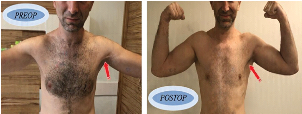
Figure 1: Irregular left anterior axillary fold with atrophy (pre-operative) and improved contour of the left anterior axillary fold with restoration of the muscular fold (post-operative, 6 months).
MRI of the left shoulder confirmed a chronic complete rupture of the PM tendon. The tendon was completely avulsed from the humeral insertion (i.e., an insertional/bony avulsion pattern rather than a mid-substance tear), with significant medial retraction of the muscle belly, moderate fatty infiltration consistent with chronic denervation and disuse, and no evidence of viable residual tendon tissue at the humeral insertion site suitable for direct repair. The long head of the biceps tendon was intact, and there were no other associated injuries to the shoulder girdle. Based on these clinical and MRI findings, the injury was classified as a Tietjen type III rupture (complete avulsion at the humeral insertion involving both the sternal and clavicular heads).
Given the chronic nature of the injury (2 years post-injury), significant muscle retraction, fatty infiltration, and the absence of viable residual tendon tissue, direct tendon-to-bone repair was not considered feasible. After discussing the surgical options, risks, benefits, and expected outcomes with the patient, we elected to proceed with reconstruction using an Achilles tendon–calcaneal allograft incorporating a calcaneal bone block, to provide immediate bone-to-bone fixation and to allow early pendulum exercises. The patient provided written informed consent and was scheduled for surgery.
The patient was operated on under general anesthesia in the beach-chair position. A standard deltopectoral incision was used. Intraoperatively, both heads of the PM tendon were found to be completely avulsed from the humeral insertion, with intense adhesions and fibrosis throughout the surgical site extending medial to the insertion site. The tendon was retracted medially to the anterior chest wall, and despite circumferential blunt dissection and release, it was not possible to bring the tendon back to its anatomical insertion site with traction sutures (Fig. 2). The need for an allograft to bridge the defect was therefore confirmed intraoperatively. An Achilles tendon allograft with a calcaneal bone block (Achilles Tendon with Bone Block; catalog code ACT-001; Arthrex, Naples, Florida, USA) was used (Fig. 3).
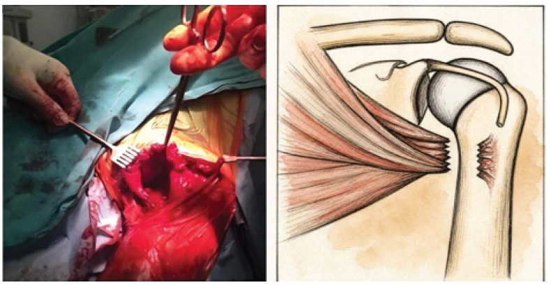
Figure 2: Intraoperative photograph and accompanying illustration demonstrating the torn and retracted pectoralis major muscle, with the retracted tendon stump unable to reach its humeral footprint despite traction sutures.
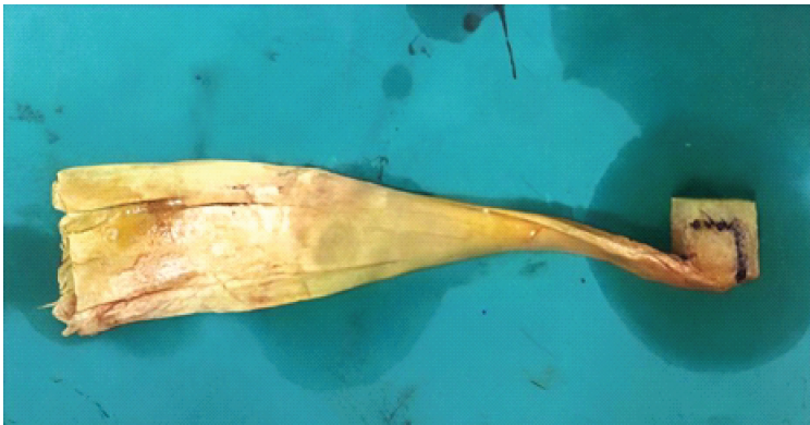
Figure 3: Achilles tendocalcaneal allograft prepared on the back table, showing the broad tendinous portion (left) and the calcaneal bone block (right) shaped to match the prepared humeral bone trough.
A rectangular bone trough measuring 3 cm in length and 1.5 cm in width was prepared at the humeral footprint located on the lateral lip of the bicipital groove, using osteotomes, curettes, and a high-speed burr (Fig. 4). The calcaneal bone block of the Achilles allograft was shaped with a burr so that it would press-fit the bone trough. Three uni-cortical holes were evenly drilled 1 cm medial to the bone trough using a 2.7 mm drill pin, and three suture anchors, each preloaded with two #2 FiberWire sutures, were inserted into these holes (Fig. 5).
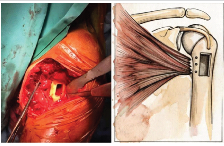
Figure 4: Preparation of the bone trough at the humeral footprint on the lateral lip of the bicipital groove (intraoperative photograph and accompanying illustration). The trough measured 3 cm in length and 1.5 cm in width.
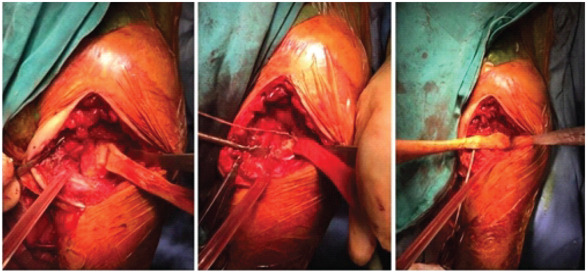
Figure 5: Application of three suture anchors medial to the trough and press-fit insertion of the calcaneal bone block of the Achilles allograft into the trough.
The retracted PM tendon remnant was secured with the anchor sutures using a Krackow locking-loop technique. A tendon-to-anchor sliding-suture technique was then used to progressively advance the tendon back toward the anatomical footprint: the sutures were serially tensioned in a pulley-like fashion while the shoulder was positioned in approximately 30° of abduction and neutral rotation, so that the retracted muscle–tendon unit could be incrementally drawn distally without cutting through the degenerated tendon tissue. Appropriate tension was determined clinically, using restoration of the anterior axillary fold and avoidance of excessive tissue tension as endpoints.
The shaped calcaneal bone block was press-fit into the trough and secured with an absorbable magnesium alloy interference screw (MAGNEZIX®, Syntellix AG, Hannover, Germany). These CE-marked implants provide initial mechanical strength comparable to titanium yet undergo gradual in vivo degradation, replaced by new bone over 12–18 months and avoiding long-term retained metallic hardware. The tendinous part of the allograft was laid over the retracted native PM tendon under appropriate tension and sutured side-to-side in a Pulvertaft-style interlocking weave with interrupted #2 non-absorbable sutures (Fig. 6). Stability of the bone block and tendon sutures was tested by gentle passive range-of-motion and rotation, with no construct failure or gapping.
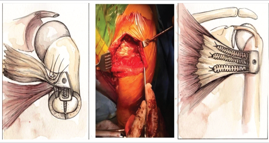
Figure 6: Fixation of the allograft with an absorbable magnesium alloy interference screw and repair of the retracted pectoralis major muscle to the tendinous portion of the allograft using a Pulvertaft-style interlocking weave (intraoperative photograph and accompanying illustrations).
Image processing disclosure
Image processing disclosure: Intraoperative photographs in Figs. 2, 4, and 6 were processed post-capture using artificial intelligence (AI)-based image enhancement (Fotor AI) for noise reduction, resolution upscaling, and stylized illustrative rendering of authentic surgical photographs; no anatomical content was generated, added, modified, or altered. Original unprocessed photographs are shown alongside the rendered versions (left panel: original; right panel: stylized rendering) to allow direct comparison. For Figs. 1, 3, 5, and 7, digital editing was limited to redaction of patient identifiers for privacy; no clinical, surgical, or radiographic content was modified.
Postoperatively, the patient was immobilized with a sling for 2 weeks, removed only to allow wrist and elbow range-of-motion exercises (Fig. 7).
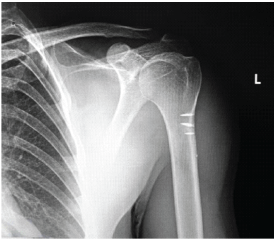
Figure 7: Post-operative radiograph of the left shoulder demonstrating the position of the absorbable magnesium alloy interference screw used to secure the calcaneal bone block at the humeral insertion site.
At the end of the 2nd post-operative week, relying on the solid primary stability provided by the bone-block press-fit, interference screw, and three medial suture anchors, pendulum exercises – forward–backward, side-to-side, and clockwise rotations – were initiated three times daily. Active shoulder exercises were begun at 4 weeks postoperatively, and active shoulder flexion reached 90° at the end of the 5th post-operative week. He returned to his routine manual labor 8 weeks after the operation and resumed weightlifting at 10 weeks.
At 6 months postoperatively, the patient demonstrated full painless range of motion in all planes, with forward elevation to 175°, external rotation to 85°, and internal rotation to the T10 vertebral level. Handheld-dynamometer testing showed adduction strength of 85% and internal-rotation strength of 87% of the contralateral side, and the anterior axillary fold had been restored with markedly improved chest-wall contour (Fig. 1).
The latest follow-up was obtained at 26 months postoperatively. The patient was continuing weightlifting – which had been the primary reason he sought surgical treatment – and was satisfied with the cosmetic appearance and function of the shoulder. Physical examination demonstrated full active range of motion without pain, with manual muscle testing revealing 5−/5 strength in resisted adduction and internal rotation, representing approximately 90% of contralateral strength. Patient-reported functional outcomes were assessed using two validated instruments: the validated Turkish version of the quick disabilities of the arm, shoulder, and hand (QuickDASH) questionnaire and the American Shoulder and Elbow Surgeons (ASES) shoulder score. The patient scored 4.55/100 on the QuickDASH (indicating minimal residual disability and near-complete functional recovery; MCID = 15.9 points) and 95/100 on the ASES (indicating excellent shoulder-specific function; scores ≥90 are conventionally classified as excellent). The two patient-reported instruments were therefore concordant in indicating excellent functional recovery; individual QuickDASH item responses are detailed in Table 1. There were no complications throughout the follow-up period, including no infection, hardware failure, nerve injury, stiffness, or re-rupture.
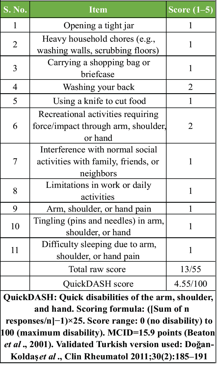
Table 1: QuickDASH item-level responses at 26-month follow-up
The clinical relevance of this technique is that it addresses the significant challenges posed by chronic PM ruptures – where muscle retraction, tendon involution, fatty infiltration, and sarcomere shortening typically preclude direct repair – by providing bone-to-bone rather than tendon-to-bone healing at the humeral footprint. This case report describes a reconstruction technique for chronic PM tendon rupture using an Achilles tendon–calcaneal allograft with a calcaneal bone block press-fitted into a humeral trough and secured with an absorbable magnesium alloy interference screw, supplemented by three medial suture anchors. The principal findings are that this construct provided clinically sufficient fixation that allowed pendulum exercises to be initiated at 2 weeks postoperatively in this patient, and resulted in excellent functional and cosmetic outcomes at 26-month follow-up, including return to recreational weightlifting, a QuickDASH score of 4.55/100, and an ASES score of 95/100.
Primary tendon-to-bone repair is usually feasible in acute PM rupture. In chronic cases, contracture of the muscle belly retracts the tendon proximally and expands the distance to the humeral insertion site; in addition, chronic tendon ruptures are associated with sarcomere shortening and loss of muscle contractile units, which further limit the ability to advance the retracted musculotendinous unit to its anatomical insertion even after extensive mobilization [4,12]. In our case, despite circumferential release, the retracted tendon could not be brought to its footprint with traction sutures alone, confirming the need for allograft augmentation. Only a few techniques have been described that specifically address the reconstruction of a chronically torn PM tendon with allografts [10,11,13,14]. Recommended graft options include hamstring autograft, fascia lata allograft, Achilles tendon allograft, and dermal allograft, with fixation options including suture anchors, bone tunnels, screws, and washer techniques [3,14,15,17]. Although reconstruction of chronic PM rupture with allograft has been reported, utilization of an allograft incorporating a bone block has not, to our knowledge, been previously reported.
Our patient is consistent with the typical PM rupture profile: A male weightlifter injured during bench press. Anabolic steroid use is a recognized risk factor; de Castro Pochini et al. reported steroid use in more than 90% of patients in their prospective series [5]. Proposed mechanisms include tendon stiffening and disproportionate gains in maximal force production [6,7]. Our patient had been using anabolic steroids in the 6 months preceding the injury, consistent with this mechanism.
PM rupture is considered chronic when surgery is performed ≥6–8 weeks after injury [1,4]. In our case, surgery was performed 2 years after injury – such late repair is rarely reported. Neumann et al. reported reconstruction with dermal allograft at a mean chronicity of 19.2 months, the closest comparator to our case [14]. Zacchilli et al. reconstructed three Achilles-allograft cases at a mean of 22.2 months using three suture anchors and a shallow anatomical trough [16]. Mardani-Kivi et al. reported 22 bodybuilding athletes (24 ruptures, 87.5% chronic) treated by Achilles allograft augmentation, with a 100% healing rate and significant improvement in Bak’s criteria and Constant scores [13]. The Achilles tendon allograft has been increasingly reported for chronic PM reconstruction in recent years. Rivera et al. described reconstruction of a chronic, retracted PM tear using an Achilles allograft secured with unicortical suture buttons [18]. DeRogatis et al. reported excellent ASES and Short Form-36 outcomes following Achilles allograft augmentation for a chronic PM muscle-belly tear at 1.5-year follow-up [19]. Wary et al. described a combined Pulvertaft-weave and onlay technique using dual Achilles allografts for a chronic injury in which a single graft was insufficient [20]. In these reports, however, the Achilles allograft was used in a tendon-only or augmentation configuration; none incorporated a press-fitted calcaneal bone block with interference-screw fixation in a humeral trough.
Achilles tendon allograft with a bone block was chosen as the allograft option in the present reconstruction. The structural and biomechanical characteristics of the Achilles tendon match well with those of the PM tendon [9,14]. The broad proximal end of the Achilles tendon can be laid over the retracted muscle belly, allowing various strong suturing techniques; the distal end of the allograft provides a native tendon-to-bone connection through the calcaneal bone block, which in our technique is used to restore the humeral footprint.
This shifts the reconstructive principle from tendon-driven augmentation toward graft- and bone-driven replacement, which we believe may represent a useful alternative reconstructive approach when chronicity has rendered the native tendon non-viable. We emphasize that this rationale is hypothesis-generating rather than a demonstrated claim of superiority. This is consistent with a recent systematic review and meta-analysis that found no significant differences in load to failure or stiffness among transosseous tunnels, suture anchors, and cortical buttons for PM repair, suggesting that the quality of the bone–tendon interface may matter as much as the specific mode of fixation [21]. Recent literature, including a narrative review of chronic PM reconstruction, underscores that no single technique has yet been established as the gold standard and that larger prospective studies with standardized outcome reporting are needed [22]. The choice of an absorbable magnesium alloy interference screw, rather than a titanium or polyetheretherketone implant, was based on three considerations: Avoidance of long-term retained metallic hardware in a young, active weightlifter; gradual stress transfer to the incorporating bone block during the 12–18-month resorption window, potentially favoring biological remodeling at the graft–host interface; and elimination of metallic artifact in any future shoulder MRI.
In a study by Tomita et al., anterior cruciate ligament reconstruction in dogs performed with flexor tendon and bone-patellar tendon-bone graft was compared: the flexor tendon was anchored to the bone at 12 weeks, whereas the bone plug of the bone-patellar tendon-bone graft showed anchorage by newly formed bone as early as 3 weeks [23]. Park et al. compared the healing of tendon-to-bone and bone-to-bone interfaces in a rabbit model in which detached patellar tendons were implanted in the proximal tibia either subperiosteally or together with a bone block; bony union was evident in the graft with a bone block at 8 weeks, whereas a bony union without a bone block required 12 weeks [17]. The native PM muscle must additionally incorporate into the allograft tendon through a process of cellular infiltration, neovascularization, and collagen remodeling, which typically takes several months. Although animal models cannot be directly extrapolated to human tendon reconstruction, these data together suggest that a bony end of the allograft, in addition to functioning as an immediate press-fit block in the bone trough, is likely to incorporate more rapidly into the host bone than a tendon-only allograft.
Given these healing timelines, one might reasonably question whether early rehabilitation could compromise the reconstruction. In our case, the primary stability provided by the press-fit bone block, the magnesium interference screw, and the three medial suture anchors was considered sufficient to tolerate the very low tensile forces generated by gravity-assisted pendulum motion. Pendulum exercises rely on gravity rather than active muscle contraction and therefore do not generate significant tensile forces across the muscle–tendon–bone interface, making them unlikely to disrupt either the bone-block fixation or the muscle-to-tendon repair. Resisted strengthening was deliberately avoided until later in the rehabilitation course, after the bone-to-bone interface had had time to incorporate. Physical therapy is an important part of PM reconstruction: graded range-of-motion exercise is crucial for shoulder mobility and eventual recovery of strength. Joseph et al. initiated their physical therapy program 4 weeks after surgery [11]; Zacchilli et al. protected the operated extremity in a sling in adduction for 6 weeks [16]. In our reconstruction, gravity-assisted pendulum exercises were initiated at 2 weeks while active shoulder exercises were deferred to the 4th post-operative week, in line with Joseph et al.’s active-rehabilitation timing. A gradual increase in motion allowed an active forward flexion of 90° by the 5th post-operative week.
Operative complications of PM repair include infection, hypertrophic scar, stiffness, re-rupture, and nerve injury [1,3,7,10]. Silverstein et al. reported a proximal humerus fracture 8 weeks after bone-trough PM repair [24], and Rabuck et al. reported one humerus fracture in a biomechanical cadaveric study using a bone-trough method [8]. Beyond these two reports, we found no additional studies describing humeral fracture after PM repair. In the present case, despite the bone-trough and early mobilization, we encountered no bone-related, hardware-related, or soft-tissue complications throughout the 26-month follow-up. The absence of bone-related complications may reflect early incorporation of the bone block into the host bone during healing, consistent with animal data [17,23].
Several specific limitations of this report should be noted. A pre-operative QuickDASH score was not formally collected, limiting our ability to quantify the magnitude of patient-reported functional improvement. Strength assessment was not performed consistently throughout follow-up: Handheld-dynamometer testing was used at 6 months, whereas only manual muscle testing was performed at the final 26-month follow-up; because manual muscle testing is examiner-dependent and less objective than isokinetic or dynamometer-based evaluation, the absence of a repeated objective dynamometer assessment limits direct quantitative comparison. Although the pre-operative MRI findings are described in the Case Report, a pre-operative MRI image is not included as a figure because the original imaging files were not available for reproduction at manuscript preparation; this limits independent radiological verification of the imaging characteristics. In addition, no post-operative MRI or other advanced imaging was obtained; consequently, biological graft incorporation and tendon-to-bone healing were inferred from clinical and functional findings rather than confirmed radiologically. Although the QuickDASH and ASES scores at 26 months confirm excellent functional recovery, additional validated shoulder-specific instruments such as the Constant–Murley score, the simple shoulder test, and isokinetic dynamometry would provide more comprehensive functional assessment in future studies.
In the present case, this construct allowed pendulum exercises from the 2nd post-operative week, return to manual labor at 8 weeks and to weightlifting at 10 weeks, and produced excellent functional and cosmetic outcomes at 26 months, with a QuickDASH score of 4.55/100, an ASES score of 95/100, and no complications. This technique therefore represents a potentially useful surgical option for chronic tears, particularly in active patients seeking restoration of strength, function, and cosmesis. We caution, however, that the early return to weightlifting reported in this case reflects the specific construct used and this individual patient’s progression and should not be generalized to other reconstruction techniques or to all patients; return to heavy upper-extremity loading should remain an individualized clinical decision based on intraoperative construct stability, rehabilitation milestones, and patient-specific risk factors.
For chronic PM ruptures with a non-viable native tendon, reconstruction with an Achilles tendon–calcaneal allograft using a press-fitted bone block, an absorbable magnesium interference screw, and three medial suture anchors offers a reliable reconstructive option that supports early rehabilitation and excellent mid-term functional outcomes.
References
- 1. Butt U, Mehta S, Funk L, Monga P. Pectoralis major ruptures: A review of current management. J Shoulder Elbow Surg 2015;24:655-62. [Google Scholar] [PubMed]
- 2. Haley CA, Zacchilli MA. Pectoralis major injuries: Evaluation and treatment. Clin Sports Med 2014;33:739-56. [Google Scholar] [PubMed]
- 3. Bak K, Cameron EA, Henderson IJ. Rupture of the pectoralis major: A meta-analysis of 112 cases. Knee Surg Sports Traumatol Arthrosc 2000;8:113-9. [Google Scholar] [PubMed]
- 4. Wolfe SW, Wickiewicz TL, Cavanaugh JT. Ruptures of the pectoralis major muscle. An anatomic and clinical analysis. Am J Sports Med 1992;20:587-93. [Google Scholar] [PubMed]
- 5. de Castro Pochini A, Ejnisman B, Andreoli CV, Monteiro GC, Silva AC, Cohen M, et al. Pectoralis major muscle rupture in athletes: A prospective study. Am J Sports Med 2010;38:92-8. [Google Scholar] [PubMed]
- 6. Manske RC, Prohaska D. Pectoralis major tendon repair post surgical rehabilitation. N Am J Sports Phys Ther 2007;2:22-33. [Google Scholar] [PubMed]
- 7. ElMaraghy AW, Devereaux MW. A systematic review and comprehensive classification of pectoralis major tears. J Shoulder Elbow Surg 2012;21:412-22. [Google Scholar] [PubMed]
- 8. Rabuck SJ, Lynch JL, Guo X, Zhang LQ, Edwards SL, Nuber GW, et al. Biomechanical comparison of 3 methods to repair pectoralis major ruptures. Am J Sports Med 2012;40:1635-40. [Google Scholar] [PubMed]
- 9. Sherman SL, Lin EC, Verma NN, Mather RC, Gregory JM, Dishkin J, et al. Biomechanical analysis of the pectoralis major tendon and comparison of techniques for tendo-osseous repair. Am J Sports Med 2012;40:1887-94. [Google Scholar] [PubMed]
- 10. Baverel L, Messedi K, Piétu G, Crenn V, Gouin F. Pectoralis major tear with retracted tendon: How to fill the gap? Reconstruction with hamstring autograft and fixation with an interference screw. Case Rep Orthop 2017;2017:2095407. [Google Scholar] [PubMed]
- 11. Joseph TA, Defranco MJ, Weiker GG. Delayed repair of a pectoralis major tendon rupture with allograft: A case report. J Shoulder Elbow Surg 2003;12:101-4. [Google Scholar] [PubMed]
- 12. Lieber RL, Fridén J. Functional and clinical significance of skeletal muscle architecture. Muscle Nerve 2000;23:1647-66. [Google Scholar] [PubMed]
- 13. Mardani-Kivi M, Maddahi A, Leili EK, Asadi K, Darabipour Z, Haghparast Ghadim-Limudahi Z. Pectoralis major rupture repair with Achilles tendon allograft in 22 bodybuilding athletes. J Orthop Sci 2023;28:1118-23. [Google Scholar] [PubMed]
- 14. Neumann JA, Klein CM, van Eck CF, Rahmi H, Itamura JM. Outcomes after dermal allograft reconstruction of chronic or subacute pectoralis major tendon ruptures. Orthop J Sports Med 2018;6:2325967117745834. [Google Scholar] [PubMed]
- 15. Schachter AK, White BJ, Namkoong S, Sherman O. Revision reconstruction of a pectoralis major tendon rupture using hamstring autograft: A case report. Am J Sports Med 2006;34:295-8. [Google Scholar] [PubMed]
- 16. Zacchilli MA, Fowler JT, Owens BD. Allograft reconstruction of chronic pectoralis major tendon ruptures. J Surg Orthop Adv 2013;22:95-102. [Google Scholar] [PubMed]
- 17. Park MJ, Lee MC, Seong SC. A comparative study of the healing of tendon autograft and tendon-bone autograft using patellar tendon in rabbits. Int Orthop 2001;25:35-9. [Google Scholar] [PubMed]
- 18. Rivera PM, Dhytadak D, Cunningham G. Reconstruction of chronic, retracted pectoralis major tendon tear with achilles tendon allograft. Arthrosc Tech 2023;12:e449-52. [Google Scholar] [PubMed]
- 19. DeRogatis MJ, De Leon RA, Smith TJ. Chronic pectoralis major muscle belly repair and reconstruction with achilles allograft augmentation. JBJS Case Connect 2024;14:???. [Google Scholar] [PubMed]
- 20. Wary JN, Bottoni CR, Zhou L. Pectoralis major tendon reconstruction with achilles tendon allografts utilizing combined pulvertaft weave and onlay techniques. Mil Med 2025;190:e851-7. [Google Scholar] [PubMed]
- 21. Sequeira SB, Sequeira LM, Wieland MD, Imbergamo CM, Tepper K. No difference in load to failure or stiffness between transosseous tunnels, suture anchors, and cortical buttons for pectoralis major tendon repair: A systematic review & meta-analysis. J Exp Orthop 2023;10:56. [Google Scholar] [PubMed]
- 22. Zhiti C, Gossuin I, Durand Y, Müller J, Daulouède PX, Mazreku S. Chronic rupture of the pectoralis major: Current concepts and various surgical repair techniques – A mini review. J Orthop Case Rep 2025;15:215-23. [Google Scholar] [PubMed]
- 23. Tomita F, Yasuda K, Mikami S, Sakai T, Yamazaki S, Tohyama H. Comparisons of intraosseous graft healing between the doubled flexor tendon graft and the bone-patellar tendon-bone graft in anterior cruciate ligament reconstruction. Arthroscopy 2001;17:461-76. [Google Scholar] [PubMed]
- 24. Silverstein JA, Goldberg B, Wolin P. Proximal humerus shaft fracture after pectoralis major tendon rupture repair. Orthopedics 2011;34:222. [Google Scholar] [PubMed]




Related Articles in Journal of Orthopaedic Case Reports
 May 10, 2022 Surgical Repair of Traumatic Rupture of Tibialis Posterior Tendon in 32-Year-Old Male
 July 1, 2025 Chronic Thigh Pain in an Adolescent: A Case Report and Literature Review of Desmoplastic Fibroma, A Rare Benign Tumor
 November 10, 2023 Case Reports and their impact on Journal Metrics, Researcher’s Indices, and other sundry issues
 August 6, 2024 A Unique Upside-down Flip-lid Type of Lateral Tibial Plateau Fracture Fragment in a Bicondylar Tibial Plateau Fracture Managed with Dual Plates and Raft Screws