

A modified volar–lateral approach, combining pulp-preserving incision with FPL retraction rather than splitting, enables complete curettage and full functional recovery in thumb distal phalanx enchondromas.
Dr. M Harshith, Department of Orthopaedic Surgery, Post Graduate Institute of Medical Education and Research, Chandigarh, India. E-mail: mharshith211@gmail.com
Abstract
Introduction: Enchondromas are the most common benign bone tumors of the hand, but distal phalanx involvement of the thumb is exceedingly rare (<4%). Surgical management is challenging due to limited exposure and risk to vital structures.
Case Report: A 19-year-old female presented with a painful swelling of the right thumb. Imaging revealed a lytic lesion in the distal phalanx consistent with enchondroma. Surgery was performed using a modified volar–lateral approach that combined the skin incision of the modified volar technique with a deep plane between the flexor pollicis longus tendon and the lateral border of the distal phalanx, preserving pulp septae and avoiding tendon splitting. Curettage and iliac crest bone grafting were performed.
Conclusion: This modified approach provided better exposure for curettage, preserved sensory and tendon integrity, and achieved complete graft incorporation with full motion and no recurrence, offering a safe, functional technique for rare thumb distal phalanx enchondromas.
Keywords: Enchondroma, benign bone tumor, lytic lesion, distal phalanx, thumb, first digit, modified volar–lateral approach.

Enchondroma is the most frequent benign bone tumor of the hand, representing nearly 90% of all cartilaginous tumors in this region and approximately 40–65% of benign hand neoplasms [1]. It arises from residual cartilaginous rests within the medullary cavity and is composed of mature hyaline cartilage [1,2]. These tumors have a predilection for the ulnar-sided tubular bones, especially the proximal and middle phalanges, whereas involvement of the distal phalanx, particularly of the thumb, remains exceptionally rare [3,4,5]. According to large case series, distal phalanx lesions account for <2–4% of all hand enchondromas [3,5,6,7]. Clinically, enchondromas are often asymptomatic, discovered incidentally on radiographs, but can present with pain, swelling, nail deformity, or pathological fracture after minor trauma [1,3,5]. Radiographically, they appear as well-defined lytic intramedullary lesions with cortical thinning or expansion, and magnetic resonance imaging (MRI) demonstrates lobulated hyperintense T2 and hypointense T1 signals consistent with their cartilaginous nature [1,3]. Although benign, the functional significance of the thumb mandates early recognition and careful surgical planning, as even small deformities or postoperative stiffness can severely impair grip and dexterity [3,4,7].
Surgical indications and treatment principles:
Curettage, with or without bone grafting, remains the gold standard for symptomatic or enlarging enchondromas, those complicated by fracture, or those that compromise the cortical integrity [1,2,8]. Several studies have demonstrated excellent results with intralesional curettage and bone grafting (autograft or allograft), with minimal recurrence and early restoration of function [1,3,5]. Alternative minimally invasive techniques, including endoscopic curettage, have been reported for proximal phalangeal lesions, allowing preservation of cortical integrity and periosteum [2]. However, their role in distal phalanx tumors is limited due to restricted working space and the proximity of neurovascular structures.
Comparison of surgical approaches:
Given the confined anatomy of the distal phalanx, the choice of surgical approach is critical to achieving complete tumor removal while minimizing soft-tissue complications. The literature describes multiple approaches, each with specific limitations:
- Lateral approach: Traditionally favored for its simplicity and minimal disruption of flexor and extensor tendons. However, it offers a narrow cortical window, restricting curettage access and increasing the risk of incomplete excision [3,5].
- Volar (palmar) approach: Provides direct exposure of the lesion but violates the pulp septae, predisposing to painful scars, sensory deficits, and loss of pulp contour, which is especially detrimental for thumb pinch mechanics [4]
- Modified volar approach: As refined by Kokubu et al. (2025), it employs a Bruner-type incision sparing the central pulp and proceeds by splitting the insertion of the flexor pollicis longus (FPL) tendon for cortical access [4]. While this method protects the pulp septae and preserves tactile sensation, splitting the FPL tendon introduces a risk of tendon weakening and post-operative stiffness.
- Dorsal approach: Described in earlier series such as Shimizu et al. (1997) and later reiterated in reviews, it offers only a small cortical window and carries a higher likelihood of wound complications, nail deformities, and extensor tendon injury [4].
- Non-surgical management: Although acceptable for small, asymptomatic lesions, as shown by Ramos-Pascua et al. (2018), conservative treatment lacks histologic confirmation and may risk recurrence or progression in functionally demanding digits such as the thumb [5].
Rationale for the present surgical approach:
Considering these limitations, a modified hybrid technique was adopted in the present case. The skin incision followed the modified volar approach of Kokubu et al., thereby avoiding violation of the pulp septae, which preserves sensation and reduces post-operative pain. However, instead of splitting the FPL tendon, the deep dissection plane was developed between the tendon and the lateral border of the distal phalanx. This modification allows for a larger cortical window for curettage compared to the lateral approach, ensuring complete tumor clearance and optimal graft placement while avoiding tendon morbidity. This anatomical corridor maintains both adequate exposure and functional preservation of the flexor and neurovascular structures. Early post-operative mobilization, as advocated by Pietramala et al. (2025) in their experience with curettage-only protocols, promotes rapid recovery without compromising healing [1].
Aim of the report:
Given the extreme rarity of enchondromas involving the distal phalanx of the thumb and the ongoing debate surrounding the ideal surgical exposure, this case highlights a modified volar–lateral approach that combines the safety of pulp preservation with the accessibility of a larger bone window. The technique aims to optimize curettage efficacy, minimize complications, and ensure early functional restoration of thumb mobility.
A 19-year-old female presented with a history of a lump over the right thumb for the past 3 years, which was occasionally associated with pain and discomfort. The swelling gradually increased in size, and the pain became more severe (VAS—8) and persistent throughout the day. Over the past 3–4 months, the symptoms had progressively interfered with her routine activities and academic work. On examination, there was a diffuse swelling involving the pulp of the right thumb, along with wasting of the thenar muscles (Fig. 1a). The nail bed showed bluish discoloration with associated clubbing (Fig. 1b).
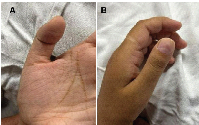
Figure 1: Clinical presentation, (a) Diffuse swelling of the pulp space and atrophy of the thenar muscles. (b) Depicts bluish discoloration of the thumbnail bed and clubbing.
There was tenderness over the nail bed and pulp of the finger. Grip strength and the range of motion of the distal interphalangeal joint were markedly reduced. Plain radiographs of the right thumb revealed an expansile lytic lesion involving the distal phalanx of the first digit (Fig. 2a). MRI demonstrated a well-defined, lobulated, hyperintense lesion on T2-weighted images without internal septations (Fig. 2b), and T1-weighted images showed a hypointense lesion (Fig. 2c), findings consistent with an enchondroma. Surgical management with curettage and bone grafting was planned.
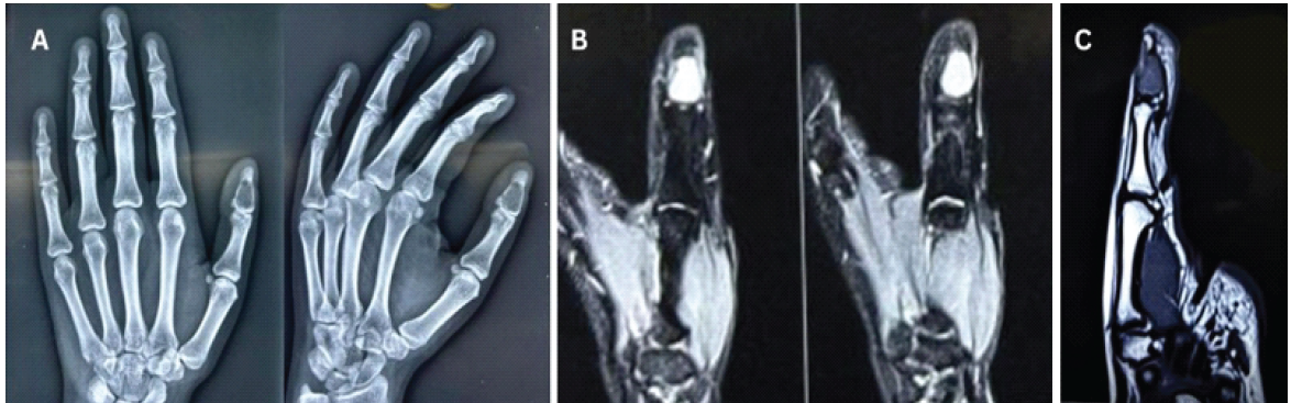
Figure 2: Pre-operative radiology, (a) plain radiograph showing a lytic lesion in the distal phalanx of the thumb. (b) T2-weighted image showing a hyperintense, well-circumscribed, geographic lesion without any marrow edema, consistent with enchondroma. (c) T1-weighted image showing a well-circumscribed hypointense lesion replacing the normal marrow in the distal phalanx of the thumb, suggestive of enchondroma.
The procedure was performed under tourniquet control. A modified volar approach, as described by Kokubu et al., was utilized (Fig. 3a). Full-thickness skin flaps were elevated on either side, taking care not to breach the lesion. The insertion of the FPL tendon was identified and retracted medially (Fig. 3b). A cortical window was created between the tendon insertion and the lateral margin of the distal phalanx (Fig. 3c). The contents of the lesion were carefully curetted using a fine scoop, avoiding fracture of the surrounding cortical walls. The cavity was packed with gauze soaked in hydrogen peroxide for 5 min, followed by thorough irrigation. The defect was then filled with cancellous bone graft harvested from the iliac crest (Fig. 3d).
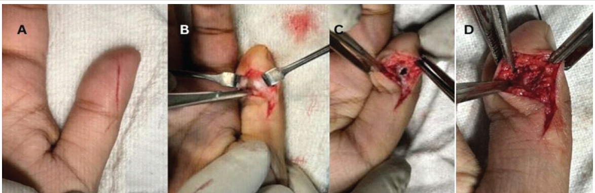
Figure 3: Intraoperative pictures, (a) Clinical picture showing the skin incision used in our novel technique. (b) Clinical picture showing retraction of the flexor pollicis longus tendon to achieve a safe corridor. (c) Clinical picture showing the cortical window made within the safe corridor. (d) Clinical picture showing bone grafting done post-curettage through the same cortical window.
Gelfoam sponges were placed over the graft, and the wound was closed in a single layer. A thumb spica splint was applied postoperatively. The immediate post-operative radiograph demonstrated a well-impacted graft within the cavity and showed no evident cortical breach (Fig. 4).
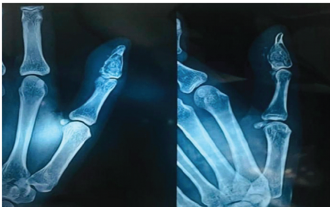
Figure 4: Plain radiograph showing immediate post-operative status, well-impacted graft within the lesion without any cortical breach.
Postoperatively, the thumb was immobilized in a splint for 3 weeks, following which range of motion exercises were initiated. The surgical wound demonstrated complete healing by the end of 3 weeks (Fig. 5a). At 2 months postoperatively, the patient had regained full range of motion at the distal interphalangeal joint (Fig. 5b) and was completely pain-free (VAS score: 0).
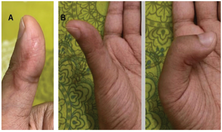
Figure 5: Post-operative status at 2 months follow-up, (a) Clinical picture showing healed scar. (b) Clinical picture showing the complete range of motion at the distal interphalangeal joint of the thumb.
At 14 months follow-up, radiographs showed complete incorporation of the bone graft within the defect, with no evidence of recurrence of the lesion (Fig. 6).
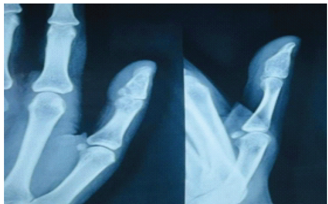
Figure 6: Plain radiograph at 14 months follow-up showing excellent graft uptake without any recurrence of the tumor.
Enchondroma is the most common benign bone tumor of the hand, yet its occurrence in the distal phalanx of the thumb remains exceedingly rare [3,4]. Large series report: < 4% involvement of this site [3,5]. Despite its benign nature, the lesion’s proximity to critical neurovascular and tendon structures poses diagnostic and surgical challenges. Accurate diagnosis also requires differentiation from low-grade chondrosarcoma and exclusion of multiple enchondromatosis syndromes such as Ollier disease and Maffucci syndrome, which carry a higher risk of malignant transformation [9,10]. Curettage with or without bone grafting remains the standard treatment, yielding excellent functional and radiologic outcomes [1,3]. Bone grafting enhances stability and osteointegration, while recurrence after adequate curettage is rare (<6%) [1,3,11]. Although non-surgical management may be considered for small asymptomatic lesions [5], symptomatic thumb lesions demand operative intervention to preserve function. Multiple approaches have been described, each with limitations. The lateral approach offers limited exposure; the volar approach violates pulp septae, risking pain and sensory loss; and the dorsal approach may cause wound and nail complications [4]. The modified volar approach of Kokubu et al. (2025) preserves pulp integrity but requires splitting the FPL tendon [4]. Our technique combined the skin incision of the modified volar approach with a deep dissection plane between the FPL and lateral border of the distal phalanx, avoiding tendon splitting while creating a larger bone window for thorough curettage. This achieved excellent exposure, early mobilization, and complete graft incorporation without recurrence—findings consistent with reports by Ampadiotaki et al. [3], Omlor et al. [11], and Pietramala et al. [1]. Thus, this modified volar–lateral approach offers a safe, functional, and reproducible method for managing rare thumb distal phalanx enchondromas.
Enchondroma of the distal phalanx of the thumb is an exceedingly rare entity that poses significant surgical challenges owing to its anatomically confined location and proximity to critical flexor tendon and neurovascular structures. The modified volar–lateral approach described herein—employing a pulp-preserving skin incision with deep dissection between the FPL tendon and the lateral cortex, without tendon splitting—provides superior cortical exposure for complete curettage compared to conventional approaches. Supplemented by autologous iliac crest bone grafting and early post-operative mobilization, this technique achieved complete graft incorporation, restoration of full painless thumb motion, and no recurrence at 14-month follow-up. We propose this modified approach as a safe, reproducible, and functionally superior technique for the surgical management of rare enchondromas involving the thumb distal phalanx.
Enchondroma of the distal phalanx of the thumb, though exceedingly rare, demands careful surgical planning given the functional importance of the thumb and the anatomically confined operative field. The modified volar–lateral approach—combining a volar skin incision with deep dissection between the FPL tendon and the lateral cortex—offers superior exposure for curettage compared to the classic lateral approach, while avoiding the tendon morbidity of FPL splitting and the sensory complications of the standard volar approach. This technique, supplemented by autologous bone grafting and early mobilization, achieves complete lesion clearance, full graft incorporation, and restoration of painless thumb motion, making it a safe and reproducible option for this challenging anatomical location.
References
- 1. Pietramala S, Rovere G, Ravaioli C, Mignano C, Smakaj A, Fidanza A, et al. Surgical treatment of enchondromas of the hand: Our experience in curettage only and early mobilization. Diseases 2025;13:84. [Google Scholar] [PubMed]
- 2. Leung CH, Lui TH. Endoscopic curettage and bone grafting of enchondroma of proximal phalanx of finger. Arthroscopy Tech 2023;12:e1335-40. [Google Scholar] [PubMed]
- 3. Ampadiotaki MM, Vlachos C, Papagrigorakis E, Galanis A, Mitsikostas P, Vasiliadis ES, et al. Thumb distal phalanx giant enchondroma. A case presentation and literature review. Maedica 2024;19:177. [Google Scholar] [PubMed]
- 4. Kokubu Y, Hirose K, Kurashige T, Ando Y, Marusugi K, Kawaguchi H. A Modified palmar approach with tendon splitting for distal phalanx enchondromas of the thumb: A report of two cases. J Hand Surg Global Online 2025;7:249-52. [Google Scholar] [PubMed]
- 5. Ramos-Pascua LR, Bárcena-Tricio V, Casas-Ramos P, Herráez SS, Izquierdo-García FM, Martín FA. Nonsurgical treatment as alternative to surgical treatment in enchondromas of the distal phalanx. Analysis of a series of 11 cases. J Hand Surg 2018;43:870-e1. [Google Scholar] [PubMed]
- 6. Shimizu K, Kotoura Y, Nishijima N, Nakamura T. Enchondroma of the distal phalanx of the hand. J Bone Joint Surg 1997;79:898-900. [Google Scholar] [PubMed]
- 7. Lu H, Chen Q, Yang H, Shen H. Enchondroma in the distal phalanx of the finger: An observational study of 34 cases in a single institution. Medicine 2016;95:e5175. [Google Scholar] [PubMed]
- 8. Wulle C. On the treatment of enchondroma. J Hand Surg Br Vol 1990;15:320-30. [Google Scholar] [PubMed]
- 9. Lubahn JD, Bachoura A. Enchondroma of the hand: Evaluation and management. J Am Acad Orthop Surg 2016;24:625-33. [Google Scholar] [PubMed]
- 10. Pansuriya TC, Kroon HM, Bovée JV. Enchondromatosis: Insights on the different subtypes. Int J Clin Exp Pathol 2010;3:557-69. [Google Scholar] [PubMed]
- 11. Omlor GW, Lohnherr V, Lange J, Gantz S, Mechtersheimer G, Merle C, et al. Outcome of conservative and surgical treatment of enchondromas and atypical cartilaginous tumors of the long bones: Retrospective analysis of 228 patients. BMC Musculoskeletal Disord 2019;20:134. [Google Scholar] [PubMed]




Related Articles in Journal of Orthopaedic Case Reports
 October 1, 2026 Clinicopathological Spectrum and Management of Aneurysmal Bone Cysts in Atypical Anatomical Location: A Comprehensive Review of Demographics, Presentation, Diagnosis, and Treatment Outcomes
 September 1, 2026 Management of Scapular Osteochondroma: An Uncommon Report of Two Cases
 August 1, 2026 Vanishing Osteochondromas of the Distal Femur and Proximal Humerus: A Two-Case Report
 July 1, 2026 Diagnostic Dilemma in a Distal Femoral Intramedullary Lesion: Enchondroma Mimicking Low-Grade Chondrosarcoma with Discordant Biopsy Findings: A Case Report