

Paprosky classification guides acetabular reconstruction – jumbo cups for contained defects, mesh cage with iliac bone grafting for uncontained type IIC defects.
Dr. Kiran Uttam Shelke, Department of Orthopaedics, Government Medical College and Hospital, Chhatrapati Sambhajinagar, Maharashtra, India. E-mail: kiranusashelke@gmail.com
Abstract
Introduction: Acetabular bone loss remains a formidable challenge in both primary and revision total hip arthroplasty (THA). Large-diameter “jumbo” acetabular cups and acetabular metal mesh with impaction bone grafting are two reconstructive strategies employed to address moderate-to-severe acetabular defects. This case series reports outcomes in four young male patients with Paprosky Type 2B, 2C, and 3A defects managed at a tertiary referral center in India, highlighting the clinical and radiological results of these techniques.
Materials and Methods: Four male patients (age range 29–39 years) presented with acetabular bone deficiency secondary to post-traumatic arthritis, neglected central fracture-dislocation, failed internal fixation, or aseptic loosening of prior cemented THA. Cases were classified using the Paprosky system. Procedures included revision THA with acetabular mesh cage and autogenous iliac crest impaction bone grafting (n = 1), primary THA with jumbo cementless acetabular cups (n = 2), and revision THA with impaction cancellous bone grafting (n = 1). Intraoperative C-arm fluoroscopy was used for cup alignment verification. Outcome assessment included Harris Hip Score (HHS), serial radiographs, and functional walking assessment at 3 months and 1 year.
Conclusion: All four patients achieved satisfactory functional recovery with improved pain relief and independent ambulation. No implant loosening, migration, infection, or early revision was observed at follow-up. Jumbo acetabular cups and acetabular mesh with impaction bone grafting represent reliable, technically feasible alternatives to complex three-dimensional cage reconstructions, delivering stable biological fixation in young patients with post-traumatic acetabular deficiency. Meticulous pre-operative planning with radiography and three-dimensional computed tomography, intraoperative decision-making, and structured rehabilitation are essential determinants of success.
Keywords: Total hip arthroplasty, jumbo acetabular cup, acetabular mesh, Paprosky classification, acetabular bone defect, revision total hip arthroplasty, impaction bone grafting, post-traumatic arthritis.

Total hip arthroplasty (THA) consistently delivers reliable pain relief and functional restoration in patients with end-stage hip disease. However, acetabular bone loss presents a formidable challenge in complex primary cases and revision surgery, with its management critically determining implant durability and patient outcome [1].
The Paprosky classification stratifies acetabular bone deficiency into Types I through IIIB based on the degree of superior migration of the hip center, rim integrity, and column competence [2]. Type I defects retain a well-supported rim and require no special reconstruction. Type II and III defects, encountered with increasing frequency as global revision rates rise, exhibit significant superior migration, rim deficiency, or segmental column loss, demanding individualized reconstructive strategies [3].
Two principal biological approaches have been established for managing moderate-to-severe acetabular bone loss: (1) Large-diameter cementless “jumbo” porous-coated acetabular cups (≥62 mm in females, ≥66 mm in males) that achieve rim fixation and osseointegration with residual host bone, and (2) acetabular metal mesh cages combined with impaction bone grafting that reconstruct the deficient medial wall and convert an uncontained defect into a contained cavity, facilitating biological incorporation [4,5].
While published series from high-volume Western centers report good mid-term survivorship for both techniques, data from Indian tertiary institutions – where the patient demographic differs significantly, with younger patients presenting with post-traumatic sequelae rather than primary degenerative disease – remain limited [6,7]. We report a case series of four young male patients managed at a Government Medical College and Hospital with Paprosky Types 2B, 2C, and 3A defects, detailing our surgical technique and early clinical and radiological outcomes.
Paprosky classification of acetabular defects (Table 1)
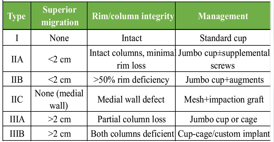
Table 1: Paprosky classification of acetabular defects
Case 1: Revision THA with acetabular mesh cage and impaction bone grafting (Paprosky type 2C)
A 36-year-old male presented with progressive right hip pain and restricted mobility. He had undergone cemented right-sided THA 6 years prior at another institution. Clinical examination revealed a shortened right lower limb with a Trendelenburg gait. Pre-operative radiographs demonstrated loosening of the cemented acetabular component with a medial wall defect consistent with Paprosky Type 2C, with preservation of the anterior and posterior columns (Fig. 1).
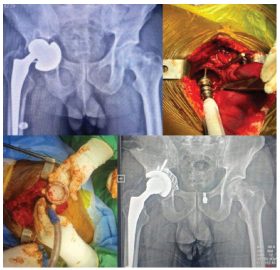
Figure 1: Case 1: Pre-operative, intraoperative photos and post-operative X-rays.
Pre-operative planning utilized weight-bearing radiographs and three-dimensional computed tomography (CT) to characterize the defect and template the reconstruction. The decision was made to perform revision THA with acetabular mesh reconstruction and impaction bone grafting. Under spinal anesthesia in the lateral decubitus position, through a posterior approach, the loosened cemented cup was carefully extracted, preserving residual acetabular rim bone. The acetabulum was debrided of fibrous membrane, and the medial wall defect was confirmed intraoperatively.
An autogenous iliac crest bone graft was harvested from the ipsilateral side. The metal mesh cage was fashioned and secured across the medial wall defect to re-establish the acetabular boundary. Morselized cancellous graft was impacted in sequential layers until a stable, well-contained neo-acetabular cavity was formed. A polyethylene liner was cemented into the reconstructed acetabulum, and a new femoral stem was implanted (Fig. 1).
Post-operative radiographs confirmed satisfactory cup position within the reconstructed acetabulum. The patient was mobilized in partial weight-bearing with a walking frame on post-operative day 2. At 3-month follow-up, radiographs demonstrated maintenance of cup position with early radiological evidence of graft consolidation, and the patient ambulated independently. At 1-year follow-up, radiographs confirmed progressive graft incorporation with no loosening or migration (Fig. 1).
Case 2: Primary THA with jumbo acetabular cup – neglected central fracture-dislocation (Paprosky Type 3A)
A 36-year-old male presented with left hip pain and severely limited walking ability. His history revealed an acetabular fracture 6 years prior, treated conservatively with a traction pin for 6 weeks, resulting in a neglected central fracture-dislocation with significant superior migration of the femoral head and loss of the true acetabular fossa. Pre-operative radiographs and CT confirmed Paprosky Type 3A defect with >2 cm superior migration of the hip center and partial anterior column deficiency (Fig. 2).
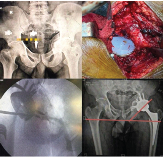
Figure 2: Case 2: Pre-operative, intraoperative and post-operative X-rays.
Given the patient’s age (36 years), high functional demand, and the nature of the defect with residual ilium and ischial fixation points, a primary THA with a large-diameter jumbo cementless porous-coated acetabular cup was planned. A 62 mm hemispherical titanium porous-coated press-fit cup was selected based on pre-operative templating.
Under spinal anesthesia in the lateral decubitus position through a posterior approach, the pseudoacetabulum was identified, debrided, and progressively reamed to restore the native hip center of rotation at the true acetabular level. The jumbo cup was impacted with a 1 mm press-fit and secured with two dome screws for supplemental fixation. Cup inclination angle of 45° and appropriate anteversion were confirmed intraoperatively with C-arm fluoroscopy (Fig. 2).
Immediate post-operative radiographs confirmed satisfactory cup positioning at an inclination angle of 45° with the hip center at the appropriate level. At the 3-month follow-up, the patient demonstrated an independent gait, and radiographs showed osseointegration in progress with no signs of migration.
Case 3: Primary THA with jumbo cup in pseudoacetabulum – failed acetabular repair (Paprosky type 3A)
A 29-year-old male, the youngest in the series, presented with significant left hip pain and functional disability. He had sustained an acetabular fracture treated with a traction pin and cannulated cancellous screw fixation, which had failed, resulting in a pseudoacetabulum formation with proximal migration of the femoral head. Pre-operative assessment confirmed a Paprosky Type 3A defect with formation of a fibrous pseudoacetabulum at a proximal and lateral location (Fig. 3).
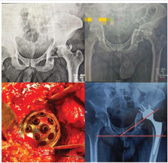
Figure 3: Case 3: Pre-operative, intraoperative photos and post-operative X-rays.
The surgical plan was primary THA with a jumbo cementless acetabular cup implanted within the existing pseudoacetabulum, aiming to maximize host bone contact across the available ilium and ischium. A 64 mm porous-coated hemispherical cup was selected. Progressive acetabular reaming through the pseudoacetabulum was performed to achieve a stable bony seat. Intraoperative C-arm guidance confirmed the appropriate cup position. Dome screw fixation was employed for supplemental stability.
Post-operative radiographs demonstrated satisfactory cup positioning within the pseudoacetabulum with the femoral head reduced concentrically (Fig. 1). At 3-month follow-up, the patient showed significant functional improvement with independent ambulation and a radiographically stable implant (Fig. 3).
Case 4: Revision THA with impaction cancellous bone grafting (Paprosky type 2B)
A 39-year-old male presented with left hip pain and functional impairment 4 years following cemented left THA performed in 2019. Clinical examination and radiographs revealed aseptic loosening of the cemented acetabular component with periprosthetic osteolysis and superior rim deficiency consistent with Paprosky Type 2B (Fig. 4). Inflammatory markers (erythrocyte sedimentation rate, C-reactive protein) were within normal limits, excluding septic loosening.
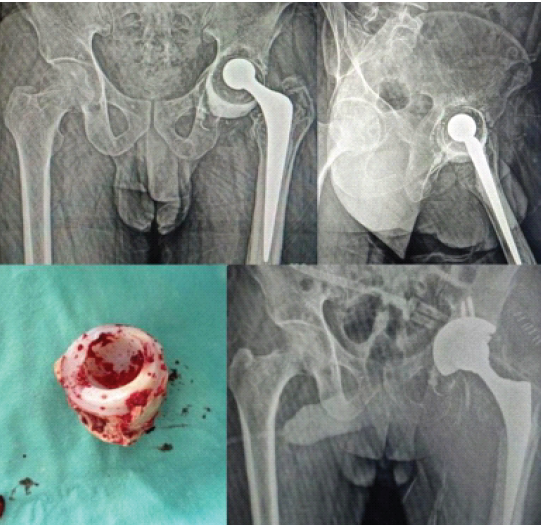
Figure 4: Case 4: Pre-operative X-rays and intraoperative photos.
Surgical planning determined revision THA with removal of the failed cemented cup and acetabular reconstruction using impaction cancellous bone grafting to restore acetabular bone stock, followed by implantation of a cementless revision shell. Under spinal anesthesia through a posterior approach, the loosened cemented cup was extracted using curved osteotomes. The fibrous membrane was excised, and the bone defects delineated.
Morselized autogenous cancellous bone graft from the iliac crest was impacted sequentially into the acetabular cavity to reconstruct the deficient areas. A cementless hemispherical revision shell was then press-fitted and secured with dome screws.
Post-operative radiographs confirmed satisfactory implant position. The patient was mobilized on post-operative day 2 with partial weight-bearing and progressed to full weight-bearing by 6 weeks. At follow-up, the patient demonstrated satisfactory functional recovery with an independent gait and reported significant pain relief.
This case series illustrates the practical application of two complementary acetabular reconstruction strategies – jumbo cementless cups and acetabular mesh with impaction bone grafting – across a spectrum of defect morphologies encountered at a tertiary center in central India. The series adds to a limited body of Indian literature on this technically demanding subject.
The predominance of young male patients (mean age 34 years) in our series contrasts markedly with Western revision arthroplasty registries, where polyethylene wear-related osteolysis in older patients prevails. In our cohort, all defects arose from post-traumatic sequelae: Neglected acetabular fractures, failed fracture fixation, or failed cemented primary THA. This reflects the epidemiology of hip disease at public sector tertiary centers in India, where resource constraints lead to delayed presentations with advanced deformity [8]. Despite this challenging substrate, our cases demonstrate that established Western reconstructive principles can be applied effectively with meticulous planning and intraoperative adaptability.
Jumbo acetabular cups exploit the remaining peripheral rim and medial wall to achieve three-point ilioischiopubic fixation without requiring structural bone graft [4]. Multiple series have documented 85–95% survivorship at 10 years for Paprosky types IIA and IIIA defects managed with hemispherical porous cups, with cup inclination angle and anteversion being key determinants of stability and wear [9,10]. In our cases, intraoperative C-arm fluoroscopy was instrumental in confirming cup inclination at 40–45°, consistent with the recommended safe zone [11]. The use of dome screws for supplemental primary stability is well supported in the literature and was employed in all jumbo cup cases [12].
For medial wall defects (Paprosky IIC), the acetabular mesh technique converts an uncontained defect into a contained defect, enabling high-quality impaction bone grafting [5]. The biological rationale is well-established: morselized autograft incorporates through creeping substitution, restoring bone stock for potential future revision [13]. The use of an autogenous iliac crest graft in Case 1 provided osteogenic, osteoconductive, and osteoinductive properties, and early graft consolidation was evident on 1-year radiographs. This approach avoids allograft-associated risks, including immunogenicity and disease transmission, which is particularly advantageous in resource-limited settings [14].
Pre-operative three-dimensional CT planning was invaluable in characterizing defect morphology beyond what is apparent on plain radiographs, enabling accurate selection of implant sizes, identification of fixation points, and templating of the reconstructed hip center [15]. This investment in planning translated to efficient intraoperative execution across all four cases.
Limitations of this series include its small sample size, exclusively male cohort, short to medium follow-up duration, and absence of formal pre- and post-operative HHS data. A prospective cohort study with validated outcome scores and a minimum 5-year follow-up is planned to build on these preliminary findings.
Jumbo cementless acetabular cups and acetabular mesh cage with impaction bone grafting are reliable surgical strategies for acetabular bone deficiency in both primary and revision THA. In this case series of four young males with Paprosky Types 2B, 2C, and 3A defects, all procedures achieved satisfactory clinical and radiological outcomes with no early complications. These techniques offer technically feasible, cost-effective alternatives to complex three-dimensional acetabular cage reconstructions, delivering stable biological fixation with appropriate pre-operative planning and intraoperative guidance. Their applicability in the Indian context – where post-traumatic young patients with delayed presentations predominate – deserves further prospective evaluation.
• The Paprosky classification guides surgical decision-making and implant selection for acetabular bone deficiency in THA
• Jumbo cementless porous-coated cups (≥62–66 mm) are effective for Paprosky types IIA, IIB, and IIIA defects with residual column integrity
• Acetabular mesh cage with autogenous impaction bone grafting effectively manages medial wall defects (Paprosky IIC)
• Intraoperative C-arm fluoroscopy is essential for confirming cup inclination angle and alignment, targeting a 40–45° safe zone
• Pre-operative three-dimensional CT planning and intraoperative adaptability are critical for successful acetabular reconstruction
• These techniques are applicable in resource-limited settings with predominantly young post-traumatic patients.
References
- 1. Paprosky WG, Perona PG, Lawrence JM. Acetabular defect classification and surgical reconstruction in revision arthroplasty. A 6-year follow-up evaluation. J Arthroplasty 1994;9:33-44. [Google Scholar] [PubMed]
- 2. Della Valle CJ, Paprosky WG. Classification and an algorithmic approach to the reconstruction of femoral deficiency in revision total hip arthroplasty. J Bone Joint Surg Am 2003;85-A Suppl 4:1-6. [Google Scholar] [PubMed]
- 3. Kurtz S, Ong K, Lau E, Mowat F, Halpern M. Projections of primary and revision hip and knee arthroplasty in the United States from 2005 to 2030. J Bone Joint Surg Am 2007;89:780-5. [Google Scholar] [PubMed]
- 4. Lachiewicz PF, Soileau ES. Jumbo acetabular components for complex primary and revision total hip arthroplasty. J Arthroplasty 2013;28:1548-51. [Google Scholar] [PubMed]
- 5. Kosashvili Y, Backstein D, Safir O, Lakstein D, Gross AE. Acetabular revision using an anti-protrusio cage and impaction bone grafting. J Bone Joint Surg Br 2009;91:742-7. [Google Scholar] [PubMed]
- 6. Siwach R, Singh R, Dahiya S. Acetabular reconstruction in complex primary and revision total hip arthroplasty. Indian J Orthop 2007;41:362-7. [Google Scholar] [PubMed]
- 7. Singh V, Jaggi N, Mago ML. Revision total hip arthroplasty: Challenges in India. J Clin Orthop Trauma 2019;10:773-9. [Google Scholar] [PubMed]
- 8. Jain AK, Srivastava A, Joshi MP. Post-traumatic arthritis of the hip: Experiences from a developing country. Int Orthop 2004;28:11-4. [Google Scholar] [PubMed]
- 9. Dearborn JT, Harris WH. High placement of an acetabular component inserted without cement in a revision total hip arthroplasty. Results after a mean of ten years. J Bone Joint Surg Am 1999;81:469-80. [Google Scholar] [PubMed]
- 10. Holt GE, Dennis DA. Use of custom triflanged acetabular components in revision total hip arthroplasty. Clin Orthop Relat Res 2004;429:209-14. [Google Scholar] [PubMed]
- 11. Lewinnek GE, Lewis JL, Tarr R, Compere CL, Zimmerman JR. Dislocations after total hip-replacement arthroplasties. J Bone Joint Surg Am 1978;60:217-20. [Google Scholar] [PubMed]
- 12. Kwon MS, Kuskowski M, Mulhall KJ, Macaulay W, Brown TE, Saleh KJ. Does surgical approach affect total hip arthroplasty dislocation rates? Clin Orthop Relat Res 2006;447:34-8. [Google Scholar] [PubMed]
- 13. Schreurs BW, Slooff TJ, Gardeniers JW, Buma P. Acetabular reconstruction with bone impaction grafting and a cemented cup: 20 Years’ experience. Clin Orthop Relat Res 2001;393:202-15. [Google Scholar] [PubMed]
- 14. Buttaro MA, Comba F, Zanotti G, Piccaluga F. Acetabular revision with impaction bone allografting and cemented cup: 7 to 17-year follow-up study. Clin Orthop Relat Res 2008;466:3073-80. [Google Scholar] [PubMed]
- 15. Steppacher SD, Kowal JH, Murphy SB. Improving cup positioning using a mechanical navigation instrument. Clin Orthop Relat Res 2011;469:423-8. [Google Scholar] [PubMed]
- 16. Harris WH. Traumatic arthritis of the hip after dislocation and acetabular fractures: Treatment by mold arthroplasty. An end-result study using a new method of result evaluation. J Bone Joint Surg Am 1969;51:737-55. [Google Scholar] [PubMed]
- 17. Nehme A, Lewallen DG, Hanssen AD. Modular porous metal augments for treatment of severe acetabular bone loss during revision hip arthroplasty. Clin Orthop Relat Res 2004;429:201-8. [Google Scholar] [PubMed]
- 18. Garbuz D, Morsi E, Gross AE. Revision of the acetabular component of a total hip arthroplasty with a massive structural allograft. Study with a minimum five-year follow-up. J Bone Joint Surg Am 1996;78:693-7. [Google Scholar] [PubMed]
- 19. Sporer SM, Paprosky WG. Acetabular revision using a trabecular metal acetabular component for severe acetabular bone loss associated with a pelvic discontinuity. J Arthroplasty 2006;21 6 Suppl 2:87-90. [Google Scholar] [PubMed]
- 20. Issack PS, Nousiainen M, Beksac B, Helfet DL, Sculco TP, Buly RL. Acetabular component revision in total hip arthroplasty. Part II: Management of major bone loss and pelvic discontinuity. Am J Orthop (Belle Mead NJ) 2009;38:550-6. [Google Scholar] [PubMed]






Related Articles in Journal of Orthopaedic Case Reports
 August 1, 2026 Metallosis Following Total Hip Arthroplasty: A Case Report
 August 1, 2026 Short-term Outcomes of Impaction Bone Grafting Technique with a Collared Fully Hydroxyapatite-Coated Stem in Revision Total Hip Arthroplasty: A Case Series
 July 1, 2026 Bone Morphogenetic Proteins-2–Augmented Acetabular Bone Stock Restoration During Two-stage Revision for Periprosthetic Joint Infection: A Case Report
 December 1, 2025 Total Hip Arthroplasty in Failed Hip Fractures, a Challenge with Fruitful Outcome: A Case Series