

Open reduction and internal fixation with cannulated cancellous screws augmented by spiked washers provides stable anti-rotational fixation for bilateral tibial tubercle avulsion fractures, enabling anatomical union, preservation of physeal integrity, early rehabilitation, and excellent long-term functional outcomes, making it a reliable surgical option for this rare and complex Tibial tuberosity avulsion injury.
Dr. Kagana Avinash, Department of Orthopaedic Surgery, All India Institute of Medical Sciences, Raipur, Chhattisgarh, India. E-mail: avinash2012.k@gmail.com
Abstract
Introduction: Bilateral tibial tuberosity avulsion fractures (TTAF) are among the rarest and most significant injuries affecting the extensor mechanism in skeletally immature patients. Successful treatment mandates acute anatomical reduction and stable internal fixation that accounts for high quadriceps tensile forces and the risk of physeal growth arrest.
Case Report: A 14-year-old male sustained an acute, displaced bilateral TTAF (Modified Watson–Jones/Ogden Type II) after a vigorous, simultaneous jump and landing. The patient presented with partial bilateral loss of active knee extension. Acute open reduction and internal fixation (ORIF) was performed. The fixation involved one 4.0 mm cannulated cancellous (CC) screw per knee. The critical technical detail was the use of a spiked washer under the head of the CC screw on both sides, ensuring enhanced fragment compression, stability, and anti-rotation against the deforming forces of the quadriceps.
Results: The patient was followed up for one year. Radiographs confirmed complete, solid bony union of both tibial tuberosities with no secondary displacement, hardware complication, or evidence of proximal tibial physeal arrest (e.g., tibia recurvatum). Clinically, the outcome was excellent, with a full, pain-free range of motion, measuring 0° extension to 135° flexion bilaterally. The patient achieved a full, unrestricted return to competitive sports activities.
Conclusion: ORIF using CC screws strategically augmented by spiked washers offers a biomechanically superior and reliable fixation strategy for displaced bilateral TTAF in young children. This approach promotes rapid and stable bony fusion, preserves proximal tibial growth, and consistently achieves excellent functional outcomes.
Keywords: Tibial tuberosity avulsion fracture, bilateral, pediatric, open reduction and internal fixation, spiked washer, physeal injury, growth arrest, knee extensor mechanism.

Tibial tuberosity avulsion fracture (TTAF) is a rare Salter–Harris equivalent injury [1-6] involving the secondary ossification center of the proximal tibia, usually caused by forceful, eccentric contraction of the quadriceps transmitted through the patellar tendon [5,7]. This injury commonly affects adolescent males aged 14–16 years during periods of rapid growth [5,8,9]. Still, its occurrence in younger children is uncommon and presents unique management challenges due to greater remaining growth potential.
Management is guided by the modified Watson–Jones/Ogden classification [1,5,10-13]. While Type I (un-displaced) and Type II (minimally displaced) fractures may be managed conservatively, Type III and Type IV injuries, characterized by complete avulsion and/or transphyseal extension, require open reduction and internal fixation (ORIF). The primary surgical objectives are anatomical reduction, stable fixation, restoration of extensor mechanism function, and prevention of premature physeal closure [3,6], which may result in angular deformities such as tibia recurvatum.
Simultaneous bilateral TTAFs are rare, with fewer than 30 cases documented in the literature [4,11,8]. Such presentations suggest a unique, high-energy, synchronous loading event affecting both lower extremities. This report details the successful surgical management of bilateral Type II TTAF in a pediatric patient, utilizing 4.0 mm cannulated cancellous (CC) screws and spiked washers to achieve stable fixation.
Patient history and examination
A previously healthy 14-year-old male, actively engaged in competitive sports, presented to the emergency department following a non-contact injury sustained during sports activity. The injury mechanism involved a forceful, simultaneous jump with an abrupt attempt to halt upon landing, resulting in intense, eccentric loading of both quadriceps muscles.
On examination, the patient exhibited marked bilateral knee effusions and localized swelling over the anterior aspects of both proximal tibias. There was pronounced tenderness and a palpable defect at the patellar tendon insertion site. The patient demonstrated complete bilateral loss of active knee extension and was unable to perform a straight leg raise (SLR). Both patellae appeared high-riding (patella Alta). Distal pulses, sensation, and motor function were preserved, and compartment pressures were within normal limits.
Imaging findings and classification
Initial anteroposterior and lateral radiographs of both knees confirmed bilateral TTAF (Fig. 1a). Lateral views revealed avulsion and proximal displacement of the entire tibial tuberosity, including the patellar tendon insertion, consistent with patella Alta. The fracture line extended into the secondary ossification center (physis), and the injury was classified as Type II according to the modified Watson–Jones/Ogden system[13]. Pre-operative computed tomography scans were performed to delineate fracture morphology, assess displacement, and exclude occult articular involvement (Fig. 1b).
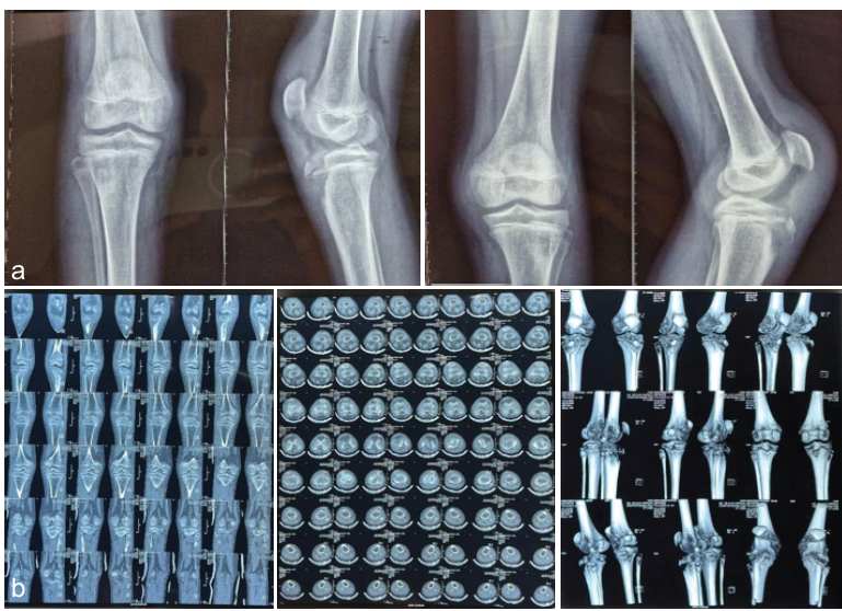
Figure 1: (a) Pre-operative bilateral knee X-rays (anteroposterior and lateral) confirming Type II tibial tuberosity avulsion fractures and patella alta. (b) Pre-operative bilateral knee computed tomography scans (sagittal and coronal reconstructions) detailing fracture displacement and physeal extension.
Surgical technique
The patient underwent acute bilateral ORIF under general anesthesia with bilateral thigh tourniquets.
- Approach and reduction: A longitudinal incision was made over each tibial tuberosity. Fracture hematomas were evacuated, and interposed periosteal flaps were removed. Anatomical reduction was achieved by applying distal traction on the patellar tendon using an Ethibond suture, drawing the fragment distally and posteriorly into its anatomical position. Reduction was confirmed both visually and fluoroscopically.
- Fixation with spiked washers: 1.6 mm guide wires for 4.0 mm CC screws were inserted under fluoroscopic guidance, parallel to the proximal tibial physis to minimize physeal injury. A single 4.0 mm CC screw was placed per knee to achieve optimal compression. The critical technical step involved “augmenting fixation with a spiked washer” beneath the screw head on each side. The washer’s serrations engaged the bone of the avulsed fragment, providing a biomechanical advantage by resisting tensile and rotational forces from the quadriceps and effectively securing the fragment. Final intraoperative fluoroscopy confirmed satisfactory reduction and hardware placement, ensuring no excessive compression across the physis.
- Closure and reinforcement: The anterior periosteum and soft-tissue sleeve were repaired over the fracture site and hardware, using absorbable sutures. Wounds were closed in layers. (Fig. 2).
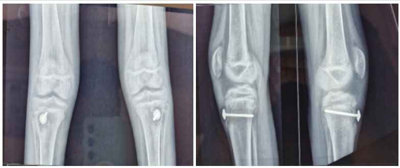
Figure 2: Immediate Post-operative bilateral knee X-rays (AP and lLateral) showing excellent reduction and stable fixation
Post-operative care and outcome
Following surgery, the patient was immobilized in an above-knee cast and maintained non-weight-bearing (NWB) for 4 weeks.
- Rehabilitation: At 4 weeks postoperatively, a controlled rehabilitation program was initiated. This included supervised, gentle passive range of motion exercises, limited to 40°, to prevent joint stiffness while protecting the fixation. At 6 weeks, with radiographic evidence of early fracture healing, the patient progressed to partial weight-bearing and began gentle active quadriceps strengthening. Full weight bearing was introduced at 8 weeks, followed by progressive resistance training to restore muscle strength and function.
- Final outcome: At one year follow-up, the patient was completely asymptomatic. Clinical examination demonstrated a stable extensor mechanism with no evidence of extensor lag. The patient achieved a full, symmetrical, pain-free range of motion (0°–135°) in both knees and successfully returned to pre-injury levels of competitive sports activity.
- Radiographic follow-up: Serial radiographs confirmed complete, solid bony union across both fracture sites, with no evidence of growth disturbance such as tibia recurvatum or limb length discrepancy. These findings confirmed successful preservation of the proximal tibial physis. Elective hardware removal was performed at 1 year post-operative, as indicated by the patient’s recovery and radiographic findings. (Fig. 3 & 4).
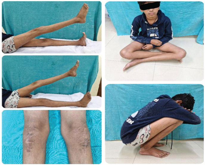
Figure 3: Clinical photo showing full, symmetrical active knee flexion and extension at 6-month follow-up.
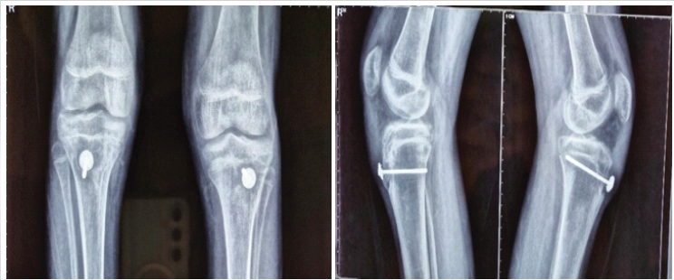
Figure 4: 6-month follow-up bilateral knee X-rays demonstrating solid fusion and confirmation of open, functional physis.
Rarity and clinical challenges
Bilateral TTAF are exceedingly rare, with only a limited number of cases reported in the literature [4,11]. The occurrence of this injury in a 14-year-old patient is particularly notable, as the secondary ossification center of the proximal tibia is still actively contributing to longitudinal growth at this age.
Biomechanical considerations and fixation strategy
The primary challenge in surgically managing TTAF lies in counteracting the substantial tensile forces generated by the quadriceps mechanism [3,5]. Traditional fixation methods, such as CC screws, provide axial compression but may be insufficient to resist rotational and shear forces, potentially leading to fixation failure. In this case, the use of spiked washers in conjunction with CC screws offered a significant biomechanical advantage. The serrated edges of the washer engaged the cortical bone, acting as a rotational stop and distributing compressive forces more evenly [10]. This construct allowed for earlier initiation of controlled passive motion, reducing the risk of joint stiffness and facilitating a more rapid return to function compared to prolonged immobilization.
Prevention of growth arrest
A critical aspect of the surgical technique was the preservation of the proximal tibial physis. By carefully placing the screws parallel to the physis and avoiding excessive compression across the growth plate, the risk of iatrogenic physeal injury was minimized. The absence of angular deformity or limb length discrepancy at follow-up supports the effectiveness of this approach in maintaining physeal integrity.
This case demonstrates that ORIF with CC screws augmented by spiked washers is a safe, reproducible, and highly effective technique for the management of bilateral tibial tubercle avulsion fractures. The construct provides rigid anti-rotational stability capable of withstanding the substantial forces of the extensor mechanism, thereby promoting anatomical healing, preserving physeal integrity, and facilitating early functional rehabilitation. The excellent radiological and clinical outcomes, culminating in an unrestricted return to competitive sports, support this technique as an optimal surgical strategy for displaced bilateral TTAFs and reinforce its value in achieving predictable long-term outcomes despite the rarity and complexity of this injury.
Open reduction and internal fixation with cannulated cancellous screws and spiked washers is a safe, reproducible, and biomechanically robust technique for bilateral tibial tuberosity avulsion fractures. This construct provides stable fixation against extensor mechanism forces, promotes anatomical healing, and supports early rehabilitation with restoration of full knee function and unrestricted return to sports.
References
- 1. </p> [Google Scholar] [PubMed]
- 2. <p>1. Watson-Jones R. Fractures and Other Bone and Joint Injuries. 4th ed. Edinburgh: Churchill Livingstone; 1955. [Google Scholar] [PubMed]
- 3. Cahill BR, Berg BC. Avulsion fracture of the tibial tubercle with avulsion of the patellar ligament. Report of two cases. J Bone Joint Surg Am. 1983;65:989-91. [Google Scholar] [PubMed]
- 4. Sponseller PD, Stanitski CL, Grogan DP. Surgical management of tibial tubercle fractures in adolescents. J Pediatr Orthop. 1987;7:121-6. [Google Scholar] [PubMed]
- 5. Mosheiff R, Ezra E, Mintz D, Mogle P. Bilateral avulsion fracture of the tibial tuberosity. J Trauma. 1988;28:1733-4. [Google Scholar] [PubMed]
- 6. Ogden JA, Hixson RL. Tibial tuberosity fractures in adolescents. J Pediatr Orthop. 1993;13:453-61. [Google Scholar] [PubMed]
- 7. Peterson HA. Physeal fractures: Part 3. Classification and management of growth plate fractures. J Bone Joint Surg Am. 1994;76:470-80. [Google Scholar] [PubMed]
- 8. Rumball KM, Finnegan MA, Raso VJ, Loehr JA. Tibial tubercle avulsion fractures: A review of 20 cases and recognition of an adolescent sporting injury. Can J Surg. 2004;47:107-11. [Google Scholar] [PubMed]
- 9. Gicquel P, Dutoit P, Karger C, Kieny P. Bilateral avulsion fracture of the tibial tuberosity in a 13-year-old boy. J Orthop Trauma. 2005;19:574-7. [Google Scholar] [PubMed]
- 10. Pandey V, Dhillon MS, Bhardwaj P, Bali K. Tibial tuberosity fractures in adolescents: A clinical and radiological review of 22 cases. J Child Orthop. 2010;4:199-204. [Google Scholar] [PubMed]
- 11. Kraus T, Rehm A, Küster M, Wipfler M. Fixation of tibial tuberosity avulsion fractures in adolescents. Eur J Trauma Emerg Surg. 2011;37:169-74. [Google Scholar] [PubMed]
- 12. Polakow I, Prevel C, Kherad O, Dujardin F, Rigoard P. Bilateral tibial tuberosity fracture in a child with no predisposing factors: A case report and review of the literature. Orthop Traumatol Surg Res. 2013;99:969-72. [Google Scholar] [PubMed]
- 13. Goudie EB, Taranu R, Naranja J. Complications of tibial tuberosity fractures in adolescents. J Pediatr Orthop B. 2015;24:111-7. [Google Scholar] [PubMed]
- 14. Preston TB, Phelan ES, Brannan A. Tibial tubercle fracture. In: StatPearls. Treasure Island, FL: StatPearls Publishing; 2024. [Google Scholar] [PubMed]




Related Articles in Journal of Orthopaedic Case Reports
 July 10, 2024 Bilateral Anterior Cruciate Ligament Reconstruction with All-Inside Technique for Adolescent: A Review of the Literature and Case Report
 October 10, 2023 Bilateral Non-traumatic Neglected Femoral Neck Fracture in a Healthy Adolescent: A Case Report and Review of Literature
 October 1, 2026 Total Hip Arthroplasty in the Adolescent Population: A Case Series
 August 1, 2026 Bisphosphonate-Related Pathological Femoral Fracture in a Child with ACTG2-Related Intestinal Failure and Metabolic Bone Disease: A Case Report