

Bisphosphonate-associated atypical femoral fractures can occur after a single annual cycle of therapy in children with severe metabolic bone disease and pre-existing intramedullary hardware, highlighting the need for pre-treatment imaging and vigilance for atypical fracture patterns in this population.
Dr. William Zhang, Max Rady College of Medicine, University of Manitoba, 37 Kings Drive, Winnipeg, MB R3T 3E6, Manitoba, Canada. E-mail: zhangw9@myumanitoba.ca
Abstract
Introduction: Atypical femoral fractures (AFFs) are stress-type insufficiency fractures classically associated with long-term bisphosphonate therapy in adults. Reports of AFFs in pediatric patients are limited primarily to osteogenesis imperfecta or other monogenic bone disorders following prolonged exposure. We report the first case of an AFF occurring after zoledronic acid administration in a child with ACTG2 gene mutation-related intestinal failure and severe metabolic bone disease.
Case Report: An 8-year-old male with ACTG2-related intestinal failure, total parenteral nutrition dependence, and multiple fragility fractures presented following initiation of annual zoledronic acid therapy. He had previously undergone right femur shaft fracture fixation via flexible intramedullary rods. Approximately 7 months later, he sustained a minimally displaced subtrochanteric proximal femoral fracture fulfilling American Society for Bone and Mineral criteria for AFF: A short oblique orientation, subtrochanteric location, cortical thickening, and minimal trauma mechanism. The fracture required conversion to open reduction and internal fixation with a plate-and-screw construct. Eight-week post-operative radiographs demonstrated progressive fracture healing with advancing periosteal callus formation and maintained hardware alignment.
Conclusion: Children with severe metabolic bone disease and underlying smooth muscle gene mutations may be vulnerable to AFF after bisphosphonate exposure. Clinicians initiating bisphosphonate therapy in children with severe metabolic bone disease and pre-existing intramedullary hardware should conduct thorough pre-treatment imaging and maintain vigilance for AFF patterns during follow-up. Level of Evidence: Level V – Expert Opinion/Case Report
Keywords: ACTG2, atypical femoral fracture, bisphosphonate, intestinal failure, metabolic bone disease, pediatric, zoledronic acid.

Atypical femoral fractures (AFFs) are stress-type insufficiency fractures classified by the American Society for Bone and Mineral (ASBMR) as characterized by a transverse or short oblique fracture line, lateral cortical thickening or beaking, minimal trauma, and a location within the subtrochanteric or diaphyseal femur [1]. In adults, AFF risk increases with duration of bisphosphonate use, with the highest rates observed after 5 or more years of therapy [2]. Reports of AFF in pediatric patients have emerged primarily in the context of long-term bisphosphonate therapy in osteogenesis imperfecta (OI) [3,4] and, more recently, in children with other monogenic bone disorders, including low-density lipoprotein receptor-related protein 5 (LRP5) mutation-related osteoporosis and spinal muscular atrophy, following months to years of exposure [5,6]. To our knowledge, no case of AFF or AFF-like fracture morphology following an initial annual cycle of bisphosphonate therapy has been reported in any age group.
Children with intestinal failure dependent on total parenteral nutrition (TPN) represent a distinct population at high risk for metabolic bone disease, in whom bisphosphonate use remains limited to a small number of case reports [7,8]. The actin gamma 2 (ACTG2) gene encodes gamma-2 smooth muscle actin; pathogenic mutations cause a spectrum of intestinal dysmotility, including megacystis-microcolon-intestinal hypoperistalsis syndrome (MMIHS), with skeletal fragility arising as a secondary consequence of malabsorption and TPN dependence rather than a primary bone structural defect [9].
We present the case of an 8-year-old male with ACTG2-related intestinal failure, TPN dependence, and a history of multiple fragility fractures who developed a femoral fracture with atypical morphological features following the initiation of zoledronic acid therapy.
Patient background
The patient is an 8-year-old male with a confirmed pathogenic ACTG2 gene mutation resulting in severe intestinal dysmotility and functional intestinal failure. TPN-dependent since infancy, he has undergone multiple gastrointestinal surgical interventions, including intestinal resection and bowel anastomosis. Chronic malabsorption and TPN-associated metabolic bone disease resulted in severe osteopenia, documented on plain radiographs, with multiple fragility fractures beginning in early childhood that were managed non-operatively.
Follow-up imaging demonstrated healing fractures and generalized osseous undermineralization, consistent with the degree of underlying metabolic bone disease. Before initiating bisphosphonate therapy, a spine radiograph confirmed no vertebral compression fractures, serving as a pre-treatment baseline.
Index fracture and initial surgical management
At age seven, the patient sustained a low-energy fall resulting in an acute oblique fracture of the distal right femoral diaphysis. CT of the right femur was performed to exclude an underlying pathological lesion, confirming the acute fracture and demonstrating cortical thickening of the femoral shaft consistent with prior fracture remodeling; no aggressive osseous lesion was identified. He underwent open reduction and internal fixation via insertion of two flexible intramedullary rods under fluoroscopic guidance, achieving satisfactory fracture alignment. Subsequent follow-up imaging demonstrated progressive fracture healing with periosteal callus formation. Hardware-related complications necessitated a rod adjustment procedure during the follow-up period.
Zoledronic acid administration
Following fracture healing, the patient was initiated on annual intravenous zoledronic acid infusions by pediatric endocrinology subspecialists for the management of TPN-associated metabolic bone disease. This represented his first exposure to bisphosphonate therapy; no antiresorptive agents had been administered at any prior point in his clinical course.
Atypical fracture event
Approximately 7 months following his initial zoledronic acid infusion, the patient presented to the emergency department with acute right thigh pain following a minimal-trauma mechanism; an audible crack was noted while ambulating on level ground. Plain radiographs of the right femur demonstrated a mildly displaced subtrochanteric proximal femur fracture with moderate anterolateral angulation, arising through the femoral shaft segment occupied by the pre-existing flexible intramedullary rods (Fig. 1). The fracture pattern fulfilled ASBMR major diagnostic criteria for an AFF: Short oblique orientation, subtrochanteric location, lateral cortical thickening on prior cross-sectional imaging, and a minimal trauma mechanism.
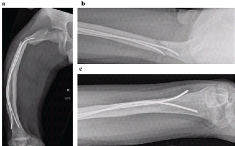
Figure 1: Pre-operative radiographs demonstrating atypical femoral fracture morphology with intramedullary rods in situ. Anteroposterior and lateral plain radiographs of the right femur were obtained at the time of fracture presentation. (a) Anteroposterior view demonstrating a mildly displaced subtrochanteric proximal femur fracture with moderate anterolateral angulation, occurring through the segment traversed by the pre-existing flexible intramedullary rods. (b) Lateral view demonstrating the same fracture with associated anterolateral angulation. (c) Distal femur view demonstrating endosteal bone formation and the diverging distal ends of the two flexible intramedullary rods. The short oblique fracture orientation, subtrochanteric location, and minimal trauma mechanism are consistent with an atypical femoral fracture per American Society for Bone and Mineral Research major diagnostic criteria.
Surgical management
The patient was brought to the operating room with a planned procedure of flexible intramedullary nail removal, corrective femoral osteotomies for prior malunions, and intramedullary fixation to reduce the risk of future fracture. The procedure was initiated with the removal of the existing flexible nails. A standard approach was then utilized for insertion of an antegrade lateral entry femoral nail.
However, the guidewire could not be advanced through the femoral canal. Intraoperative assessment demonstrated marked obliteration of the medullary canal by dense cortical bone along much of the diaphysis (Fig. 1a-c), likely secondary to prior bisphosphonate therapy and multiple fragility fractures.
Given the inability to establish intramedullary access, an intraoperative decision was made to proceed with plate fixation. Through an extended lateral approach, a proximal femoral locking plate was applied, achieving satisfactory reduction (Fig. 2). Intraoperative fluoroscopy confirmed appropriate implant positioning and cortical apposition at the osteotomy site.
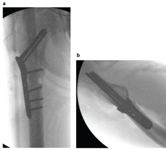
Figure 2: Intraoperative fluoroscopic images demonstrating open reduction and internal fixation of the right proximal femoral fracture. Fluoroscopic images obtained intraoperatively during open reduction and internal fixation of the right proximal femoral atypical fracture. (a) Anteroposterior fluoroscopic image demonstrating plate-and-screw fixation with a lateral proximal femoral locking plate. (b) Lateral fluoroscopic image demonstrating the same construct, confirming maintained reduction and appropriate hardware positioning.
A subsequent attempt was made to introduce flexible intramedullary nails in a retrograde fashion through the prior entry sites to provide supplemental full-length stability and mitigate the risk of distal stress riser–related periprosthetic fracture. This was abandoned due to persistent lack of canal patency and endosteal bone formation.
Postoperatively, the patient was mobilized with crutches under a partial weight-bearing protocol. Two-week follow-up radiographs demonstrated maintained fracture alignment with early periosteal callus formation (Fig. 3). Eight-week follow-up radiographs demonstrated progressive fracture healing with advancing periosteal callus formation and maintained alignment of the proximal femoral locking plate (Fig. 4).
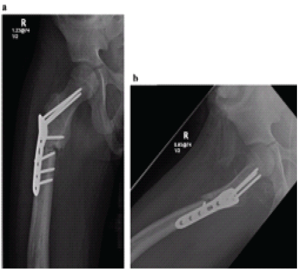
Figure 3: Two-week post-operative radiographs demonstrating proximal femoral fracture fixation. Plain radiographs of the right femur were obtained 2 weeks following open reduction and internal fixation. (a) Anteroposterior radiograph demonstrating a proximal femoral locking plate in situ with maintained fracture alignment. (b) Lateral radiograph confirming satisfactory hardware positioning without evidence of malposition.
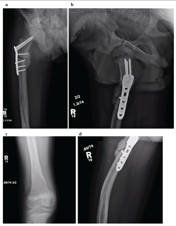
Figure 4: Eight-week post-operative radiographs demonstrating progressive proximal femoral fracture healing. Plain radiographs of the right femur obtained 8 weeks following open reduction and internal fixation, demonstrating progressive fracture healing. (a) Anteroposterior radiograph of the right femur demonstrating advancing periosteal callus formation and maintained proximal femoral locking plate alignment. (b) Anteroposterior radiograph of the right hip demonstrating the proximal extent of the plate construct with maintained reduction at the fracture site. (c) Anteroposterior radiograph of the distal femur and knee demonstrating no evidence of distal stress riser-related periprosthetic fracture. (d) Lateral radiograph of the proximal femur demonstrating the plate-and-screw construct in profile, confirming cortical apposition at the fracture site and progressive callus bridging.
This case presents what we believe to be the first reported instance of AFF-consistent fracture morphology following an initial annual cycle of bisphosphonate therapy in any patient, pediatric or adult. All previously reported pediatric AFFs have occurred in the context of months to years of bisphosphonate exposure: The earliest pediatric AFF report involved a 16-year-old girl after 2 years of pamidronate therapy; [5] a child with spinal muscular atrophy developed an AFF after 3 years of intravenous bisphosphonate treatment; [6] and twin 9-year-olds with LRP5 mutation sustained AFFs following 3 years of zoledronic acid [10]. In adult literature, the risk of AFF increases proportionally with treatment duration, with meaningful risk generally recognized after 2 or more years of use [2].
The pathophysiology of AFFs involves suppression of bone remodeling, leading to accumulation of microdamage and propagation of cortical microcracks that cannot be repaired in the absence of normal remodeling activity [11]. In otherwise healthy adults, this process requires years of bisphosphonate-induced remodeling suppression. We propose that the patient’s unique bone biology dramatically lowered the threshold for this mechanism. His skeleton was, in effect, already “primed” for atypical fracture: Years of TPN-associated metabolic bone disease and malabsorption had produced severe undermineralization documented on plain radiography; prior fractures had generated areas of cortical remodeling and periosteal reaction; and existing flexible intramedullary rods concentrated mechanical stress at the implant-bone interface. In this context, even an initial annual infusion of zoledronic acid a potent bisphosphonate with prolonged skeletal retention may have been sufficient to tip the balance toward impaired microcrack repair, particularly at a site of existing implant-related stress concentration.
The role of intramedullary hardware as a stress concentrator deserves specific attention. Lee et al. described a series of bisphosphonate-associated peri-implant fractures occurring at the ends of plate constructs, proposing that differential stiffness concentrates dynamic tensile strains at implant boundaries in antiresorptive-treated bone [12]. Duarte Simoes et al. reported the first cases of AFF-type fractures occurring around intramedullary nails specifically, noting that even intramedullary implants, which distribute loads more homogeneously than plates, are not exempt from this phenomenon [13]. Hegazy et al. described six OI children who developed AFF-pattern fractures through or adjacent to existing intramedullary rods after long-term pamidronate therapy [3]. In the patient’s case, the fracture occurred precisely within the segment of femoral shaft traversed by two flexible intramedullary rods, raising the possibility that hardware-related stress concentration contributed to fracture localization.
The ACTG2 mutation introduces an additional layer of complexity. ACTG2 encodes gamma-2 smooth muscle actin, expressed in the smooth muscle of the gastrointestinal tract. The primary consequence of pathogenic ACTG2 variants is intestinal dysmotility leading to functional intestinal failure, with skeletal pathology arising as a secondary consequence of malabsorption and TPN dependence rather than a primary bone structural defect. The closest published analog is the case reported by Duke et al. of a child with MMIHS, a condition sharing genetic and phenotypic overlap with ACTG2-related disease, who developed progressive metabolic bone disease and pathological fractures on TPN [7]. Pastore et al. subsequently demonstrated that pamidronate improves bone mineral density (BMD) in pediatric intestinal failure, and that children with congenital gut dysmotility disorders are at particularly high risk for metabolic bone disease [8].
Whether ACTG2 or related smooth muscle gene mutations independently affect bone biology beyond the metabolic consequences of intestinal failure remains unknown. However, emerging evidence suggests that genetic variants affecting the Wingless-related integration site signaling pathway and other bone remodeling pathways may confer susceptibility to AFF independent of bisphosphonate use [14].
The residual diaphyseal bowing deformity distal to the fixation construct, and its potential to act as a stress riser for future periprosthetic fracture, was considered a potential stress riser for future periprosthetic fracture. Given the patient’s history of significant post-operative complications, including a hospital admission exceeding 8 months secondary to post-operative ileus, the intraoperative decision was made to limit operative duration to mitigate the risk of recurrent perioperative morbidity. A corrective osteotomy spanning the full femoral length would have substantially increased operative time and was felt to carry unacceptable risk in this medically fragile patient. This decision was made with acceptance of the potential for post-operative periprosthetic fracture, and options for deformity correction were discussed with the family as part of long-term management planning.
Several important limitations must be acknowledged. First, establishing causality between bisphosphonate administration and the subsequent fracture is inherently difficult. The fracture could represent another fragility fracture in the context of ongoing severe metabolic bone disease, independent of the zoledronic acid exposure. However, the short oblique subtrochanteric fracture pattern, cortical thickening documented on prior imaging, minimal trauma mechanism, and occurrence at the implant-bone interface are morphological features that collectively favor an AFF-consistent interpretation rather than a simple fragility fracture. Second, we do not have dual-energy X-ray absorptiometry scan data to quantify the degree of bone fragility at baseline formally. Third, this is a single case report, and no causal inference can be drawn at the population level.
Clinically, this case highlights the need for caution when initiating bisphosphonate therapy in children with severe metabolic bone disease and pre-existing intramedullary hardware. We suggest that in such patients, a thorough assessment of fracture history, imaging characteristics of existing fractures, and hardware configuration should precede bisphosphonate initiation. Contralateral femoral screening for cortical thickening and prodromal stress reactions, as recommended in adults and pediatric patients with monogenic bone disorders, may be appropriate in this population [15]. The decision to continue, modify, or discontinue bisphosphonate therapy following an AFF-pattern fracture should weigh the potential benefits of improved BMD against the risk of further atypical fractures, and should involve shared decision-making with the family and a multidisciplinary team including endocrinology, gastroenterology, and orthopedic surgery. We recommend pre-operative advanced imaging, such as computed tomography or magnetic resonance imaging, to assess for endosteal bone formation that may progressively narrow or obliterate the medullary canal. Identification of these changes preoperatively may facilitate surgical planning, including selection of fixation strategy and implant choice.
We describe the first reported case of an AFF following an initial annual cycle of zoledronic acid in a child with ACTG2-related intestinal failure, TPN dependence, and severe metabolic bone disease. The severity of pre-existing bone compromise, combined with stress concentration at the site of pre-existing intramedullary hardware, likely lowered the threshold for atypical fracture pathophysiology below that typically associated with prolonged antiresorptive therapy. This case highlights that monogenic and metabolic conditions affecting bone biology may modify individual susceptibility to AFF, and underscores the importance of careful patient selection, pre-treatment imaging, and close clinical follow-up when bisphosphonates are initiated in medically complex pediatric patients.
Children with severe metabolic bone disease secondary to intestinal failure and pre-existing intramedullary hardware may be susceptible to AFF even after a single cycle of bisphosphonate therapy. Clinicians should obtain pre-treatment advanced imaging to assess medullary canal patency and maintain vigilance for atypical fracture morphology during follow-up in this population.
References
- 1. Shane E, Burr D, Abrahamsen B, Adler RA, Brown TD, Cheung AM, et al. Atypical subtrochanteric and diaphyseal femoral fractures: Second report of a task force of the American Society for bone and mineral research. J Bone Miner Res 2014;29:1-23. [Google Scholar] [PubMed]
- 2. Dell RM, Adams AL, Greene DF, Funahashi TT, Silverman SL, Eisemon EO, et al. Incidence of atypical nontraumatic diaphyseal fractures of the femur. J Bone Miner Res 2012;27:2544-50. [Google Scholar] [PubMed]
- 3. Hegazy A, Kenawey M, Sochett E, Tile L, Cheung AM, Howard AW, et al. Unusual femur stress fractures in children with osteogenesis imperfecta and intramedullary rods on long-term intravenous pamidronate therapy. J Pediatr Orthop 2016;36:757-61. [Google Scholar] [PubMed]
- 4. Vasanwala RF, Sanghrajka A, Bishop NJ, Högler W. Recurrent proximal femur fractures in a teenager with osteogenesis imperfecta on continuous bisphosphonate therapy: Are we overtreating? J Bone Miner Res 2016;31:1923-8. [Google Scholar] [PubMed]
- 5. Boyce AM, Collins MT, Tosi LL, Gafni RI. A subtrochanteric femoral stress fracture following bisphosphonate treatment in an adolescent girl. Horm Res Paediatr 2017;87:69-72. [Google Scholar] [PubMed]
- 6. Nasomyont N, Hornung LN, Wasserman H. Intravenous bisphosphonate therapy in children with spinal muscular atrophy. Osteoporos Int 2020;31:563-9. [Google Scholar] [PubMed]
- 7. Duke JL, Jones DP, Frizzell NK, Chesney RW, Hak EB. Pamidronate in a girl with chronic renal insufficiency dependent on parenteral nutrition. Pediatr Nephrol 2003;18:714-7. [Google Scholar] [PubMed]
- 8. Pastore S, Londero M, Barbieri F, Di Leo G, Paparazzo R, Ventura A. Treatment with pamidronate for osteoporosis complicating long-term intestinal failure. J Pediatr Gastroenterol Nutr 2012;55:615-8. [Google Scholar] [PubMed]
- 9. Wangler MF, Gonzaga-Jauregui C, Gambin T, Penney S, Moss T, Chopra A, et al. Heterozygous de novo and inherited mutations in the smooth muscle actin (ACTG2) gene underlie megacystis-microcolon-intestinal hypoperistalsis syndrome. PLoS Genet 2014;10:e1004258. [Google Scholar] [PubMed]
- 10. Sarhan M, Aly M, Williams P, Humphry S. Bisphosphonate-induced atypical femoral fractures in pediatric patients with lipoprotein receptor-related protein 5 (LRP5) gene mutation: A case report of twin patients. Cureus 2025;17:e87984. [Google Scholar] [PubMed]
- 11. Chapurlat RD, Delmas PD. Bone microdamage: A clinical perspective. Osteoporos Int 2009;20:1299-308. [Google Scholar] [PubMed]
- 12. Lee JY, Soh T, Howe TS, Koh JS, Kwek EB, Chua DT, et al. Bisphosphonate-associated peri-implant fractures: A new clinical entity? Acta Orthop 2015;86:622-6. [Google Scholar] [PubMed]
- 13. Duarte Simoes N, Goncalves Z, Moreno J, Paiva F, Pinho S, Varzielas M. Peri-implant atypical fractures associated with bisphosphonates: Should this clinical entity be included in the definition of atypical femoral fracture? J Orthop Case Rep 2018;8:66-69. [Google Scholar] [PubMed]
- 14. Zhou W, Van Rooij JG, Ebeling PR, Verkerk AJ, Zillikens MC. The genetics of atypical femur fractures: A systematic review. Curr Osteoporos Rep 2021;19:123-30. [Google Scholar] [PubMed]
- 15. Van de Laarschot DM, Zillikens MC. Atypical femur fracture in an adolescent boy treated with bisphosphonates for X-linked osteoporosis based on PLS3 mutation. Bone 2016;91:148-51. [Google Scholar] [PubMed]





Related Articles in Journal of Orthopaedic Case Reports
 August 1, 2025 Fracture Occurrence following Prophylactic Nailing in a Case of Bisphosphonate-induced Atypical Femoral Fracture – A Rare Case Report
 October 1, 2026 Delayed Metachronous Multicentric Giant Cell Tumor of Bone after a 16-Year Disease-Free Interval: A Case Report
 October 1, 2026 Total Hip Arthroplasty in the Adolescent Population: A Case Series
 August 1, 2026 Spiked Washer–Augmented Screw Fixation for Bilateral Tibial Tuberosity Avulsion Fractures in an Adolescent: A Case Report