

Spinoxiety helps identify whether spine-related anxiety is internally amplified or externally reinforced, enabling tailored communication, better expectation-setting, and improved patient-centered spine care
Dr. Bharatkumar R Dave, Department of Spine Surgery, Stavya Spine Hospital and Research Institute, Nr Nagari Hospital, I P Mission Compound, Mithakhali, Ellis’s Bridge, Ahmedabad - 380006, Gujarat, India. E-mail: brd_172@yahoo.com
Abstract
Introduction: Anxiety, catastrophic interpretation, and fear-based illness beliefs can amplify pain and disability in patients with spine-related symptoms, particularly when imaging findings, online information, or previous medical opinions are interpreted as evidence of serious damage. We proposed Spinoxiety as a non-diagnostic clinical communication framework describing anxiety-driven amplification of spine-related symptoms. We evaluated its potential clinical utility in adults presenting with neck and/or back pain.
Materials and Methods: In this prospective observational study, 200 adults with spine-related pain were categorized as Type 1 Spinoxiety (predominantly self-amplified anxiety through internet-based interpretation, self-diagnosis, cognitive hypervigilance, and catastrophic thinking) or Type 2 Spinoxiety (predominantly externally reinforced anxiety arising from doctors, relatives, friends, or social sources). Anxiety (generalized anxiety disorder-7), pain catastrophizing (PCS), pain intensity (Visual Analog Scale), and functional disability (Oswestry disability index/neck disability index) were assessed at baseline and after 6 weeks of routine clinical care and communication.
Results: All clinical and psychological outcomes improved significantly at 6 weeks. However, patients with Type 1 Spinoxiety demonstrated significantly higher residual anxiety and PCS than those with Type 2 Spinoxiety, despite broadly comparable improvements in pain intensity and functional disability. Type 2 patients exhibited greater psychological recovery during follow-up.
Conclusion: Spinoxiety may provide a practical clinical communication framework for identifying the dominant source of fear, tailoring patient and family communication, and improving expectation-setting in routine spine care. Further multicenter studies and formal psychometric validation are required before Spinoxiety can be considered an established psychological construct.
Keywords: Spinoxiety, health anxiety, spine-related pain, symptom amplification, patient communication.

Spine-related pain is one of the leading causes of disability worldwide and represents a major source of healthcare utilization, work loss, and personal distress [1,2]. However, the severity of pain and disability in patients with spine-related symptoms cannot always be explained by structural pathology alone. Degenerative imaging findings are common even in asymptomatic individuals, and therefore imaging abnormalities require careful interpretation within the patient’s clinical and psychological context [3]. These observations support a biopsychosocial approach in which biological pathology, emotional response, cognitive appraisal, illness behavior and social influences interact to shape symptom experience and recovery [2,4].
Psychological factors are important determinants of pain perception, disability and chronicity in musculoskeletal disorders. Psychological distress, depressive symptoms, somatization, fear-avoidance beliefs and maladaptive coping have been associated with persistence of low back pain and transition to chronic disability [5]. The fear-avoidance model further explains how catastrophic interpretation of pain can generate fear, hypervigilance, avoidance of activity, deconditioning and persistent disability [6]. Pain catastrophizing (PCS) has also been identified as a prognostic factor for poorer outcomes in patients with low back pain [7]. These findings highlight the need to assess not only the anatomical diagnosis, but also the patient’s interpretation of symptoms, perceived threat and expectations regarding recovery.
The concept of psychological “yellow flags” was introduced to identify psychosocial factors that increase the risk of prolonged pain and disability after musculoskeletal symptom onset [8]. These include fear, unhelpful beliefs, catastrophic thinking, low confidence in recovery, emotional distress and maladaptive illness behavior. In routine spine practice, such factors are commonly encountered when patients interpret terms such as “disc prolapse,” “degeneration,” “nerve compression,” or “stenosis” as evidence of irreversible damage, impending paralysis or inevitable surgery. This fear may persist even when clinical findings are stable or imaging changes are age-related.
Modern healthcare environments have introduced additional sources of symptom amplification. Patients frequently encounter online medical information, social media narratives, surgical videos, imaging terminology and anecdotal accounts of poor outcomes. Excessive or repeated online searching for health information may increase distress, a pattern described as cyberchondria [9]. Cyberchondria has been linked conceptually and empirically to health anxiety and anxiety-spectrum phenomena [10]. In spine care, such information may intensify fear of neurological deterioration, disability, or surgery when interpreted without clinical context.
Clinical communication may also influence symptom interpretation. Negative expectations and threatening explanations can contribute to nocebo responses, whereas empathic communication and positive expectation framing have been shown to reduce pain and anxiety in healthcare consultations [11,12]. This is particularly relevant in spine practice, where patients and relatives often seek certainty about imaging findings, future disability and the need for surgery. A practical framework that identifies the dominant source of anxiety may therefore help clinicians tailor communication, reduce fear-based interpretation and improve communication with both patients and families.
We used the term Spinoxiety as a non-diagnostic clinical descriptor for anxiety-driven amplification of spine-related symptoms, illness perception, disability and treatment concern. Type 1 Spinoxiety refers to predominantly internally amplified anxiety, usually driven by self-diagnosis, internet-based interpretation, cognitive hypervigilance and catastrophic thinking. Type 2 Spinoxiety refers to predominantly externally reinforced anxiety arising from information provided by doctors, family members, friends, social circles or previous healthcare encounters. The term was not intended to create a new psychiatric diagnosis, but to provide a clinically usable language for identifying the source of fear and guiding communication.
The present prospective observational study evaluated the relationship between anxiety, PCS, pain intensity, and functional disability in adults presenting with neck and/or back pain. It also examined whether Type 1 and Type 2 Spinoxiety differed in short-term psychological and functional recovery after routine clinical care and communication.
This prospective observational study was conducted at Stavya Spine Hospital and Research Institute, Ahmedabad, India. Adult patients presenting with spine-related pain were enrolled and followed for 6 weeks. The study was designed to evaluate the relationship between anxiety-driven symptom amplification, PCS, pain intensity and functional disability and to examine whether different sources of anxiety amplification were associated with different short-term recovery patterns.
The study was approved by the Institutional Ethics Committee of Stavya Spine Hospital and Research Institute, Ahmedabad, India, under protocol number SSHRI/CS/NS/Spinoxiety/BRD/95/12.25. Written informed consent was obtained from all participants before enrolment. The study was conducted in accordance with the principles of the Declaration of Helsinki [13].
Consecutive adult patients presenting with neck pain, back pain, radicular pain, or spine-related symptoms were screened for inclusion. Patients were eligible if they were 18–70 years of age, able to complete the study questionnaires, and willing to undergo baseline and 6-week follow-up assessments. Patients were excluded if they had severe psychiatric illness requiring active treatment, major neurological deficit requiring urgent intervention, traumatic spine pathology, spine surgery within the preceding 6 months, inability to complete the questionnaires, unwillingness to provide consent, or age below 18 years.
At baseline, demographic details, clinical history, symptom region, neurological findings, available imaging findings, and prior treatment history were recorded. Pain intensity was assessed using the Visual Analog Scale (VAS), scored from 0 to 10, with higher scores indicating greater pain intensity. Functional disability was assessed using the Oswestry disability index (ODI) for patients with predominant lumbar symptoms and the neck disability index (NDI) for patients with predominant cervical symptoms [14,15]. Because the study population included both lumbar and cervical presentations, disability was analyzed as a region-specific percentage disability score. In mixed cervical-lumbar presentations, the disability score corresponding to the clinically dominant symptom region was used, so that each patient contributed only one disability score to the analysis.
Spinoxiety was defined as anxiety-driven amplification of spine-related symptoms, illness perception, disability and treatment concern. The term was used as a non-diagnostic clinical descriptor and not as a formal psychiatric diagnosis. The purpose of the construct was to identify the dominant source of fear amplification and to guide communication with patients and relatives.
Type 1 Spinoxiety was defined as predominantly internally amplified anxiety, usually arising from self-diagnosis, repeated internet-based interpretation, cognitive hypervigilance and catastrophic thinking. Type 2 Spinoxiety was defined as predominantly externally reinforced anxiety, arising from information or opinions provided by doctors, relatives, friends, social circles, previous consultations or conflicting explanations.
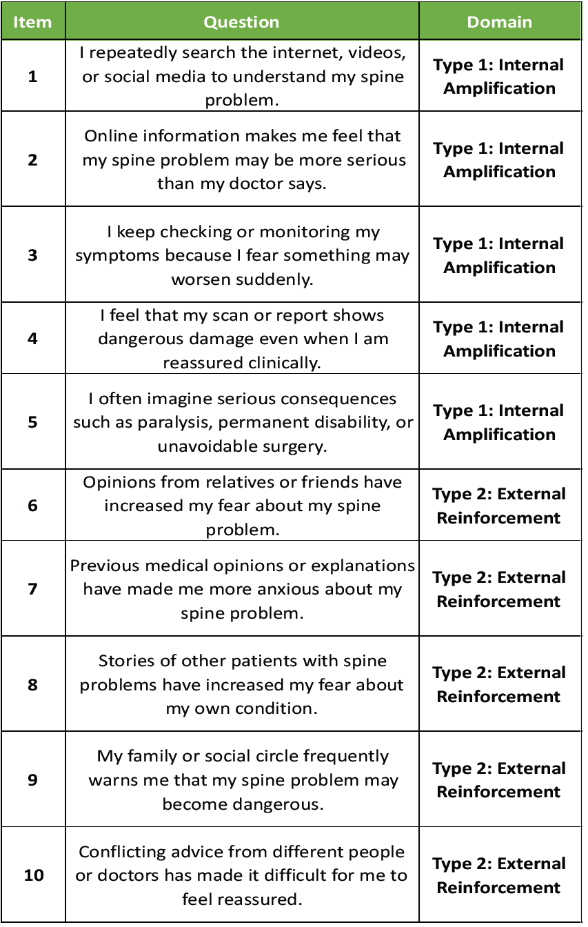
Because no established instrument specifically evaluates anxiety amplification related to spine symptoms, a study-specific Spinoxiety Identification Questionnaire-10 (SIQ-10), abbreviated as SIQ-10, was used at baseline. SIQ-10-style responses were collected prospectively as part of the structured clinical assessment. The questionnaire was designed as a clinical classification tool to identify the dominant source of anxiety amplification, rather than as an independently validated psychiatric diagnostic scale.
The SIQ-10 included ten items scored from 0 to 4: 0 = not at all, 1 = occasionally, 2 = sometimes, 3 = often, and 4 = almost always. Items 1–5 assessed internally amplified anxiety, including internet-based interpretation, self-diagnosis, symptom monitoring, hypervigilance, and catastrophic appraisal. Items 6–10 assessed externally reinforced anxiety, including fear arising from relatives, friends, previous medical opinions, social narratives and conflicting advice.
The Type 1 subscore was calculated by summing items 1–5, giving a score range of 0–20. The Type 2 subscore was calculated by summing items 6–10, also giving a score range of 0–20. The total SIQ-10 score ranged from 0 to 40, with higher scores indicating greater anxiety-driven amplification of spine-related symptoms.
A patient was considered to have probable Spinoxiety when all three of the following criteria were present: Spine-related symptom concern; evidence of psychological amplification; and clinically relevant illness concern. Evidence of psychological amplification was defined as the presence of at least one of the following: Generalized Anxiety Disorder-7 (GAD-7) score ≥10, PCS score ≥30, or SIQ-10 total score ≥12 [16,17]. Clinically relevant illness concerns included repeated reassurance-seeking, excessive symptom monitoring, activity avoidance, fear of paralysis, fear of irreversible spinal damage, fear of unavoidable surgery, or persistent worry despite routine clinical reassurance.
A patient was classified as having Type 1 Spinoxiety when internally amplified anxiety was the dominant pattern, reflected by higher SIQ-10 Type 1 domain responses and supported by clinical interview evidence of self-diagnosis, repeated internet-based interpretation, symptom hypervigilance, or catastrophic appraisal. A patient was classified as having Type 2 Spinoxiety when externally reinforced anxiety was the dominant pattern, reflected by higher SIQ-10 Type 2 domain responses and supported by clinical interview evidence that fear was mainly driven by relatives, friends, social narratives, previous medical opinions or conflicting explanations.
For operational classification, Type 1 Spinoxiety was assigned when the SIQ-10 Type 1 subscore was ≥10 and at least 3 points higher than the Type 2 subscore. Type 2 Spinoxiety was assigned when the SIQ-10 Type 2 subscore was ≥10 and at least 3 points higher than the Type 1 subscore. Where both patterns were present, the final classification was based on the dominant source of anxiety amplification identified during the structured clinical interview. Mixed Spinoxiety was not analyzed as a separate subgroup in the present exploratory study.
All patients received routine clinical care according to the treating clinician’s assessment. This included clinical evaluation, explanation of symptoms, interpretation of imaging findings where available, pharmacological treatment when indicated, physiotherapy advice, activity guidance and follow-up. Communication focused on correcting fear-based misinterpretation of spine symptoms. Patients were informed that imaging abnormalities do not always indicate severe or progressive disease and that pain intensity does not necessarily imply irreversible damage.
The Spinoxiety classification was used to guide communication. In patients with Type 1 Spinoxiety, communication emphasized correction of internet-derived misconceptions, reduction of repetitive symptom checking, clarification of radiology-symptom mismatch, and reassurance regarding feared consequences such as paralysis or inevitable surgery. In patients with Type 2 Spinoxiety, communication emphasized clarification of externally reinforced fears, reconciliation of previous medical opinions and, when appropriate, discussion with relatives or caregivers to ensure consistent messaging. This approach was intended to improve communication by identifying whether the patient’s fear was primarily self-amplified or externally reinforced.
Patients were reassessed 6 weeks after baseline evaluation. At follow-up, VAS, region-specific functional disability score, GAD-7 and PCS were repeated. Change in score between baseline and 6 weeks was calculated for each outcome. Higher delta values indicated greater improvement.
The primary outcome was the association between psychological distress and clinical symptom burden, assessed through the relationship of GAD-7 and PCS scores with pain intensity and functional disability. Secondary outcomes were changes in anxiety, PCS, pain intensity and functional disability from baseline to 6 weeks. Exploratory outcomes included comparison of baseline scores, 6-week residual scores and delta recovery between Type 1 and Type 2 Spinoxiety patients.
Data were analyzed using the Statistical Package for the Social Sciences software. Continuous variables were expressed as mean and standard deviation or median and interquartile range, depending on data distribution. Categorical variables were expressed as frequencies and percentages. Normality was assessed using the Shapiro-Wilk test. Baseline and 6-week follow-up scores were compared using paired t-tests for normally distributed variables and Wilcoxon signed-rank tests for non-normally distributed variables. Between-group comparisons of Type 1 and Type 2 Spinoxiety patients were performed using independent-sample t-tests or Mann-Whitney U tests for continuous variables, and Chi-square or Fisher’s exact test for categorical variables, as appropriate.
Correlation analysis was performed to evaluate associations between psychological variables and clinical symptom burden. Pearson correlation coefficient was used for normally distributed variables and Spearman correlation coefficient for non-normally distributed variables. Because Type 1 and Type 2 subgroups were unequal in size, adjusted analyses were planned to reduce the risk of confounding. Linear regression models were used to assess whether Spinoxiety subtype independently predicted 6-week residual GAD-7, PCS, VAS and functional disability scores after adjustment for baseline score, age, sex, symptom region and baseline pain intensity. Effect sizes and 95% confidence intervals were calculated where appropriate. A P < 0.05 was considered statistically significant.
As the SIQ-10 was developed for this study, internal consistency of the total questionnaire and its two domains was assessed using Cronbach’s alpha. If classification was performed by more than one assessor, inter-rater agreement for Type 1 versus Type 2 categorization was assessed using Cohen’s kappa.
A total of 200 patients presenting with spine-related pain were included in the final analysis. Complete baseline and 6-week follow-up data were available for all participants. The mean age of the cohort was 45.03 ± 13.51 years. There were 116 female patients and 84 male patients. Based on SIQ-10-style responses and structured clinical interview, 166 patients were classified as having Type 1 Spinoxiety and 34 as having Type 2 Spinoxiety.
All patients met the operational criteria for probable Spinoxiety. The SIQ-10 total score was 20.86 ± 1.44. Type 1 and Type 2 patients had similar total SIQ10 scores, suggesting that the classification reflected the dominant source of anxiety amplification rather than the overall amount of anxiety-driven symptom concern. However, the domain profile differed strongly between groups. Type 1 patients had higher internal amplification scores, whereas Type 2 patients had higher external reinforcement scores. The Type 1 and Type 2 SIQ-10 domains showed high internal consistency, with Cronbach’s alpha values of 0.897 and 0.893, respectively, as shown in Table 1.
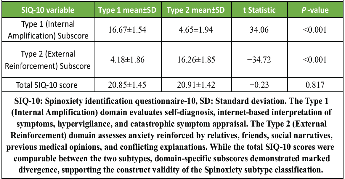
Table 1: SIQ-10 domain profile according to spinoxiety subtype
At baseline, the cohort showed substantial psychological and clinical symptom burden. The mean GAD-7 score was 15.87 ± 2.07, the mean PCS score was 42.13 ± 5.43, the mean VAS score was 7.15 ± 0.84, and the mean region-specific functional disability score was 56.47 ± 9.29. A GAD-7 score of 10 or more was present in 199 patients, and a PCS score of 30 or more was present in 195 patients.
At 6 weeks, significant improvement was observed in all measured outcomes. Mean GAD-7 improved from 15.87 ± 2.07 to 10.33 ± 2.87, PCS from 42.13 ± 5.43 to 27.26 ± 9.51, VAS from 7.15 ± 0.84 to 3.18 ± 1.02, and functional disability from 56.47 ± 9.29 to 19.42 ± 7.72. All changes were statistically significant, as illustrated in Tables 2 and 3.
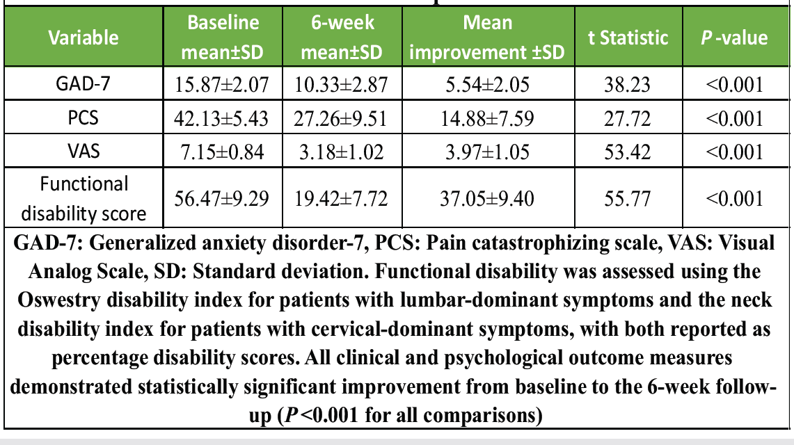
Table 2: Change in clinical and psychological scores from baseline to 6-week follow-up
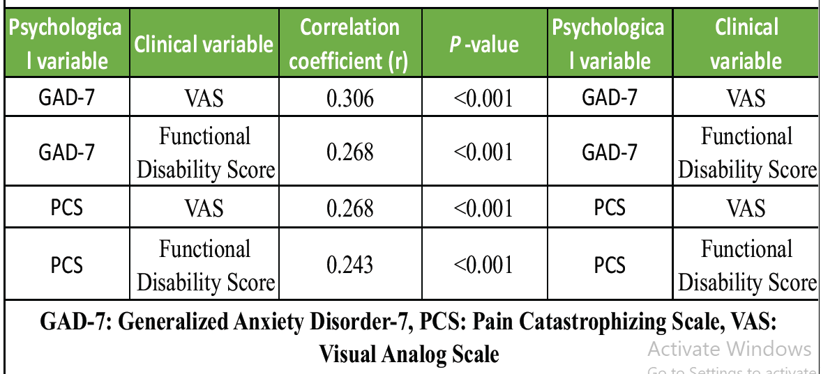
Table 3: Baseline correlations between psychological and clinical variables
At baseline, Type 1 and Type 2 patients did not differ significantly in age, GAD-7 score, or PCS score. However, Type 1 patients had significantly higher baseline pain intensity and functional disability. Mean baseline VAS was 7.20 ± 0.82 in Type 1 patients and 6.85 ± 0.89 in Type 2 patients. Mean baseline functional disability was 57.25 ± 9.03 in Type 1 patients and 52.65 ± 9.71 in Type 2 patients.
At 6 weeks, both groups improved; however, Type 1 patients retained a substantially higher residual psychological burden. Mean 6-week GAD-7 was 11.24 ± 2.13 in Type 1 patients and 5.88 ± 1.63 in Type 2 patients. Mean 6-week PCS was 30.30 ± 7.21 in Type 1 patients and 12.41 ± 3.40 in Type 2 patients. Persistent GAD-7 score ≥10 at 6 weeks was present in 133 patients, all of whom belonged to the Type 1 group. Persistent PCS score ≥30 was present in 115 patients, all within the Type 1 group, as shown in Table 4.
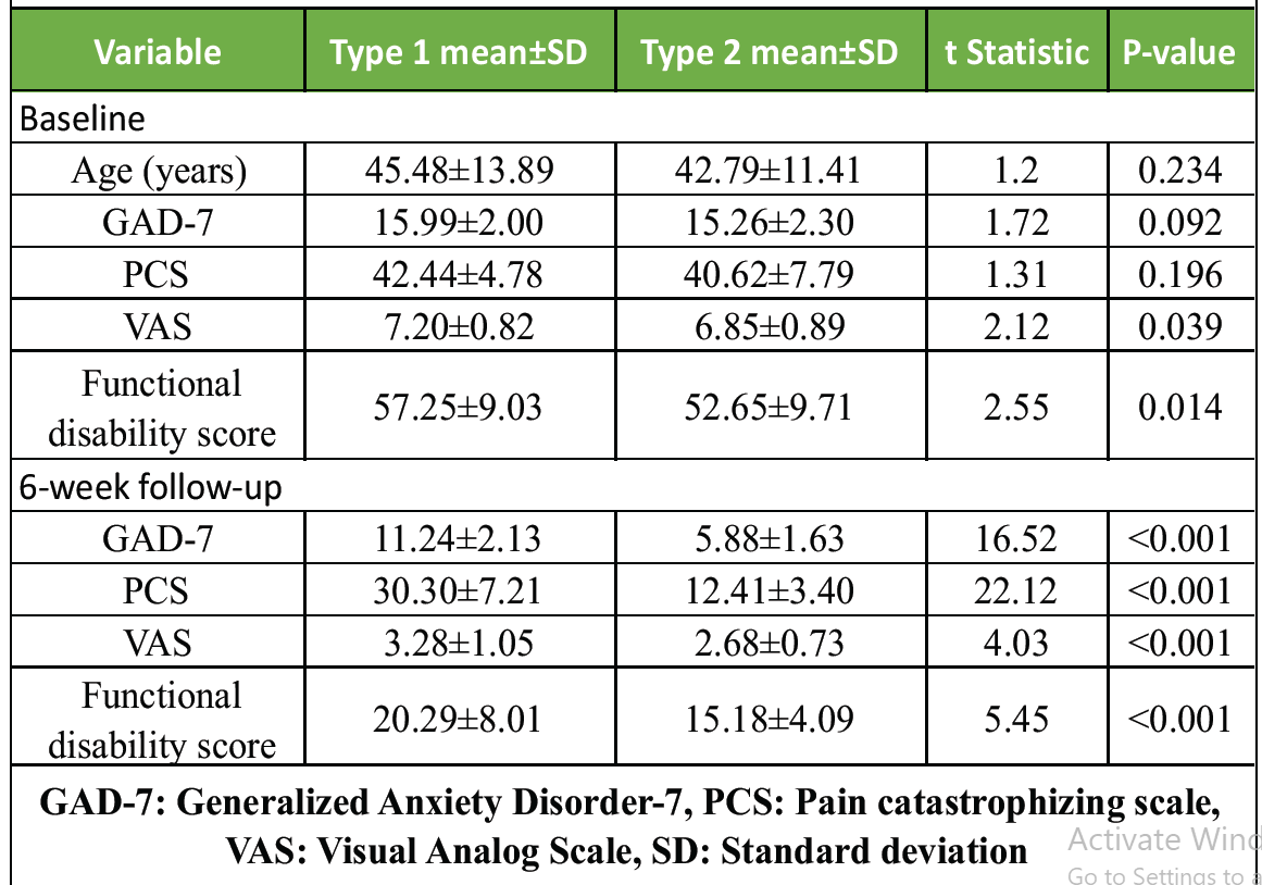
Table 4: Comparison between Type 1 and Type 2 Spinoxiety
Delta recovery analysis showed that Type 2 patients had significantly greater psychological recovery than Type 1 patients. The mean reduction in GAD-7 was 9.38 ± 1.28 in Type 2 patients and 4.75 ± 1.04 in Type 1 patients. The mean reduction in PCS was 28.21 ± 6.11 in Type 2 patients and 12.14 ± 4.23 in Type 1 patients. In contrast, improvement in pain intensity and functional disability was comparatively similar between the two groups, as shown in Tables 5 and 6.
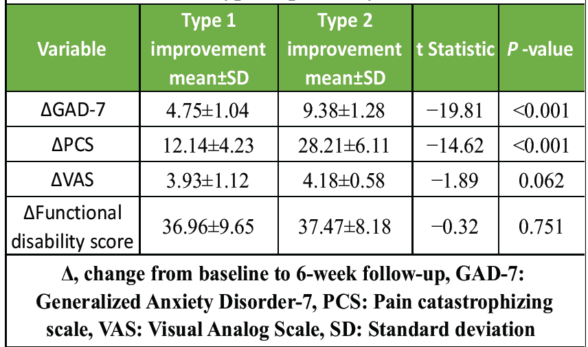
Table 5: Delta recovery comparison between type 1 and type 2 spinoxiety
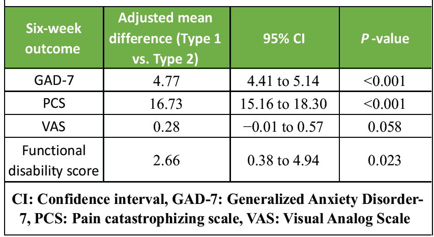
Table 6: Adjusted association of type 1 spinoxiety with 6-week residual outcomes
Adjusted linear regression was performed to assess whether Spinoxiety subtype independently predicted 6-week residual scores. Type 2 Spinoxiety was used as the reference group. After adjustment for baseline score, age, sex, symptom region and baseline pain intensity, Type 1 Spinoxiety remained independently associated with higher residual GAD-7 and PCS scores at 6 weeks. The adjusted association with residual VAS was no longer statistically significant, whereas the association with residual functional disability remained modest but significant.
Models were adjusted for baseline value of the corresponding outcome, age, sex, symptom region and baseline VAS. For the VAS model, baseline VAS was included as the baseline outcome variable and was not duplicated.
Overall, the results show that anxiety and catastrophizing were significantly associated with greater pain and disability at presentation. Although routine care and communication were associated with significant improvement across all measured outcomes, Type 1 Spinoxiety was associated with persistent residual anxiety and catastrophizing at 6 weeks. Type 2 Spinoxiety showed greater psychological recovery, whereas pain and disability improvement were broadly comparable between the two groups. Clinically, this supports the value of identifying whether spine-related fear is primarily self-amplified or externally reinforced, as this distinction may help tailor communication and improve communication with patients and relatives.
Appendix S1
Clinically, subtype identification helped the treating team frame discussions with patients and relatives. Type 1 patients more commonly required clarification of self-diagnosis, internet-derived fears and catastrophic interpretation of imaging findings, whereas Type 2 patients more often required clarification of externally reinforced fears arising from relatives, previous opinions, social narratives or conflicting advice. This was used as part of routine clinical explanation and expectation-setting, not as a structured psychological intervention.
This prospective observational study suggests that anxiety-driven symptom amplification is clinically relevant in patients presenting with spine-related pain. Anxiety and PCS were significantly associated with greater pain intensity and functional disability at presentation. Although pain, disability, anxiety, and catastrophizing improved significantly after routine care and communication, patients with Type 1 Spinoxiety showed more persistent residual anxiety and catastrophizing than patients with Type 2 Spinoxiety. Importantly, improvement in pain and disability was broadly comparable between the two groups, whereas psychological recovery differed substantially. This indicates that physical symptom improvement and psychological recovery may not always progress in parallel.
The findings support the broader biopsychosocial understanding of spine-related pain, in which symptom burden is shaped not only by structural pathology but also by cognitive appraisal, emotional response, illness behavior and social context [2,4]. Degenerative spinal imaging findings are common in asymptomatic individuals, and therefore, imaging abnormalities may acquire harmful meaning when interpreted without clinical context [3]. In this setting, anxiety and catastrophizing may increase perceived threat, symptom monitoring and fear-based avoidance, thereby amplifying pain and disability. This is consistent with the fear-avoidance model, which proposes that catastrophic interpretation of pain can lead to fear, hypervigilance, avoidance and persistent disability [6].
The concept of Spinoxiety was developed to provide a clinically usable language for this anxiety-driven amplification. It is important to emphasize that Spinoxiety is not proposed as a new psychiatric diagnosis. Rather, it is a structured clinical descriptor that helps identify whether spine-related fear is predominantly self-amplified or externally reinforced. This distinction may be useful because the same level of anxiety can arise from different sources and may therefore require different communication strategies.
Type 1 Spinoxiety appeared to represent a more internally sustained cognitive-emotional pattern. These patients showed high internal amplification scores on SIQ-10 and had persistent anxiety and catastrophizing at 6 weeks despite meaningful improvement in pain and disability. Clinically, these patients often required repeated correction of internet-derived misconceptions, explanation of radiology-symptom mismatch, and reassurance that pain did not necessarily indicate progressive damage, paralysis, or inevitable surgery. This pattern overlaps conceptually with health anxiety, cyberchondria, symptom hypervigilance and catastrophizing [7,9,10].
Type 2 Spinoxiety appeared to represent a more externally reinforced anxiety pattern. These patients had high external reinforcement scores on SIQ-10, suggesting that their fear was shaped mainly by relatives, friends, previous medical opinions, social narratives, or conflicting advice. Their greater psychological recovery may indicate that externally reinforced anxiety is more responsive to clear explanation, reconciliation of prior opinions and consistent messaging from the treating team. In many such cases, communication was most effective when relatives or caregivers were included, because the source of fear was not limited to the patient alone.
A practical implication of the Spinoxiety framework is improved clinical communication. In routine spine practice, patients and relatives may interpret terms such as “disc prolapse,” “degeneration,” “stenosis,” or “nerve compression” as evidence of irreversible damage, impending paralysis, or inevitable surgery. Identifying whether fear was predominantly self-amplified or externally reinforced helped the treating team communicate more specifically. Type 1 patients required clarification of self-generated and internet-driven fears, whereas Type 2 patients required clarification of externally reinforced concerns, often involving relatives or caregivers. This approach remained within the scope of routine clinical explanation, education and expectation-setting by spine clinicians and was not intended to replace formal psychological or psychiatric care when needed.
This communication aspect is clinically important because a clinician’s language can influence symptom expectations. Threatening explanations, alarming diagnostic labels, and poorly contextualized imaging findings may contribute to negative expectations and nocebo responses [11]. Conversely, empathic and positive communication has been associated with reductions in pain and anxiety during healthcare consultations [12]. The Spinoxiety framework may therefore help clinicians avoid both dismissal and overmedicalization: the patient’s symptoms are acknowledged as real, whereas the role of anxiety-driven amplification is explained in a practical and non-blaming manner.
The SIQ-10 findings provide preliminary support for the proposed Type 1 and Type 2 distinction. The two domains showed high internal consistency, and the total SIQ-10 scores were similar between subtypes, suggesting that the classification reflected the dominant source of anxiety rather than merely the total severity of fear. In adjusted analysis, Type 1 Spinoxiety remained independently associated with higher residual GAD-7 and PCS scores at 6 weeks, even after accounting for baseline score, age, sex, symptom region, and baseline pain intensity. This supports the possibility that internally amplified anxiety may be a marker of delayed psychological recovery.
These findings have practical implications for spine care and liaison psychiatry. Routine screening for anxiety and catastrophizing using brief tools such as GAD-7 and PCS may help identify patients at risk of persistent psychological distress [16,17]. The SIQ-10 may add a clinically useful layer by identifying the source of fear amplification. Type 1 patients may benefit from structured education, correction of internet-based misconceptions, reduction of repeated reassurance seeking, and, when needed, referral for psychological intervention. Type 2 patients may benefit from family-inclusive communication, clarification of previous advice and consistent communication across treating professionals.
The study has limitations. First, it was conducted at a single tertiary spine center, which may limit generalizability. Second, although the total sample size was 200 patients, the Type 2 subgroup was smaller than the Type 1 subgroup, and subgroup comparisons should therefore be interpreted with caution. Third, the follow-up duration was limited to 6 weeks, and a longer follow-up is needed to determine whether persistent Spinoxiety predicts chronic disability, repeated consultations, unnecessary imaging, treatment dissatisfaction, or surgical decision-making.
Fourth, SIQ-10 was developed for this study and should be considered a structured clinical classification tool rather than a validated psychiatric scale. Although the domain-level internal consistency was high, future studies should evaluate its test-retest reliability, inter-rater reliability, construct validity, and predictive validity. Validation should include comparison with established measures of health anxiety, somatic symptom burden, fear-avoidance beliefs, depression, PCS and generalized anxiety. Fifth, formal psychiatric diagnostic interviews were not performed, and the study did not assess comorbid depressive disorders, illness anxiety disorder or somatic symptom disorder in detail.
Additional limitations include possible confounding by diagnosis, symptom duration, radiological severity, prior consultations, treatment type and family involvement. Functional disability was analyzed as a region-specific percentage score using ODI for lumbar-dominant symptoms and NDI for cervical-dominant symptoms [14,15]. Although this approach allowed patient-level analysis across cervical and lumbar presentations, future studies should analyze lumbar and cervical cohorts separately. Finally, because all patients received routine care and communication, the study cannot determine whether Spinoxiety-guided communication is superior to standard communication without a comparative intervention design.
Despite these limitations, the study provides preliminary evidence that identifying the source of spine-related anxiety may have clinical value. Type 1 Spinoxiety may represent a more persistent internally amplified pattern requiring focused psychological support, whereas Type 2 Spinoxiety may be more responsive to clarification of external messages and family-inclusive communication. The concept may help clinicians move beyond simply asking whether a patient is anxious, toward understanding why the patient is anxious and how that anxiety is being reinforced.
In summary, Spinoxiety may serve as a practical communication framework for anxiety-driven symptom amplification in spine-related pain. It may help clinicians identify patients who require more focused communication, improve communication with patients and relatives, and reduce fear-based interpretation of spine symptoms. Further multicentre validation and longer follow-up are required before Spinoxiety can be considered an established psychological construct or prognostic tool.
Spinoxiety is proposed as a practical clinical communication framework for identifying anxiety-driven amplification of spine-related symptoms rather than a psychiatric diagnosis. In this prospective observational study, patients with Type 1 Spinoxiety demonstrated greater residual anxiety and PCS than those with Type 2 Spinoxiety despite comparable improvement in pain intensity and functional disability. Identifying the predominant source of fear may help clinicians tailor communication, improve patient and family understanding, and optimize expectation-setting during routine spine care. Further multicenter studies with long-term follow-up and formal psychometric validation of the SIQ-10 are required before Spinoxiety can be considered an established psychological construct or prognostic tool.
Identifying whether spine-related anxiety is internally amplified or externally reinforced may help clinicians tailor communication, improve patient reassurance, and support psychological recovery.
References
- 1. GBD 2021 Low Back Pain Collaborators. Global, regional, and national burden of low back pain, 1990-2020, its attributable risk factors, and projections to 2050: A systematic analysis of the global burden of disease study 2021. Lancet Rheumatol 2023;5:e316-29. [Google Scholar] [PubMed]
- 2. Hartvigsen J, Hancock MJ, Kongsted A, Louw Q, Ferreira ML, Genevay S, et al. What low back pain is and why we need to pay attention. Lancet 2018;391:2356-67. [Google Scholar] [PubMed]
- 3. Brinjikji W, Luetmer PH, Comstock B, Bresnahan BW, Chen LE, Deyo RA, et al. Systematic literature review of imaging features of spinal degeneration in asymptomatic populations. AJNR Am J Neuroradiol 2015;36:811-6. [Google Scholar] [PubMed]
- 4. Engel GL. The need for a new medical model: A challenge for biomedicine. Science 1977;196:129-36. [Google Scholar] [PubMed]
- 5. Pincus T, Burton AK, Vogel S, Field AP. A systematic review of psychological factors as predictors of chronicity/disability in prospective cohorts of low back pain. Spine (Phila Pa 1976) 2002;27:E109-20. [Google Scholar] [PubMed]
- 6. Vlaeyen JW, Linton SJ. Fear-avoidance model of chronic musculoskeletal pain: 12 years on. Pain 2012;153:1144-7. [Google Scholar] [PubMed]
- 7. Wertli MM, Eugster R, Held U, Steurer J, Kofmehl R, Weiser S, et al. Catastrophizing-a prognostic factor for outcome in patients with low back pain: A systematic review. Spine J 2014;14:2639-57. [Google Scholar] [PubMed]
- 8. Nicholas MK, Linton SJ, Watson PJ, Main CJ. Early identification and management of psychological risk factors (“yellow flags”) in patients with low back pain: A reappraisal. Phys Ther 2011;91:737-53. [Google Scholar] [PubMed]
- 9. Starcevic V, Berle D. Cyberchondria: Towards a better understanding of excessive health-related Internet use. Expert Rev Neurother 2013;13:205-13. [Google Scholar] [PubMed]
- 10. Schenkel SK, Jungmann SM, Gropalis M, Witthöft M. Conceptualizations of cyberchondria and relations to the anxiety spectrum: Systematic review and meta-analysis. J Med Internet Res 2021;23:e27835. [Google Scholar] [PubMed]
- 11. Colloca L, Miller FG. The nocebo effect and its relevance for clinical practice. Psychosom Med 2011;73:598-603. [Google Scholar] [PubMed]
- 12. Howick J, Moscrop A, Mebius A, Fanshawe TR, Lewith G, Bishop FL, et al. Effects of empathic and positive communication in healthcare consultations: A systematic review and meta-analysis. J R Soc Med 2018;111:240-52. [Google Scholar] [PubMed]
- 13. World Medical Association. World Medical Association Declaration of Helsinki: Ethical principles for medical research involving human subjects. JAMA 2024;310:2191-4. [Google Scholar] [PubMed]
- 14. Fairbank JC, Pynsent PB. The oswestry disability index. Spine (Phila Pa 1976) 2000;25:2940-52. [Google Scholar] [PubMed]
- 15. Vernon H, Mior S. The neck disability index: A study of reliability and validity. J Manipulative Physiol Ther 1991;14:409-15. [Google Scholar] [PubMed]
- 16. Spitzer RL, Kroenke K, Williams JB, Löwe B. A brief measure for assessing generalized anxiety disorder: The GAD-7. Arch Intern Med 2006;166:1092-7. [Google Scholar] [PubMed]
- 17. Sullivan MJL, Bishop SR, Pivik J. The pain catastrophizing scale: Development and validation. Psychol Assess 1995;7:524-32. [Google Scholar] [PubMed]






Related Articles in Journal of Orthopaedic Case Reports
 May 1, 2026 Femoral Nerve Paralysis Associated with Traumatic Iliacus Hematoma: A Case Report
 August 10, 2023 Total Scapular Prosthesis, an Option for the Treatment of Scapular Chondrosarcoma
 August 6, 2024 Total Hip Arthroplasty Infection Due to Yersinia enterocolitica: Report of Two Cases and Comprehensive Review of the Literature
 July 14, 2012 Greater Trochanter Reconstruction in Unstable Intertrochanteric Fractures Treated With Cemented Bipolar Hemiarthroplasty: A Technical Note