

Loop Endobutton fixation provides stable anatomical reduction with excellent functional recovery, significant pain relief, and minimal complications, making it an effective surgical option for acute Rockwood type III–VI acromioclavicular joint dislocations.
Dr. Abhishek Chauhan, Department of Orthopaedics, Gandhi Medical College and Associated Hamidia Hospital, Bhopal, Madhya Pradesh, India. E-mail: abhichn66@gmail.com
Abstract
Introduction: Acromioclavicular (AC) joint dislocations represent a frequent injury among young- and middle-aged adults, often resulting from high-energy trauma such as road traffic accidents and falls. While surgical intervention is standard for high-grade (Rockwood III–VI) injuries, the optimal technique remains debated. This study evaluates the clinical and functional outcomes of the Endobutton loop system in the management of acute AC joint dislocations.
Materials and Methods: This prospective observational study was conducted at the Department of Orthopaedics, Gandhi Medical College, Bhopal, over 18 months. A cohort of 25 skeletally mature patients presenting with acute Rockwood Grade III to VI AC joint dislocations underwent surgical reconstruction using a closed-loop Endobutton device. Patients were monitored at 2 weeks, 6 weeks, 3 months, and 6 months postoperatively. Outcomes were assessed using the Constant–Murley Shoulder Score (CMS) and Visual Analog Scale (VAS) for pain.
Results: The mean age of participants was 41.16 ± 12.25 years, with a marked male predominance (84%). Rockwood Type III was the most frequent presentation (52%). Functional recovery demonstrated a statistically significant progressive improvement, with the mean CMS increasing from 54.68 ± 2.65 at 2 weeks to 90.64 ± 3.59 at 6 months. Simultaneously, mean VAS scores showed a significant reduction from 8.12 ± 0.92 to 1.52 ± 0.50 over the same period. The overall complication rate was low (8%), with no hardware-related failures requiring revision in 92% of cases.
Conclusion: The Endobutton loop system is a safe, reliable, and effective surgical method for managing acute AC joint dislocations. It provides stable anatomical reduction, leads to excellent functional recovery, and is associated with minimal post-operative complications across all Rockwood grades.
Keywords: Acromioclavicular Joint dislocation, Endobutton fixation, coracoclavicular reconstruction, rockwood classification, shoulder injury, Functional outcome, constant murley score, visual analogue

The acromioclavicular (AC) joint acts as a crucial diarthrodial connection within the shoulder girdle [1]. Its stability relies heavily on the surrounding soft tissues. The AC ligaments primarily restrict horizontal translation, whereas the robust coracoclavicular (CC) ligaments, specifically the conoid and trapezoid, provide vital vertical stability [2]. AC joint dislocations are a frequent clinical encounter in orthopedic practice, accounting for 9–12% of all shoulder injuries, and disproportionately affecting young, active males [1,3]. These injuries typically result from high-velocity trauma, such as road traffic accidents or direct-impact sports. The classic mechanism of injury involves a direct blow to the lateral aspect of the shoulder with the arm held in adduction [2]. This downward force sequentially disrupts the AC and CC ligaments. Clinicians routinely rely on the Rockwood classification to guide decision-making [3]. It is standard practice to manage Rockwood Types I and II non-operatively, while Types IV, V, and VI invariably require surgical stabilization [4]. However, the ideal management strategy for Rockwood Type III injuries remains highly debated and challenging [5]. Historically, a wide array of surgical techniques has been used, including Kirschner wire transfixation and clavicular hook plates [6,7]. While they achieve reduction, these rigid traditional constructs often fail to respect the native biomechanics of the joint. In clinical settings, they are notoriously tied to hardware-related complications such as implant migration, subacromial impingement, chronic shoulder pain, and the added morbidity of a mandatory secondary surgery for implant removal [6]. To address these specific limitations, modern surgical trends have shifted toward dynamic, suspensory fixation [8,9]. The loop endobutton system has emerged as a highly reliable technique because it biologically mimics the function of the native CC ligament complex [10,11]. This knotless approach offers robust intraoperative stability, allows for aggressive early rehabilitation, and completely bypasses the need for routine hardware removal [10]. Despite the clear biomechanical advantages and growing popularity of suspensory devices, comprehensive regional evidence detailing the long-term functional recovery and complication rates remains limited [12]. Therefore, this study aims to evaluate the clinical efficacy of loop endobutton repair in acute AC joint dislocations, with a specific focus on functional shoulder recovery, radiological maintenance of joint alignment, and the incidence of post-operative complications [13,14].
Study Design
This was a prospective observational study conducted at the Department of Orthopaedics, Gandhi Medical College and associated Hamidia Hospital, Bhopal, over a period of 18 months.
Sample size and selection
The required sample size was statistically calculated to be approximately 30 patients, assuming a 3% injury prevalence with a 7% margin of error and a 95% confidence interval. During the study, a total of 25 consecutive patients who met the criteria were enrolled and evaluated.
Inclusion and exclusion criteria
We included skeletally mature individuals presenting with acute AC joint dislocations that were radiologically classified as Rockwood Grades III, IV, V, or VI. Patients were strictly excluded if they had chronic AC dislocations, pre-existing degenerative shoulder disease, concomitant upper limb fractures, rotator cuff tears, or associated neurovascular and brachial plexus injuries.
Ethical consideration
The study adhered to institutional ethical guidelines. Thorough informed consent was obtained from all participants before their enrollment and subsequent surgical intervention.
Clinical and functional assessment
Patient outcomes were objectively quantified utilizing the Constant–Murley Shoulder Score (CMS) to evaluate functional shoulder mechanics. Subjective pain intensity was concurrently measured using the Visual Analog Scale (VAS).
Operative procedure
The patient is placed under general anesthesia in the beach-chair position. A saber incision is made from the distal clavicle extending towards the region inferior to the coracoid process. The supraclavicular nerves are identified and protected. The trapeziodeltoid fascia is incised, and the deltoid muscle is elevated subperiosteally from the anterosuperior clavicle to expose the AC joint and coracoid process. The AC joint is reduced and temporarily maintained with manual pressure or a clamp. A 3.65-mm drill bit is used to create a tunnel from the superior surface of the clavicle through the coracoid process, ensuring the drill passes through the center of the coracoid. A guide wire with the endobutton system is then passed through the tunnel. The inferior oval button is advanced through the coracoid and flipped against its undersurface, while the superior button rests on the clavicle. The sutures are tightened alternately to approximate the clavicle to the coracoid and restore the CC distance. Reduction and implant position are confirmed under fluoroscopy. The key operative steps are illustrated in Fig. 1.
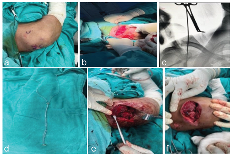
Fig. 1: (a) Draping and bony landmarks for incision, (b) Drilling and passing guide wire through clavicle to coracoid, (c) C-arm image of passage of guide wire, (d) Double loop endobutton (implant), (e) Tightening of endobutton, (f) Final picture after placement of endobutton.
Finally, the sutures are secured, and the CC ligaments, AC ligament, and trapeziodeltoid fascia are repaired before wound closure.
Post-operative rehabilitation
The operated shoulder was protected in a standard arm sling for 6 weeks. Sutures were removed after 2 weeks. The rehabilitation protocol was graduated: Gentle pendulum exercises were initiated between 3 and 6 weeks. From 6 to 10 weeks, patients progressed to active range-of-motion exercises, including forward flexion, abduction, and internal/external rotation. Patients were cleared to resume full, unrestricted functional activities at 6 months. Representative radiographic outcomes following loop endobutton fixation are shown in Fig. 2.
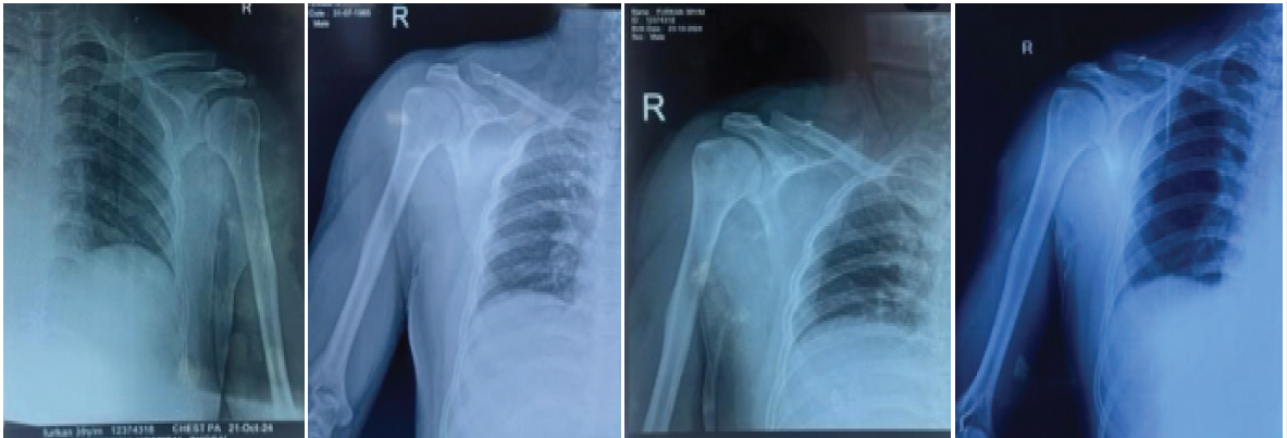
Fig. 2: Case showing acromiclavicular joint treated with endobutton. (a) Pre-op radiograph showing acromioclavicular joint disruption left. (b) Immediate post-operative radiograph. (c) Radiograph at 1 month of follow. (d) Radiograph at 6 months of follow-up.
Follow-up and assessment
Patients were prospectively followed for a minimum of 6 months. Standardized clinical evaluations were conducted at specific intervals: 2 weeks, 6 weeks, 3 months, and 6 months postoperatively. At each interval, functional recovery (CMS), pain levels (VAS), and any post-operative complications were actively documented. Clinical outcome at final follow-up is demonstrated in Fig. 3.
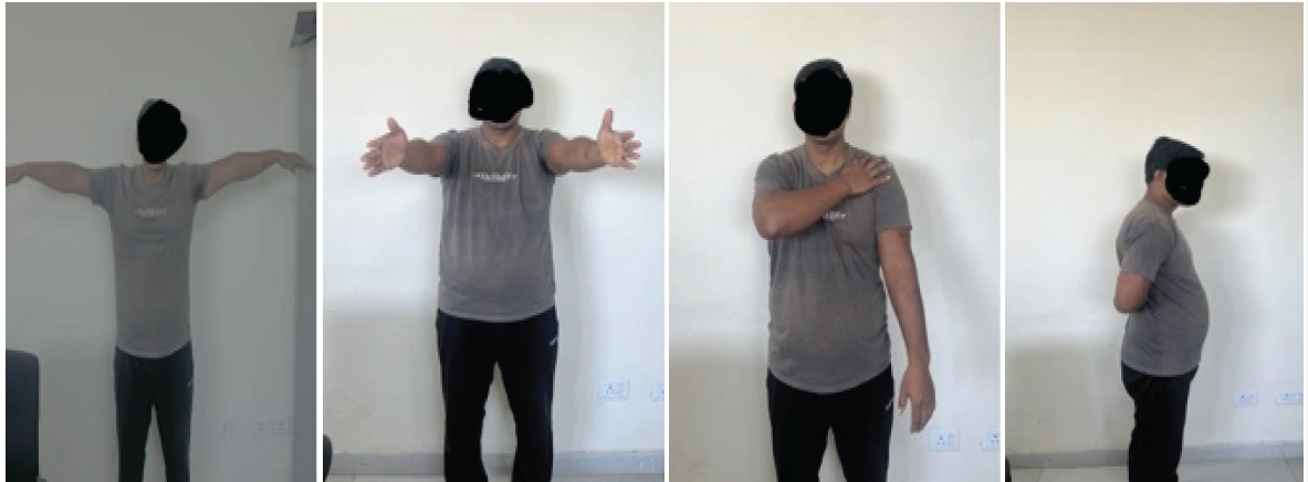
Fig. 3: Clinical pictures at 6 months of follow up.
A total of 25 patients completed the minimum 6-month follow-up protocol. The cohort demonstrated a strong male predilection, comprising 21 males (84.0%) and 4 females (16.0%). The mean age at the time of surgical intervention was 41.16 ± 12.25 years, with the majority of the patient population (52.0%) falling into the 31–45-year age. The demographic and injury characteristics are summarized in Table 1.
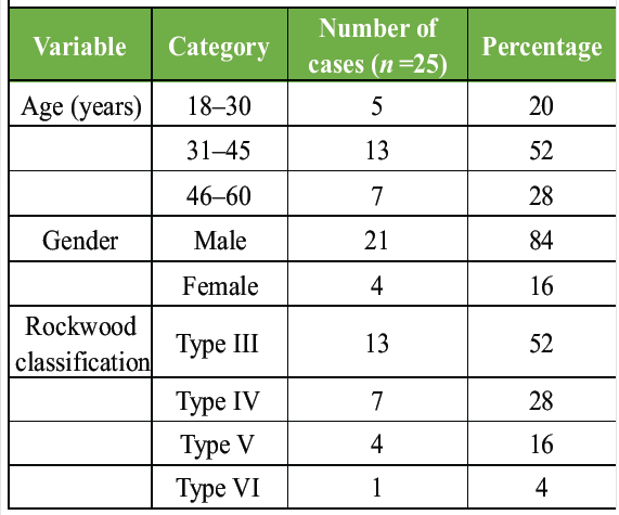
Table 1: Patient demographics and injury characteristics
On radiographic evaluation, injuries were categorized using the Rockwood classification system. Type III dislocations were the most prevalent clinical entity, accounting for 13 cases (52.0%). This was followed by Type IV injuries in 7 patients (28.0%), Type V in 4 patients (16.0%), and a single documented Type VI injury (4.0%). Regarding the timing of surgical intervention, 40.0% of the cohort underwent the procedure within the first 7 days following their initial trauma.
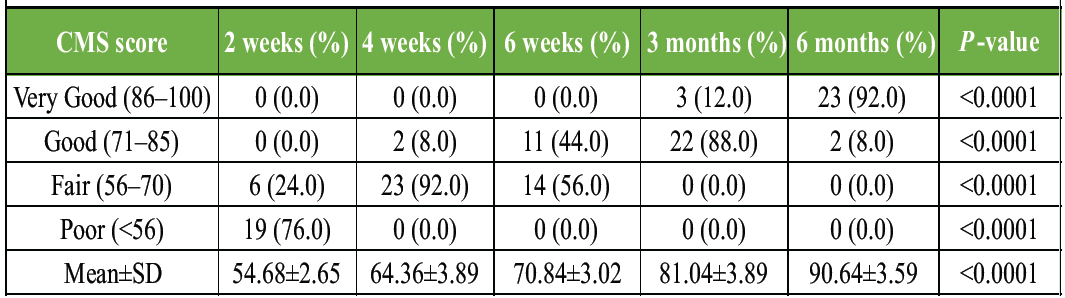
Table 2: Functional outcome assessed by Constant–Murley score at different follow-up intervals
Table 2 summarizes the progression of functional recovery as assessed by the CMS at different follow-up intervals. Progression of functional recovery as assessed by the CMS at different follow-up intervals. At 2 weeks, the majority of patients (76.0%) had poor scores, while 24.0% had fair scores, and none had good or very good scores. By 4 weeks, most patients (92.0%) had fair scores, and 8.0% had good scores. At 6 weeks, 44.0% had good scores, and 56.0% had fair scores. At 3 months, 88.0% of patients had good scores, and 12.0% had very good scores. At 6 months, the majority of patients (92.0%) achieved very good scores, and 8.0% had good scores. The mean CMS improved progressively from 54.68 ± 2.65 at 2 weeks to 90.64 ± 3.59 at 6 months, with a statistically significant improvement over time (P < 0.0001), indicating excellent functional recovery following loop endobutton repair.
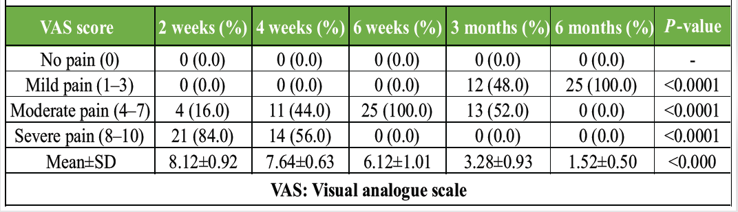
Table 3: Post-operative pain assessment using VAS at different follow-up intervals
Table 3 shows the progression of post-operative pain as assessed by the VAS at different follow-up intervals. At 2 weeks, the majority of patients (84.0%) experienced severe pain, and 16.0% had moderate pain. At 4 weeks, 56.0% of patients had severe pain, 44.0% had moderate pain, and none had mild or no pain. By 6 weeks, all patients (100.0%) experienced moderate pain. At 3 months, 52.0% had moderate pain and 48.0% had mild pain. At 6 months, all patients (100.0%) reported mild pain. The mean VAS score decreased progressively from 8.12 ± 0.92 at 2 weeks to 1.52 ± 0.50 at 6 months, showing a statistically significant reduction in pain over time (P < 0.0001) following loop endobutton repair.
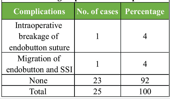
Table 4: Post-operative complications following loop endobutton repair
Table 4 shows the post-operative complications observed in the study population. One patient (4.00%) experienced intraoperative breakage of the endobutton suture, and another patient (4.00%) had migration of the endobutton along with surgical site infection. No complications were observed in the remaining 23 patients (92.00%). This indicated that loop endobutton repair was associated with minimal complications in the present study.
The optimal surgical management of displaced AC joint injuries, specifically Rockwood Types III to VI, remains an area of active debate within orthopedic literature. The primary goal of any intervention is to achieve a stable, anatomical reduction that restores native shoulder biomechanics and allows for early functional rehabilitation. Our prospective evaluation of the loop endobutton technique demonstrates that it is a highly effective, minimally invasive solution, yielding excellent functional recovery and reliable pain relief with a minimal complication profile.
Demographic and injury profile
Our study population exhibited a marked male predominance (84.0%) and a mean age of 41.16 ± 12.25 years, with the majority of patients falling into the 31–45-year age bracket. This strong male preponderance closely mirrors the broader demographic trends associated with high-energy trauma, such as road traffic accidents and contact sports. Our findings align with Bansal et al. [14], who similarly reported that AC joint dislocations predominantly affect young- to middle-aged males. Teodoro et al. [15] noted an even higher male incidence at 91.3% in a slightly younger cohort (mean age 31.04 years). Shukla et al. [16] and Ahmad et al. [17] also documented male-dominated cohorts, reinforcing that our patient profile accurately represents the typical clinical presentation of this injury.
Regarding injury severity, Rockwood Type III dislocations were the most frequently encountered in our series, representing 52.0% of cases. This distribution reflects a common institutional pattern where moderate-grade, borderline injuries are increasingly managed surgically to optimize functional outcomes. In contrast, Ahmad et al. [17] evaluated a cohort consisting entirely of Rockwood Type V injuries, while Bansal et al. [14] reported a broader distribution skewed toward more severe disruptions (18 Type V cases vs. 10 Type III cases). These variations likely stem from differences in regional referral patterns and institutional indications for operative management.
Functional and pain outcomes
The most critical metric of surgical success in AC joint reconstruction is the restoration of shoulder function. We observed a progressive, statistically significant improvement in the CMS, escalating from a mean of 54.68 ± 2.65 at 2 weeks to an excellent 90.64 ± 3.59 at 6 months. This functional recovery trajectory is strongly supported by existing literature. For instance, Shukla et al. [16] noted that while only 10% of their patients achieved excellent CMS scores at 6 weeks, 56.67% reached the 86–100 range by 9 months, reflecting a similarly delayed but substantial clinical improvement.
Our final 6-month functional outcomes are highly comparable to several significant studies. Ahmad et al. [17] reported a nearly identical mean CMS of 90.62 ± 5.36 specifically for AC joint dislocations. Furthermore, Zhang et al. [5] and Lu et al. [12] reported mean scores of 92.33 and 93.5, respectively, utilizing similar double-endobutton techniques. Importantly, while Sautet et al. [18] reported a commendable mean CMS of 91%, their cohort experienced a 28% rate of clavicular button irritation, with some requiring subsequent implant removal. In our series, we bypassed this issue entirely, observing zero cases of implant-related soft-tissue irritation.
Parallel to functional return, post-operative pain dropped precipitously. The mean VAS score in our cohort decreased from 8.12 ± 0.92 to a minimal 1.52 ± 0.50 over 6 months. This profound reduction in pain confirms the biomechanical stability of the knotless suspensory construct, which avoids the subacromial impingement and chronic discomfort historically associated with rigid hook plates.
Complication profile
The safety profile of the loop endobutton repair in our study was excellent, with an overall complication rate of just 8.0% (one intraoperative suture breakage and one post-operative button migration with a superficial infection). The remaining 92.0% of our patients experienced an entirely uneventful recovery. This low incidence of adverse events reinforces the reliability of the technique. Our findings parallel those of Bansal et al. [14], who reported a 15% rate of minor complications (such as stitch granulomas and transient stiffness) and an 85% completely complication-free rate. Similarly, Yadav et al. [19] emphasized that endobutton fixation consistently yields minimal complications due to its less invasive nature compared to traditional open rigid fixation.
Limitations of the study
While our results are encouraging, certain limitations inherent to this study must be acknowledged. First, the sample size (n = 25) is relatively modest; however, it remains statistically robust for a prospective single-center clinical assessment. Second, the follow-up period of 6 months, while sufficient for assessing acute functional recovery and early hardware complications, does not capture the long-term potential for late-onset osteoarthritis or potential late-stage reduction loss. Finally, as a prospective observational study, we did not employ a randomized control group (such as a cohort managed with hook plates or conservative protocols), which limits our ability to make direct comparative superiority claims. Future multi-center randomized controlled trials with extended follow-up durations are recommended to solidify these findings and evaluate long-term joint health.
Our prospective study demonstrates that the loop endobutton technique is a highly effective, safe, and reliable surgical option for managing acute AC joint dislocations (Rockwood Types III–VI). By successfully replicating the natural biomechanics of the CC ligament complex, this knotless suspensory construct provides robust immediate stability that allows for early functional rehabilitation without the morbidity often associated with rigid traditional implants. Our findings indicate that patients achieve excellent functional outcomes and significant pain resolution by the six-month mark with a notably low complication rate. Given its efficacy and the avoidance of routine hardware removal, we believe the loop endobutton technique represents an optimal choice for treating acute AC joint disruptions in young, active patient populations.
The loop endobutton system offers a compelling “clinical pearl” for orthopedic surgeons: It effectively bridges the gap between biological anatomical reconstruction and the requirement for stable, early post-operative mobilization. Surgeons should consider this technique as a first-line operative approach for high-grade AC joint dislocations, as it provides a predictable pathway to functional recovery while minimizing hardware-related patient distress. In our experience, adherence to the described technical steps, specifically, precise tunnel placement and secure button flipping, is the cornerstone of achieving these excellent clinical and radiological results.
References
- 1. Papadakis M. Wound irrigation for preventing surgical site infections. World J Methodol 2021;11:222. [Google Scholar] [PubMed]
- 2. Iorio R, Yu S, Anoushiravani AA, Riesgo AM, Park B, Vigdorchik J, et al. Vancomycin powder and dilute povidone-iodine lavage for infection prophylaxis in high-risk total joint arthroplasty. J Arthroplasty 2020;35:1933-6. [Google Scholar] [PubMed]
- 3. Dial BL, Lampley AJ, Green CL, Hallows R. Intrawound vancomycin powder in primary total hip arthroplasty increases rate of sterile wound complications. Hip Pelvis 2018;30:37. [Google Scholar] [PubMed]
- 4. Buchalter DB, Teo GM, Kirby DJ, Schwarzkopf R, Aggarwal VK, Long WJ. Does the organism profile of periprosthetic joint infections change with a topical vancomycin powder and dilute povidone-iodine lavage protocol? J Arthroplasty 2021;36:S314-9. [Google Scholar] [PubMed]
- 5. Otte JE, Politi JR, Chambers B, Smith CA. Intrawound vancomycin powder reduces early prosthetic joint infections in revision hip and knee arthroplasty. Surg Technol Int 2017;30:284-9. [Google Scholar] [PubMed]
- 6. Johnson JD, Nessler JM, Horazdovsky RD, Vang S, Thomas AJ, Marston SB. Serum and wound vancomycin levels after intrawound administration in primary total joint arthroplasty. J Arthroplasty 2017;32:924-8. [Google Scholar] [PubMed]
- 7. Brown NM, Cipriano CA, Moric M, Sporer SM, Della Valle CJ. Dilute betadine lavage before closure for the prevention of acute postoperative deep periprosthetic joint infection. J Arthroplasty 2012;27:27-30. [Google Scholar] [PubMed]
- 8. Wilson AP, Treasure T, Sturridge MF, Grüneberg RN. A scoring method (ASEPSIS) for postoperative wound infections for use in clinical trials of antibiotic prophylaxis. Lancet 1986;1:311-3. [Google Scholar] [PubMed]
- 9. Shohat N, Goh GS, Harrer SL, Brown S. Dilute povidone-iodine irrigation reduces the rate of periprosthetic joint infection following hip and knee arthroplasty: An analysis of 31,331 cases. J Arthroplasty 2022;37:226-31.e1. [Google Scholar] [PubMed]
- 10. Riesgo AM, Park BK, Herrero CP, Yu S, Schwarzkopf R, Iorio R. Vancomycin povidone-iodine protocol improves survivorship of periprosthetic joint infection treated with irrigation and debridement. J Arthroplasty 2018;33:847-50. [Google Scholar] [PubMed]
- 11. Winkler C, Dennison J, Wooldridge A, Larumbe E, Caroom C, Jenkins M, et al. Do local antibiotics reduce periprosthetic joint infections? A retrospective review of 744 cases. J Clin Orthop Trauma 2018;9 Suppl 1:S34-9. [Google Scholar] [PubMed]
- 12. Patel NN, Guild GN, Kumar AR. Intrawound vancomycin in primary hip and knee arthroplasty: A safe and cost-effective means to decrease early periprosthetic joint infection. Arthroplast Today 2018;4:479-83. [Google Scholar] [PubMed]
- 13. Ahmad MA, Rahman SA, Islam MA. Prevalence and risk of infection in patients with diabetes following primary total knee arthroplasty: A global systematic review and meta-analysis of 120,754 knees. J Clin Med 2022;11:3752. [Google Scholar] [PubMed]
- 14. Slullitel PA, Dobransky JS, Bali K, Poitras S, Bhullar RS, Gofton WT, et al. Is There a Role for preclosure dilute betadine irrigation in the prevention of postoperative infection following total joint arthroplasty? J Arthroplasty 2020;35:1374-8. [Google Scholar] [PubMed]
- 15. Calkins TE, Culvern C, Nam D, Gerlinger TL, Levine BR, Sporer SM, et al. Dilute betadine lavage reduces the risk of acute postoperative periprosthetic joint infection in aseptic revision total knee and hip arthroplasty: A randomized controlled trial. J Arthroplasty 2020;35:538-43.e1. [Google Scholar] [PubMed]
- 16. Yavuz IA, Oken OF, Yildirim AO, Inci F, Ceyhan E, Gurhan U. No effect of vancomycin powder to prevent infection in primary total knee arthroplasty: A retrospective review of 976 cases. Knee Surg Sports Traumatol Arthrosc 2020;28:3055-60. [Google Scholar] [PubMed]
- 17. Erken HY, Nusran G, Karagüven D, Yilmaz O, Kuru T. No decrease in infection rate with the use of local vancomycin powder after partial hip replacement in elderly patients with comorbidities. Cureus 2020;12:e10296. [Google Scholar] [PubMed]
- 18. Matziolis G, Brodt S, Böhle S, Kirschberg J, Jacob B, Röhner E. Intraarticular vancomycin powder is effective in preventing infections following total hip and knee arthroplasty. Sci Rep 2020;10:13053. [Google Scholar] [PubMed]
- 19. Tahmasebi MN, Vaziri AS, Vosoughi F, Tahami M, Khalilizad M, Rabie H. Low post-arthroplasty infection rate is possible in developing countries: Long-term experience of local vancomycin use in Iran. J Orthop Surg Res 2021;16:1-7. [Google Scholar] [PubMed]
- 20. Buchalter DB, Kirby DJ, Teo GM, Iorio R, Aggarwal VK, Long WJ. Topical vancomycin powder and dilute povidone-iodine lavage reduce the rate of early periprosthetic joint infection after primary total knee arthroplasty. J Arthroplasty 2021;36:286-90.e1. [Google Scholar] [PubMed]






Related Articles in Journal of Orthopaedic Case Reports
 October 1, 2026 Functional Outcome of Operative Titanium Elastic Nailing System Versus Non-Operative Figure-of-Eight Bandage in Displaced Mid-Shaft Clavicle Fractures: A Randomized Controlled Study
 October 1, 2026 Clinical and Functional Outcomes of Closed Intramedullary Interlocking Nailing in Extra-articular Distal Tibial Fractures: A Prospective Observational Study
 October 1, 2026 Functional Outcome of Lucerne Cast in Patients with Metacarpal Fracture with or without Proximal Phalanx Fracture: An Observational Prospective Study
 October 1, 2026 A Functional Outcome of Closed Intra-articular Fracture of Proximal Interphalangeal Joint of Hand Managed By Suzuki Frame – A Longitudinal Study