


Introduction
The ankle and subtalar joints are formed by the talus bone articulating with the tibia, fibula, calcaneus, and navicular. The talus bone is the second-largest bone after the calcaneus bone in size, and it is made up of three parts (body, neck, and head) [1], which is an essential weight-bearing structure in the ankle.
The calcaneus sustentaculum tali is a shelf that protrudes from the medial calcaneus and is the bulk support for the Hindfoot’s medial column [2]. Both are very important for maintaining the structure and function of the foot.
Concurrent fractures of both components of the talus and the calcaneus are rare [3,4,5] and usually result from high-energy trauma, such as vehicular accidents or a fall from a height. Our patient presented after sustaining a pedestrian injury with isolated foot trauma.
Fractures of the talus and calcaneus are challenging due to their intricate anatomy and high propensity for joint involvement, post-traumatic arthritis, talus avascular necrosis (AVN), or hindfoot malalignment [6]. These fractures require meticulous management to restore anatomy and function. We present one of the rare cases of a combined fracture of the talus and the sustentaculum tali of the calcaneus.
Case Report
A 38-year-old male patient, not known to have any medical illness, case of pedestrian injury, was referred from another hospital to our emergency department after 1 week from the accident, complaining of left ankle pain with severe swelling. The primary and secondary surveys were unremarkable.
On examination, he was conscious, alert, and vitally stable; there was severe swelling at the left ankle with blisters, no open wound, and ecchymosis. The hindfoot was in varus malalignment, tenderness was around the left ankle and the medial side of the calcaneus, and the ankle range of motion was restricted. He was Unable to bear his weight.
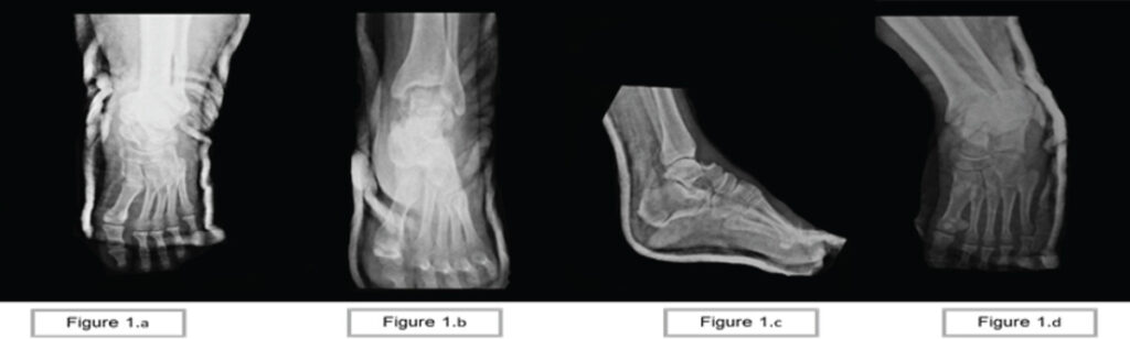
X-ray of the left ankle shows left talus fracture at the neck, and sustentaculum tali fracture of the calcaneus (Fig. 1).
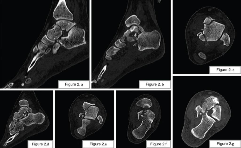
Computed tomography scan shows the left talus neck, head, body, and posterior process of the left talus with talonavicular and subtalar joint extension, and a comminuted sustentaculum tali fracture of the left calcaneus (Fig. 2).
As a further investigation, preoperatively to rule out deep vein thrombosis, since the patient presented late, an ultrasound of the left lower extremity was done and reported negative. Back slab, ice packing, and leg elevation were the initial management measures upon admission, with a conservative approach for swelling and blisters to improve before surgical management. After swelling subsided and the skin condition improved, the patient underwent surgery.
Surgical technique
The patient was in a supine position, and a tourniquet was applied to the left thigh. We utilized a modified anteromedial approach. A modified anteromedial approach offers access to both talar neck, body, and the sustentaculum tali, as well as safe management of soft tissue. Our landmarks were the tip of the medial malleolus and the tibial posterior tendon. This approach involves a longitudinal incision 1 cm distal to the tip of the medial malleolus. It extended to the anterior border of the posterior tibial tendon, as identified (Fig. 3), until reaching the navicular bone. Tibialis posterior and flexor digitorum longus were identified and protected. The plan between those two tendons was used to approach the sustentaculum. Care was taken not to violate the neurovascular structures, which they normally course posteriorly and inferiorly to the flexor digitorum longus. The saphenous vein was in our surgical site and was ligated by a vicryl suture.
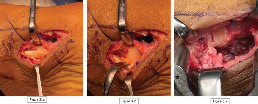
During dissection, the intact posterior tibial tendon, spring ligament, and superficial deltoid ligament were shown. The deep deltoid ligament had an avulsed tear; a stay suture was taken for repair later (Fig. 3).
We started to identify the fracture of the sustentaculum tali of the calcaneus and reduced it under direct visualization of the medial cortex of the calcaneus and stabilized it with two Kirschner wires, and then fixed it with two Herbert screw 4.0 mm (Fig. 3).
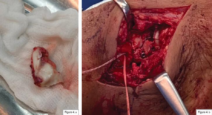
After fixation of the sustentaculum tali of the calcaneus, we addressed the fracture of the talus. There was a comminuted fracture, and fragment curing and washing were performed at the fracture site. The fragment was then reduced and stabilized with two Kirschner wires. Then it’s fixed by two Herbert screws 4.0 mm. The deltoid ligament was repaired by an anchor suture 5 mm (Fig. 4).
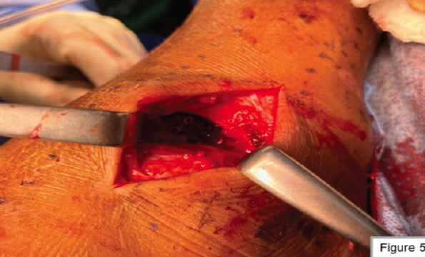
After that, we augmented our fixation by applying a mini fragment plate from the lateral side of the ankle utilizing a lateral approach (Fig. 5).
Drains were placed medially and laterally, and the surgical wounds were closed in layers. After a sterile dressing, the back slab was applied.
On post-operative day 3, the drains were removed, and the patient was discharged in good health. He was instructed to avoid weight-bearing on the left lower limb and to have follow-up at 2 and 6 weeks.
At the 2-week follow-up, the clips were removed, and the wound was clean and healed with no complications.
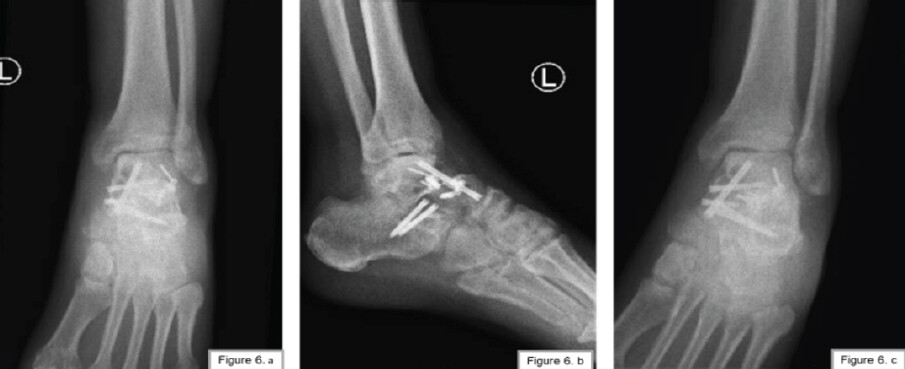
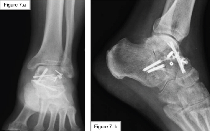
The patient presented later at the 5th (Fig. 6) and 10th months (Fig. 7), followed up postoperatively with a full range of motion of the left ankle, and only complained of mild pain while walking.
Discussion
Midfoot fractures are uncommon due to the anatomical position and close connection of the tarsal bones to the ligaments; they are often associated with edema and soft-tissue injury, necessitating early surgical intervention [7]. Sustentaculum tali fractures are uncommon and frequently associated with other foot injuries; however, isolated talus and calcaneus fractures occur in 6% and 60% of all tarsal fractures, respectively [8]. Fractures of the talus body are rare, accounting for 0.62% of all fractures and 7–38% of all talar fractures [8,9]. The fracture pattern in our study included the neck, head, body, and posterior process of the talus, as well as the talonavicular and subtalar joints, and the sustentaculum tali of the calcaneus.
The most common causes of combined talus and calcaneus fractures have been reported as small-height falls [7], whereas in our case, the patient was a pedestrian accident victim.
The medial foot approach has been described as the best way to reduce, provide better direct visualization with a small incision, and fix these fractures. A longitudinal incision approximately 5 cm long, 2 cm distal to the tip of the medial malleolus, aiming at the tuberosity of the navicular bone, is recommended.
The tibialis posterior is identified and retracted dorsally. In contrast, the flexor digitorum longus and the flexor hallucis longus are mobilized and retracted plantarly, as shown in (Fig. 2), thereby protecting the neurovascular bundle that runs between them. Periosteal striping will reveal the fracture, which can then be reduced anatomically and fixed with either interfragmentary screws and/or plate [3,10]. Moreover, we used, in our case, for fixation of the sustentaculum tali of calcaneus, two Herbert screws.
The complications of inadequate treatment for sustentaculum tali fractures can be divided into early and late complications. Tarsal tunnel syndrome and flexor hallucis longus tendon damage are the common early complications, while post-traumatic arthritis is often recognized as the late complication [11,12]. Surgical reduction of the fracture fragment accelerates bone healing and prevents these complications [12].
For a talus fracture, the fracture pattern dictates the appropriate approach, and malleolar osteotomy may be required for fixation. However, we did not use it in our case. We fixed the talus using a previous medial incision with two Herbert screws, and we augmented the fixation for the talus by applying a plate from the lateral side without an osteotomy of the lateral malleolus.
Postoperatively, a non-weight-bearing ankle cast is applied for 6 weeks, followed by another 6 weeks of non-weight-bearing ankle range-of-motion exercises. Full weight bearing is allowed 12 weeks post-surgery. The same post-operative care is found in the literature [13]. It is well reported in the literature that fractures of the body of the talus are associated with several early and late complications. Wound dehiscence occurs frequently, especially if swelling has not subsided before surgery. Superficial skin infections occur in 6.2%, and deep infections in 3.1%. Tibiotalar or subtalar joint arthritis ranges from 27% to 100%. AVN ranges from 27% to 50% [4].
The patient was successfully managed, resulting in an excellent outcome. Ideally, the treatment requires at least 1 year of follow-up to evaluate complications.
Conclusion
Ipsilateral fractures of the talus and sustentaculum tali are rare but challenging injuries that require meticulous surgical planning and execution. The modified anteromedial approach provides excellent access to the medial talus and sustentaculum tali while preserving critical neurovascular structures. This case highlights the importance of advanced imaging for accurate diagnosis, the role of anatomic reduction in optimizing outcomes, and the need for a multidisciplinary approach to rehabilitation.
Future studies should focus on long-term outcomes of combined talus and sustentaculum fractures, as well as the development of standardized treatment protocols to guide clinical decision-making.
Clinical Message
Meticulous surgical planning and tailored approaches, such as the modified anteromedial technique, are crucial for the successful management of complex ipsilateral talus and sustentaculum tali fractures, enabling optimal anatomical and functional outcomes.
Conflict of Interest:
Nil
Source of Support:
Nil
Consent:
The authors confirm that informed consent was obtained from the patient for publication of this article
References
- Khan IA, Varacallo MA. Anatomy, bony pelvis and lower limb, foot talus. In: StatPearls. Treasure Island, FL: StatPearls Publishing; 2025. [Google Scholar] | [PubMed]
- He B, Neiman R. Talar neck and sustentaculum fracture fixation. J Orthop Trauma 2021;35:S56-7. [Google Scholar] | [PubMed]
- Dürr C, Zwipp H, Rammelt S. Fractures of the sustentaculum tali. Oper Orthop Traumatol 2013;25:569-78. [Google Scholar] | [PubMed]
- Sundararajan S, Badurudeen AA, Ramakanth R, Rajasekaran S. Management of talar body fractures. Indian J Orthop 2018;52:258-68. [Google Scholar] | [PubMed]
- Bachhal V, Bhagwat K, Bali K, Dhillon MS. Ipsilateral type IV closed talus and calcaneal fractures: A case report and review of literature. J Postgrad Med Educ Res 2013;47:156-8. [Google Scholar] | [PubMed]
- Howard JL, Buckley R, McCormack R, Pate G, Leighton R, Petrie D. Complications following management of displaced intra-articular calcaneal fractures: A prospective randomized trial comparing open reduction internal fixation with nonoperative management. J Orthop Trauma 2003;17:241-9. [Google Scholar] | [PubMed]
- Stavrakakis IM, Magarakis GE, Christoforakis Z, Daskalakis I. Case report: Concurrent sustentaculum tali and lateral talar body fracture in a 19 year old patient. Int J Foot Ankle 2020;4:042. [Google Scholar] | [PubMed]
- Seybold D, Schildhauer TA, Muhr G. Combined ipsilateral fractures of talus and calcaneus. Foot Ankle Int 2008;29:318-24. [Google Scholar] | [PubMed]
- Bulut G, Colak I, Mik G, Kilic Z, Tasdemir Z. Bilateral Talus and navicular fractures accompanied with unilateral calcaneal fracture: A case report. Malays Orthop J 2018;12:47-51. [Google Scholar] | [PubMed]
- Della Rocca GJ, Nork SE, Barei DP, Taitsman LA, Benirschke SK. Fractures of the sustentaculum tali: Injury characteristics and surgical technique for reduction. Foot Ankle Int 2009;30:1037-41. [Google Scholar] | [PubMed]
- Mu H, Xiangyang X, Gang B. Isolated fractures of the sustentaculum tali: Injury characteristics and surgical technique for reduction management. J Foot Ankle Surg (Asia-Pacific) 2014;1:48-51. [Google Scholar] | [PubMed]
- Moonot P, Sharma G, Kadakia AR. Mal-union of sustentaculum tali fracture with talo-calcaneal coalition leading to tarsal tunnel syndrome: A case report. Foot (Edinb) 2021;47:101797. [Google Scholar] | [PubMed]
- Early JS. Management of fractures of the talus: Body and head regions. Foot Ankle Clin 2004;9:709-22. [Google Scholar] | [PubMed]
Related Articles in Journal of Orthopaedic Case Reports
 March 10, 2022 A Rare Case of Open 3A VTB2W Periprosthetic Fracture around a Total Hip Arthroplasty
 July 26, 2013 Post-traumatic Median Nerve Neuroma in Wrist. A Case Report and brief review of Literature
 October 10, 2020 Bilateral Transcervical Femur Neck Fracture in a Case of Pseudohypoparathyroidism: A Rare Case Report and Review of Literature
 May 1, 2026 Spinal Aneurysmal Bone Cyst in a Pregnant Patient Presenting with Neurological Symptoms: A Case Report