


Introduction
The volar rim of the distal radius helps maintain carpal stability. A fracture involving this area can compromise stability and load-bearing function, while an associated scaphoid injury causes reduced wrist mobility. Post-traumatic arthritis may occur as a result of untreated radiocarpal injuries. Prolonged stress results in chondral damage, causing progressive cartilage degeneration [1].
The volar lunate facet is a small yet biomechanically critical component of the distal radius. It acts as the volar buttress, preventing volar translation of the carpus. Failure to recognize or adequately stabilize this fragment can result in loss of radiocarpal congruity, carpal subluxation, abnormal load transmission, and accelerated cartilage degeneration [2].
In the present case, inadequate fixation of the volar rim fragment led to early radiocarpal arthritis despite previous treatment, ultimately necessitating a salvage procedure.
Management of post-traumatic wrist arthritis presents practical challenges requiring salvage procedures that focus on pain relief rather than wrist mobility. Surgical procedures are complicated by the wrist’s complex anatomy [3]. Wrist arthrodesis is one such procedure that provides pain relief while sacrificing motion [4]. Proximal row carpectomy (PRC) offers an alternative that preserves motion is preserved in appropriate patients by reducing radiocapitate articulation and stabilizing it using a K-wire. Arthroscopic management also minimizes soft-tissue damage and preserves ligament integrity with the use of small skin incisions.
PRC was selected as a motion-preserving salvage option because it removes the arthritic proximal carpal row and creates a new articulation between the capitate head and the lunate fossa of the distal radius.
Although the capitate head has a smaller radius of curvature than the distal radial articular surface, biomechanical studies have demonstrated that satisfactory congruity is maintained throughout the arc of wrist motion. This neo-articulation distributes load effectively, preserves a functional range of motion, and provides durable pain relief.
For young and active patients with radiocarpal arthritis secondary to missed or malunited volar rim fractures, PRC offers several advantages:
- Preservation of wrist motion
- Reliable pain relief
- Avoidance of total wrist arthrodesis
- Earlier rehabilitation and return to function
- Biomechanically stable radiocapitate articulation.
We present a case of established post-traumatic radiocarpal arthritis developing within 4 months of combined distal radius volar rim and scaphoid fractures, successfully managed with PRC, posterior interosseous nerve neurectomy, capsular interposition arthroplasty, and radiocapitate K-wire stabilization.
Case Report
A 32-year-old male presented with difficulty with fine motor activities, reduced grip strength, and difficulty lifting heavy weights with his right wrist. The patient reported pain during wrist flexion and extension. Pain was aggravated by wrist movement and relieved with rest. The patient had a history of a fall from a motorcycle 4 months ago.
Prior medical history of the patient includes a distal radius fracture in the left hand, which had been surgically treated. He had also sustained a right scaphoid fracture and fractures involving the fourth and fifth metacarpophalangeal joints, which were treated with a K-wire fixation.
On clinical examination, it was found that there was a bilateral reduction in grip strength. Scarring was found over the right wrist and left forearm. The Visual Analog Scale was 2/10 at rest and increased during movement.
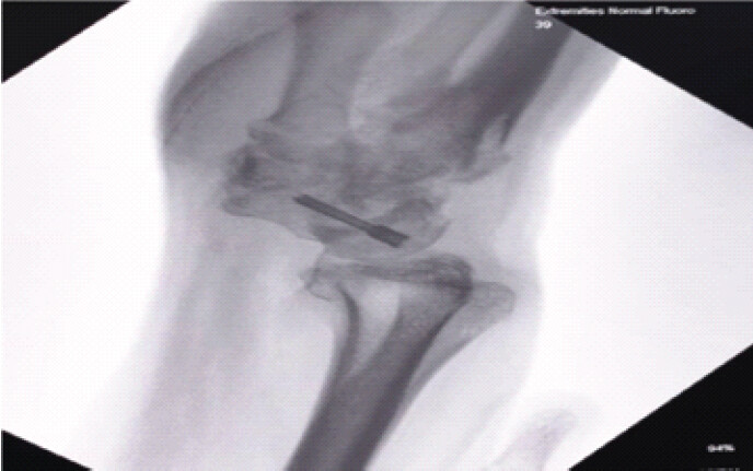
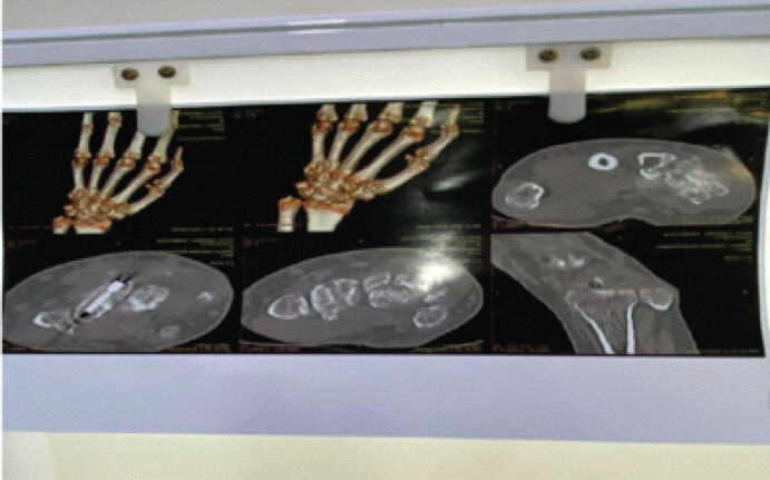
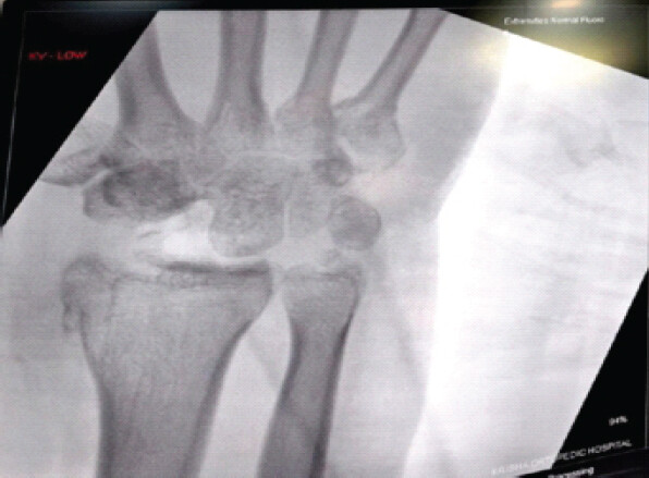
Radiological evaluation (Figs. 1 and 2) demonstrated established radiocarpal arthritis secondary to a distal radius volar rim fracture associated with a scaphoid fracture. Based on persistent symptoms and degenerative changes, operative intervention was planned.
Surgical technique
The procedure was performed under regional anesthesia with the patient in the supine position and the affected upper limb prepared under tourniquet control.
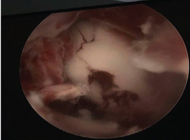
Diagnostic wrist arthroscopy was first performed through standard 3–4 and 6R portals. Arthroscopic assessment demonstrated significant chondral damage and confirmed post-traumatic arthritic changes involving the radiocarpal joint (Fig. 3). The triangular fibrocartilage complex and midcarpal joint were also evaluated arthroscopically.
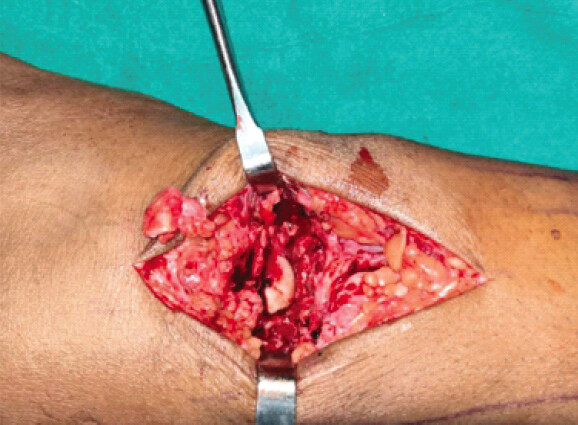
A longitudinal dorsal wrist incision was then made to expose the radiocarpal joint (Fig. 4). The scaphoid, lunate and triquetrum were excised completely using osteotomes, rongeurs and curettes. The remaining articular surfaces were contoured, and the radiocapitate articulation was assessed.
A posterior interosseous nerve was identified, and a segment was excised for wrist denervation. A distally based dorsal capsular flap was fashioned and interposed between the capitate head and the lunate fossa of the distal radius and secured using absorbable sutures. The radiocapitate articulation was reduced and stabilized using K-wire fixation under fluoroscopic guidance. Position was confirmed on anteroposterior and lateral views before layered closure and immobilization.
Post-operative management
A sterile dressing was applied, and a well-padded dorsal plaster slab was used to maintain the wrist in a neutral position. Standard post-operative analgesia, antibiotics, and rehabilitation protocols were initiated (Fig. 5).
Discussion
Treatment of radiocarpal arthritis is mostly directed at the relief of pain and may cause a reduction in wrist mobility [5]. Post-traumatic arthritis following combined distal radius volar rim and scaphoid fractures may cause accelerated cartilage damage with prolonged strain and can result in profound functional impairment [6].
PRC has traditionally been used for degenerative wrist conditions such as scapholunate advanced collapse and scaphoid non-union advanced collapse. However, its indications have expanded to include selected cases of post-traumatic radiocarpal arthritis with preserved cartilage over the capitate head and the lunate fossa of the distal radius [7]. In the present case, PRC was selected as the salvage procedure because it removes the diseased proximal carpal row while preserving wrist motion through the creation of a new radiocapitate articulation.
The addition of capsular interposition arthroplasty improves radiocapitate congruity and potentially reduces contact pressures [8]. Posterior interosseous nerve neurectomy serves as an adjunctive pain-relieving procedure without compromising motor function. Temporary radiocapitate stabilization with K-wire fixation can further support early healing and maintenance of alignment [9].
This case not only highlights the success of PRC, but also illustrates the consequences of failing to adequately address the volar lunate facet fracture at the time of the original injury. The volar rim fragment serves as the primary restraint to volar carpal subluxation [10]. Inadequate fixation may lead to persistent radiocarpal incongruity and rapid degenerative arthritis, as observed in this patient. Once arthritis becomes established, reconstructive options are limited. In such circumstances, PRC serves as a biomechanically sound salvage procedure that preserves motion while providing a congruent and pain-free radiocapitate articulation.
Conclusion
PRC represents a biomechanically sound salvage option for radiocarpal arthritis secondary to a missed or malunited volar rim fracture. In carefully selected patients, PRC combined with capsular interposition arthroplasty, posterior interosseous nerve neurectomy, and radiocapitate stabilization can provide a viable motion-preserving salvage option by maintaining satisfactory congruity and providing a functional and pain-relieving articulation. Long-term follow-up is required to evaluate the durability of outcomes and progression of degenerative changes.
Clinical Message
Missed or malunited volar rim (volar lunate facet) fractures of the distal radius can result in persistent radiocarpal incongruity, leading to early post-traumatic radiocarpal arthritis and significant functional impairment. Early recognition and stable fixation of this critical fragment are essential to preserve wrist biomechanics. When degenerative changes become established, PRC serves as a biomechanically sound, motion-preserving salvage procedure by creating a functional radiocapitate articulation, providing reliable pain relief while maintaining a useful range of wrist motion.
Conflict of Interest:
Nil
Source of Support:
Nil
Consent:
The authors confirm that informed consent was obtained from the patient for publication of this article
References
- MacLean SB, Bain GI. Volar marginal rim fractures of the distal radius. J Orthop Rep 2024;3:100307. Google Scholar | PubMed
- Clarnette J, Silva AD, Eardley-Harris N, MacLean S, Bain GI. Volar Lunate facet fractures of the distal radius: Fracture mapping using 3D CT scans. J Wrist Surg 2022;11:484-92. Google Scholar | PubMed
- Hayat Z, Varacallo MA. Scaphoid Wrist Fracture. In: StatPearls. Treasure Island, FL: StatPearls Publishing; 2026. Available from: https://www.ncbi.nlm.nih.gov/books/nbk536907. [Last accessed on 2026 Jun 28]. Google Scholar | PubMed
- Wagner ER, Barras LA, Harstad C, Elhassan BT, Moran SL. Proximal row carpectomy in young patients. JBJS Essent Surg Tech 2021;11:e19.00054. Google Scholar | PubMed
- Tang CQ, Fok MW, Chung SR, Choudhury MM, Chin AY. Managing the arthritic wrist: Complication prevention and management. J Clin Orthop Trauma 2025;62:102877. Google Scholar | PubMed
- Lee A, Klyce W, Webber M. Management of post-traumatic wrist arthritis with proximal row carpectomy. Oper Tech Orthop 2022;32:100945. Google Scholar | PubMed
- Clarke AD, Gozzard C. Management of 'radiocarpal arthritis.'. Surg (Oxford) 2019;37:270-5. Google Scholar | PubMed
- Gilheany M, Edwards SR, Cotchett M. Capsular interposition arthroplasty with percutaneous suture anchoring: A pragmatic and novel surgical technique for hallux rigidus. Cureus 2020;12:e8556. Google Scholar | PubMed
- Meng H, Xu B, Xu Y, Niu H, Liu N, Sun D. Treatment of distal radius fractures using a cemented K-wire frame. BMC Musculoskelet Disord 2022;23:591. Google Scholar | PubMed
- Heifner JJ, Orbay JL. Assessment and management of acute volar rim fractures. J Wrist Surg 2021;11:214-8. Google Scholar | PubMed
© 2026 Journal of Orthopaedic Case Reports - Published by Indian Orthopaedic Research Group
About the Authors
-

Karn Maheshwari
-

Happy Sathwara
-

Hiren Patel
-

Kana Musar
-

Nikita Bhimani
Related Articles in Journal of Orthopaedic Case Reports
 September 1, 2026 Concomitant Distal Radius Fracture with Scaphoid Fracture-Dislocation and Carpal Instability: A Case Report
 June 1, 2026 Case Report of Simultaneous Galeazzi Fracture Dislocation and Ipsilateral Scaphoid
 June 1, 2026 Nonvascularized Iliac Crest Autograft for Wrist and Hand Reconstructions: A Report of Two Clinical Cases and Literature Review
 June 1, 2026 Kienbock’s Disease in a Young Female without Classical Risk Factors: A Case Report with 1-year Functional Outcome Following Proximal Row Carpectomy