


Introduction
A significant contributor to the functional ability of the thumb is the extensor pollicis longus (EPL) tendon. This extrinsic thumb muscle is responsible for extension and adduction at the 1st metacarpophalangeal and interphalangeal joints, and assists in radial deviation at the wrist. Passing through the 3rd extensor compartment around the Lister’s tubercle [1], ruptures of the EPL can occur commonly after distal radius fractures [2], or without prior injury in patients with rheumatic disease, carpometacarpal osteoarthritis, or following local corticosteroid injection [3]. Iatrogenic tendon attrition can occur following plate and screw osteosynthesis after surgical fixation of a distal radius fracture, either by the prominent lengthy screws of a volar plate [4] or against the dorsal plate [2]. EPL injuries occurring secondary to distal radius fractures and their management, whether surgical or conservative, have been reported in the literature with an incidence of up to 17% [5].
Surgical fixation of distal radius fractures, among the most common injuries encountered by orthopedic surgeons, can require the use of dorsal plate fixation, especially in cases with posterior comminution or displacement [6]. However, delayed rupture of the extensor tendons around the Lister’s tubercle following such fixation is a rare and potentially devastating complication that requires meticulous attention to detail during the procedure for its prevention. Furthermore, it should be strongly considered during pre-operative evaluation and planning in patients with medical comorbidities (diabetes mellitus, rheumatoid arthritis) that predispose toward tendon attrition and rupture [7].
Management of such pathologies begins with the diagnosis, which is unfortunately usually delayed due to the subtle clinical findings in the early stages, which may be mistaken for sequelae of the bony injury. Ultrasonography is a reliable imaging modality that can assist in the diagnosis [8], and treatment of the described tendon damage usually requires extensor indicis proprius (EIP) tendon transfer if direct repair is not possible [9].
We report a case of a delayed EPL tendon rupture following attritional damage after dorsal plate and screw osteosynthesis of a distal radius fracture in a patient with rheumatoid arthritis, which was successfully managed with implant removal and EIP tendon transfer.
Case Report
A 49-year-old female presented with inability to actively extend the left thumb for the past 3 months. The patient had no significant history of recent trauma to the involved part, fever, or local infection.
The patient gave a history of surgery for a distal radius fracture of the affected side 1.5 years prior, which had been managed with dorsal plate and screw fixation.
On clinical examination, loss of active extension at the metacarpophalangeal and interphalangeal joints of the 1st digit was noted. A healed surgical scar over the posterior distal wrist was noted, and no tenderness was elicited upon palpation of the scar. No local warmth or signs of infection were noted.
The post-operative period following the index surgery was reported by the patient to have be uneventful.
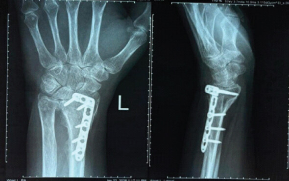
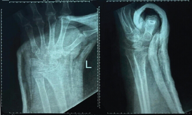
Radiographs were ordered, and fracture union was noted on the anteroposterior and lateral views (Fig. 1). In addition, the radiograph also appeared to show dorsal plate prominence, with the distal borders of the plate appearing to be extending beyond the joint line distally and posteriorly.
The patient reported being a known case of rheumatoid arthritis on regular medical treatment (tablet methotrexate 10 mg once weekly and tablet hydroxychloroquine sulfate 200 mg OD with tablet folic acid 5 mg supplementation) for 3 years, as well as a type 2 diabetic on medications (tablet metformin 500 mg OD) for glycemic control for 5 years.
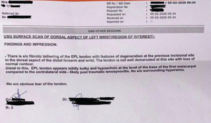
An ultrasonography examination was performed on the affected wrist, with the area of importance being localized to the extensor compartments. The EPL tendon was poorly demarcated proximally with loss of normal contour and fibrotic tethering associated with features of degeneration. Distally, the tendon appeared bulky and hypoechoic at the level of the 1st metacarpal with features of tenosynovitis (Fig. 2).
Based on the clinical and radiographic evaluation, the patient was counseled regarding the further management options and was planned for exploration and tendon repair/transfer to EIP.
Following pre-operative anesthetic evaluation, the patient was taken up for surgery a week later. A dorsal approach to the distal radius through the existing surgical scar was utilized. The dorsal radius plate and screws were identified and removed in toto.
Exploration of the extensor compartments, the 3rd in particular, was performed. Careful and meticulous dissection showed discontinuity of the EPL at the extensor retinaculum level (Fig. 3 and 4). The distal tendon stump was identified within the 3rd compartment below the extensor retinaculum, and its integrity was confirmed upon passive extension of the 1st interphalangeal with application of tension.
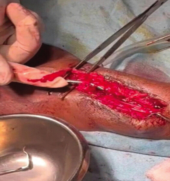
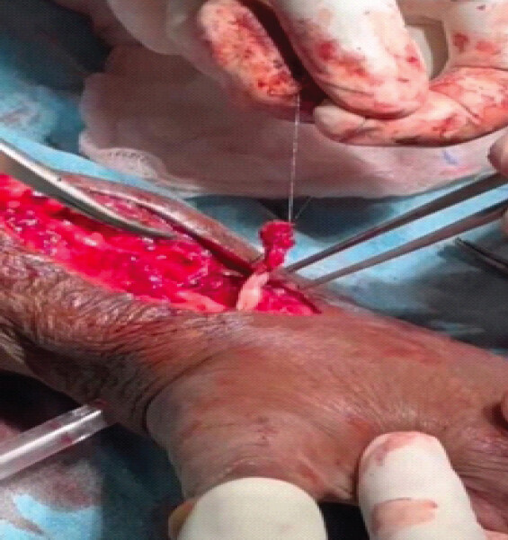
A significant intraoperative challenge was noted while attempting to locate the proximal end of the cut EPL tendon, which could not be identified within the surgical field. Primary repair of the tendon ends was ruled out as unfeasible owing to the inability to visualize the proximal end.
Reconstruction was planned and performed using an EIP to EPL tendon transfer (Fig. 5).
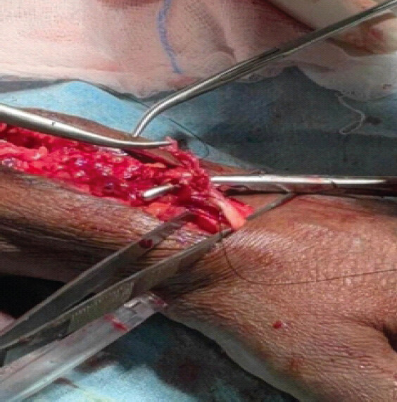
The EIP was identified through the same incision ulnar to the 1st slip of the extensor digitorum communis and divided close to the 2nd Metacarpophalangeal joint. With the thumb held in extension to maintain appropriate tension, the EIP was sutured to the distal end of the EPL tendon with end-to-end fish-mouth sutures (Pulvertaft weave), following which the extensor retinaculum was repaired.
A sterile dressing was done, and a post-operative radiograph was taken (Fig. 6).
The limb was immobilized in a thumb spica splint for 4 weeks, following which she was started on a planned and structured rehabilitation program under regular follow-up.
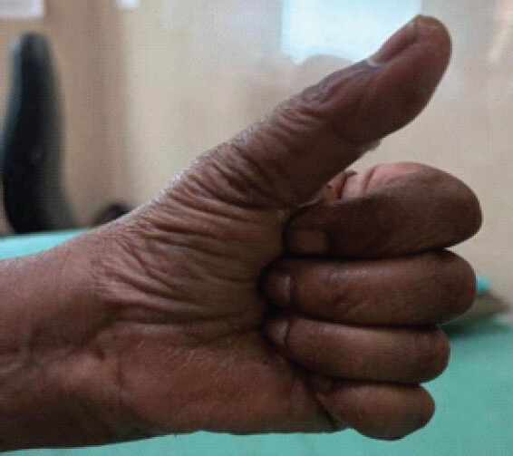
Stitches were removed at 14 days, and the wound was noted to be healthy. At 4 weeks, the patient was started on gentle range-of-motion exercises, with gradual resistance training of the transferred tendon, the results of which were assessed weekly on follow-up. At 8 weeks, the patient demonstrated a good range of motion of the 1st and 2nd digits with satisfactory extension of the thumb without support and had no complaints of pain over the operated site (Fig. 7).
Discussion
The pathogenesis of EPL rupture has been widely described in the literature, with local and systemic causes identified [2]. The incidence of rupture following a traumatic injury to the lower end of the radius has been suggested as a result of the poor vascularization of the tendon sheath; a hematoma within the sheath can jeopardize the blood flow to the already poorly supplied regions of the tendon [10]. The course of the EPL tendon, ranging around a vascular watershed area at the Lister’s tubercle, predisposes it to significant mechanical friction [5]. At this region, an absent mesotenon combined with a narrowed compartment leads to a compromise in the circulation and diffusion of synovial fluid, leading to tendon attrition and rupture as a result [11].
Furthermore, systemic comorbidities such as rheumatoid arthritis or diabetes mellitus can cause an increase in the synovial pressure and inflammatory mediators, which contribute to weakening of the tendon and its degeneration [12,13]. The microvascular compromise and glycation-related tendon degeneration, which are features of diabetes mellitus, are significant predictors of poor vascularization at anatomically compromised areas. Even in the absence of obvious implant prominence, the combination of systemic disorders in the presence of a local mechanical irritation can result in tendon attrition and damage; the result of a multifactorial pathogenesis involving ischemia, metabolic and vascular insult, synovial inflammation, and local mechanical tendon attrition [14].
The importance of maintaining a high clinical suspicion and a thorough clinical examination of such cases must be emphasized. Weakness and pain in the early stages of such an injury are easily missed, due to the focus on the fracture of the lower end of the radius. Sudden inability of extension of the digits should raise a suspicion of extensor tendon rupture, regardless of the condition of the fracture and its management.
High-resolution ultrasonography is a routine imaging modality in confirming tendon damage, identifying characteristic features of rupture at the level of the Lister’s tubercle as well as the extent of proximal and distal tendon ends retraction [15]. Furthermore, sonography can also identify anatomical variants of Lister’s tubercle, which could be critical due to its implication of potential future risk to the tendon transfer or graft, during and after repair [8].
However, its value being dependent on operator skill, can result in differing findings during surgery as compared to the report.
Surgical management remains the mainstay of treatment and can involve primary repair, tendon transfer and/or intercalary/intertendinous grafting [9,11]. Tendon transfer is among the most popular choices for the management of EPL ruptures, with EIP being the most common donor [16].
Conclusion
Delayed degenerative EPL rupture can be a major late complication of distal radius fractures, especially in patients with pre-existing risk factors, regardless of the management. Early identification and accurate diagnosis require a high degree of suspicion and clinical acumen, and are essential for a satisfactory functional recovery.
Clinical Message
Delayed EPL tendon rupture should be suspected in patients with medical comorbidities presenting with loss of thumb extension after distal radius fracture fixation and requires thorough evaluation, surgical management, and rehabilitation.
Conflict of Interest:
Nil
Source of Support:
Nil
Consent:
The authors confirm that informed consent was obtained from the patient for publication of this article
References
- Khan IA, Launico MV. Anatomy, shoulder and upper limb, hand extensor pollicis longus muscle. In: StatPearls. Florida: StatPearls Publishing. 2026. Google Scholar | PubMed
- Björkman A, Jörgsholm P. Rupture of the extensor pollicis longus tendon: A study of aetiological factors. Scand J Plast Reconstr Surg Hand Surg 2004;38:32-5. Google Scholar | PubMed
- McCann PA. Comment on: Minimising the risk of extensor pollicis longus rupture following volar plate fixation of distal radius fractures: A cadaveric study. Ann R Coll Surg Engl 2011;93:494. Google Scholar | PubMed
- Apard T, Marcucci L, Jarriges J. Spontaneous rupture of extensor pollicis longus in isolated trapeziometacarpal arthritis. Chir Main 2011;30:349-51. Google Scholar | PubMed
- Hirasawa Y, Katsumi Y, Akiyoshi T, Tamai K, Tokioka T. Clinical and microangiographic studies on rupture of the E. P. L. tendon after distal radial fractures. J Hand Surg Br Eur Vol 1990;15:51-7. Google Scholar | PubMed
- Disseldorp DJ, Hannemann PF, Poeze M, Brink PR. Dorsal or volar plate fixation of the distal radius: Does the complication rate help us to choose?. J Wrist Surg 2016;5:202-10. Google Scholar | PubMed
- Alsaedi OF, Alshahir AA, Alzahim AF, Alharbi WM, Alsaygh EF, Alhamaid YA. Etiology of traumatic causes of extensor pollicis longus tendon rupture: A systematic review. J Hand Microsurg 2024;16:100038. Google Scholar | PubMed
- Ghazal L, Nabi M, Little C, Teh J. Ultrasound assessment of extensor pollicis longus tendon rupture following distal radius fracture: A sonographic and surgical correlation. J Ultrason 2020;20:e1-5. Google Scholar | PubMed
- Ganon S, Bellity J, Zbili D, Boccara D. Reconstruction strategies after rupture of the extensor pollicis longus tendon: A systematic review. Hand Surg Rehabil 2020;39:502-7. Google Scholar | PubMed
- Engkvist O, Lundborg G. Rupture of the extensor pollicis longus tendon after fracture of the lower end of the radius--a clinical and microangiographic study. Hand 1979;11:76-86. Google Scholar | PubMed
- Bhardwaj P, Varadharajan V, Jain D, Sabapathy SR. Management of extensor tendon injuries concerning distal radius fractures. J Orthop 2024;48:96-102. Google Scholar | PubMed
- Lister RC, Bradford HC 4th, Joo A, Carr CW, Delancy A, Naram A. Spontaneous rupture of the extensor pollicis longus tendon: A systematic review. Hand (N Y) 2024;19:1314-20. Google Scholar | PubMed
- Lee JY, Roh YT, Jeong C, Jung S, Park IJ. Systemic risk factors of extensor pollicis longus tendon rupture after distal radius fracture. Arch Hand Microsurg 2017;22:240-6. Google Scholar | PubMed
- Hu CH, Fufa D, Hsu CC, Lin YT, Lin CH. Revisiting spontaneous rupture of the extensor pollicis longus tendon: Eight cases without identifiable predisposing factor. Hand (N Y) 2015;10:726-31. Google Scholar | PubMed
- Santiago FR, Plazas PG, Fernández JM. Sonography findings in tears of the extensor pollicis longus tendon and correlation with CT, MRI and surgical findings. Eur J Radiol 2008;66:112-6. Google Scholar | PubMed
- Magnussen PA, Harvey FJ, Tonkin MA. Extensor indicis proprius transfer for rupture of the extensor pollicis longus tendon. J Bone Joint Surg Br 1990;72:881-3. Google Scholar | PubMed
© 2026 Journal of Orthopaedic Case Reports - Published by Indian Orthopaedic Research Group
About the Authors
-

C Abhijith
-

Adarsh Varghese Mathew
Related Articles in Journal of Orthopaedic Case Reports
 June 1, 2026 Delayed Spontaneous Pectoralis Major Tendon Rupture Following Anterior Latissimus Dorsi Transfer for Irreparable Subscapularis Tear: A Case Report
 January 1, 2026 Dual Reconstruction, Combined Anterior Latissimus Dorsi with Teres Major and Middle Trapezius Tendon Transfer, Reduces Anterior Superior Humeral Subluxation: Case Report
 December 1, 2025 Flexor Tendon Injuries in Zone 3: A Comprehensive Review and Case Report on Flexor Digitorum Profundus Rupture of the Middle Finger
 February 1, 2025 Medial Epicondyle Fracture Dislocation, Multiple Failed Reductions, and a Median Nerve Entrapment: A Case Report