


Introduction
Concomitant fractures of the distal radius and scaphoid are uncommon injuries, representing a small proportion of wrist fractures and typically resulting from high-energy trauma [1,2]. The distal radius fracture is frequently intra-articular, while the associated scaphoid fracture may be overlooked on initial radiographs, particularly when displacement is minimal [2]. Advanced imaging with computed tomography (CT) is therefore valuable when the injury mechanism or radiographic appearance raises suspicion of an associated carpal injury [2, 3].
The combination of a comminuted intra-articular distal radius fracture with a scaphoid fracture-dislocation is considerably more unusual and presents a challenging injury pattern due to the potential for carpal malalignment and instability. High-energy mechanisms have been associated with greater fracture displacement and more complex intra-articular patterns in patients sustaining concomitant distal radius and scaphoid fractures [4].
Although the optimal management of simultaneous distal radius and scaphoid fractures remains incompletely established, early operative fixation has been reported to provide satisfactory radiological and functional outcomes in appropriately selected patients [2,4,5]. Recent clinical evidence suggests that operative management of the scaphoid in this setting may facilitate earlier radiographic union and earlier restoration of wrist motion [4, 5].
We present a rare case of a high-energy Arbeitsgemeinschaft für Osteosynthesefragen/Orthopaedic Trauma Association (AO/OTA) 23-B3 intra-articular distal radius fracture associated with scaphoid fracture-dislocation and carpal instability, managed with combined volar and dorsal plating of the distal radius and Kirschner wire (K-wire) stabilization of the carpal injury.
Case Report
This case report describes a 32-year-old right-hand-dominant male who sustained a high-energy injury to the left wrist following a motorcycle road traffic accident. The patient was thrown from the motorcycle and landed on his outstretched left hand. On presentation, he had severe pain, swelling, deformity, and marked restriction of wrist movements. There was no open wound, and distal neurovascular status was intact.
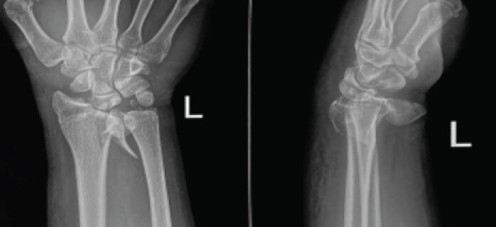
Initial anteroposterior and lateral radiographs demonstrated a comminuted intra-articular distal radius fracture (AO/OTA 23-B3) associated with a displaced scaphoid fracture and disruption of normal carpal alignment. Given the high-energy mechanism and radiographic evidence of carpal instability, careful assessment of the associated scaphoid and carpal injury was undertaken, as concomitant scaphoid fractures may be overlooked on initial assessment [1, 2]. (Fig. 1).
The wrist was initially immobilized in a below-elbow slab to allow subsidence of soft-tissue swelling. Definitive surgical management was performed on the 3rd day following injury under regional anesthesia. Operative stabilization was selected due to the intra-articular, comminuted distal radius fracture and associated displaced scaphoid fracture-dislocation with carpal instability. Previous literature supports operative management in selected patients with displaced concomitant distal radius and scaphoid fractures [2, 3, 6].
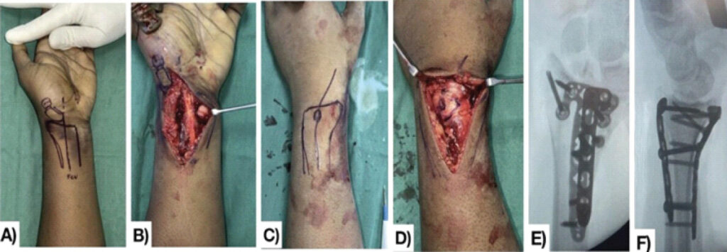
A modified Henry approach was used for exposure and reduction of the distal radius. Following restoration of the articular anatomy, the distal radius was stabilized using a volar locking plate. Due to the fracture comminution and residual instability, supplementary dorsal plating was performed (Fig. 2).
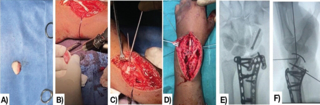
The associated scaphoid fracture-dislocation was reduced under fluoroscopic guidance and stabilized with a Herbert screw. The associated carpal instability was reduced and stabilized using K-wire fixation. Intraoperative fluoroscopy confirmed satisfactory reduction and restoration of distal radius alignment, scaphoid position, and carpal relationships. The wrist was immobilized in a functional slab postoperatively. Following 4 weeks of immobilization, the slab was removed, and wrist range-of-motion exercises were initiated. The carpal K-wires were removed at 6 weeks, with radiographs demonstrating maintained carpal alignment (Fig. 3).
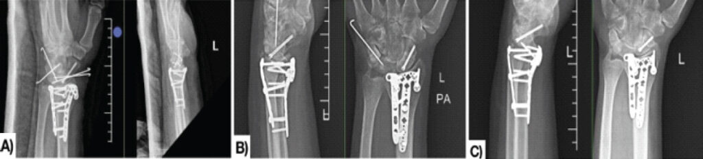
Serial post-operative clinical and radiological assessments were performed to evaluate fracture union, maintenance of carpal alignment, wrist range of motion, and functional recovery. Approximately 40° of flexion and 60° of extension at the end of 8 months in final follow-up (Fig. 4).
Results
At 8 months postoperatively, radiographs demonstrated complete union of the distal radius and scaphoid fractures with maintained reduction and carpal alignment. The patient achieved 40° of wrist flexion and 60° of extension, providing a total flexion–extension arc of approximately 100°. The Visual Analog Scale (VAS) pain score was 3/10, and the patient reported no limitation in activities of daily living. There was no residual neurovascular deficit or clinically evident carpal instability at final follow-up. Overall, the patient demonstrated satisfactory radiological and functional recovery despite the complexity of the initial injury.
Discussion
Concomitant ipsilateral fractures of the distal radius and scaphoid are uncommon, with reported prevalence ranging from approximately 0.5% to 5% among distal radius fractures [1, 2]. These injuries are predominantly associated with high-energy trauma. In a systematic review of 178 patients with 182 simultaneous fractures, most injuries resulted from high-energy mechanisms, and the majority of distal radius fractures were intra-articular [2].
The present case represents an unusual injury pattern due to the combination of an AO/OTA 23-B3 comminuted intra-articular distal radius fracture with a displaced scaphoid fracture-dislocation and carpal instability. Simultaneous distal radius and scaphoid fractures may occur as part of a greater-arc carpal injury, including perilunate fracture-dislocation patterns [2, 4]. Consequently, careful assessment of carpal alignment is essential, particularly following high-energy trauma.
The management of these combined injuries remains challenging because treatment must restore distal radius anatomy while simultaneously addressing scaphoid stability and associated carpal malalignment. A systematic review by Blackburn et al. found that the available literature is predominantly composed of retrospective case series, with no high-level comparative evidence establishing a single optimal treatment strategy [2]. Nevertheless, the review reported high union rates following both operative and non-operative treatment, while acknowledging that operative fixation is increasingly favored for displaced or unstable injuries [2, 7, 8, 9].
More recent evidence further supports operative stabilization in selected cases. Verlinsky et al. [4] reviewed 31 concomitant distal radius and scaphoid fractures and found that 87.2% were associated with high-energy trauma, while 71% involved AO/OTA Type C distal radius fractures [4]. Among patients treated operatively for the scaphoid fracture, radiological union occurred significantly earlier than in the non-operative group (6.0 ± 2.0 versus 13.7 ± 11.7 weeks) [4]. These findings support consideration of operative stabilization when the scaphoid fracture is displaced or associated with instability [4, 6]. Similar favorable outcomes following surgical fixation of concomitant distal radius and scaphoid fractures have been reported in clinical series, supporting early stabilization in appropriately selected unstable injuries [6, 10].
In the present case, the distal radius was stabilized using combined volar and dorsal plating, while the associated carpal instability was addressed with K-wire fixation. This allowed restoration and maintenance of the distal radius and carpal alignment during healing. At 8 months, radiological union was achieved with a 100° flexion–extension arc, a VAS pain score of 3/10, and no limitation in activities of daily living.
The favorable outcome in this case emphasizes the importance of identifying associated scaphoid and carpal injuries in high-energy distal radius fractures. Advanced imaging, particularly CT, can assist in defining complex scaphoid fracture morphology, carpal alignment, and associated instability when plain radiographs are inconclusive [5, 9, 10].
Conclusion
Concomitant distal radius fracture with scaphoid fracture-dislocation is a rare and potentially unstable high-energy wrist injury. Accurate assessment of the distal radius, scaphoid, and carpal alignment is essential for appropriate treatment planning. Combined stabilization addressing both the comminuted distal radius fracture and associated carpal instability can restore and maintain anatomical alignment. In this case, combined volar and dorsal plating with K-wire stabilization resulted in radiological union at 8 months, satisfactory wrist motion, low residual pain, and unrestricted activities of daily living. Early recognition and comprehensive stabilization of this complex injury pattern may help achieve satisfactory radiological and functional outcomes.
Clinical Message
High-energy distal radius fractures with abnormal carpal alignment should prompt careful evaluation for associated scaphoid and intercarpal injuries, as early stabilization of both the fracture and carpal instability can restore and maintain wrist alignment.
Conflict of Interest:
Nil
Source of Support:
Nil
Consent:
The authors confirm that informed consent was obtained from the patient for publication of this article
References
- Fowler TP, Fitzpatrick E. Simultaneous fractures of the ipsilateral scaphoid and distal radius. J Wrist Surg 2018;7:303-11. Google Scholar | PubMed
- Blackburn J, Johnson N, Pocnetz S, Lindau TR. Effective treatment of simultaneous distal radius and scaphoid fractures. J Wrist Surg 2021;11:89-94. Google Scholar | PubMed
- Komura S, Yokoi T, Nonomura H, Tanahashi H, Satake T, Watanabe N. Incidence and characteristics of carpal fractures occurring concurrently with distal radius fractures. J Hand Surg Am 2012;37:469-76. Google Scholar | PubMed
- Verlinsky L, Ulmer C, Rose A, Brady C, Rose R. Improved outcomes in operative management of concomitant distal radius and scaphoid fractures. Hand (N Y) 2024;19:1114-9. Google Scholar | PubMed
- Gürbüz Y, Sügün TS, Kayalar M. Combined fractures of the scaphoid and distal radius: Evaluation of early surgical fixation (21 patients with 22 wrists). J Wrist Surg 2018;7:11-7. Google Scholar | PubMed
- Clementson M, Björkman A, Thomsen NO. Acute scaphoid fractures: Guidelines for diagnosis and treatment. EFORT Open Rev 2020;5:96-103. Google Scholar | PubMed
- Tountas AA, Waddell JP. Simultaneous fractures of the distal radius and scaphoid. J Orthop Trauma 1987;1:312-7. Google Scholar | PubMed
- Chang CH, Tsai YS, Sun JS, Hou SM. Ipsilateral distal radius and scaphoid fractures. J Formos Med Assoc 2000;99:733-7. Google Scholar | PubMed
- Herzberg G, Comtet JJ, Linscheid RL, Amadio PC, Cooney WP, Stalder J. Perilunate dislocations and fracture-dislocations: A multicenter study. J Hand Surg Am 1993;18:768-79. Google Scholar | PubMed
- Singh HP, Forward DP, Davis TR. Scaphoid fractures: Current concepts and management. Injury 2024;55:111-9. Google Scholar | PubMed
© 2026 Journal of Orthopaedic Case Reports - Published by Indian Orthopaedic Research Group
About the Authors
-

Pranay Choudhary
-

Praneeth Chowdhary
-

Sayam Ghosh
Related Articles in Journal of Orthopaedic Case Reports
 September 1, 2026 Proximal Row Carpectomy for Established Post-Traumatic Radiocarpal Arthritis Following Distal Radius Volar Rim and Scaphoid Fractures: A Case Report
 August 1, 2025 Modified Minimally Invasive Reduction and Osteosynthesis System Technique for Fixation of Distal Radius
 July 1, 2025 Missed Volar Intercalated Segment Instability with Distal Radius Fracture: Challenges in Diagnosis, Surgical Management, and Clinical Outcome – Case Report
 July 1, 2026 Fracture Fixation Sequencing in Polyskeletal Trauma: Clinical Experience from a Five-Patient Retrospective Series