


Introduction
Traumatic spinal cord injury (SCI) remains a major cause of global disability; a 2025 analysis of the Global Burden of Disease study reported that the global incidence of SCI more than doubled between 1990 and 2019, with falls and road-traffic injuries as the leading risk factors and young-to-middle-aged men bearing the greatest burden [1]. Traumatic thoracolumbar spondylolisthesis is a rare consequence of this broader pattern of high-energy spinal trauma, distinct from degenerative or isthmic spondylolisthesis because acute translation reflects failure of multiple osteoligamentous stabilizers rather than chronic pars or disc degeneration. The thoracolumbar junction is biomechanically vulnerable to this pattern of injury because it sits at the transition between the relatively rigid, rib-supported thoracic spine and the more mobile lumbar spine, concentrating flexion, rotation, and shear forces during high-energy falls and vehicular trauma. Meyerding Grade IV denotes 75–100% vertebral translation, while displacement exceeding 100% constitutes Grade V, or spondyloptosis [2,3]. This distinction correlates with the degree of mechanical instability and, in many series, with the severity of the accompanying neurological injury.
Traumatic spondyloptosis and near-spondyloptotic injuries carry extreme mechanical instability and a high rate of neurological injury; small series have documented complete deficits in most patients with sagittal-plane injury [4], although case reports describe meaningful recovery after reduction and stabilization, including patients who progressed from American Spinal Injury Association Impairment Scale (AIS) A or C to AIS D [5,6,7,8]. A systematic review of 125 cases of traumatic lumbar spondylolisthesis found that roughly half resulted from vehicular accidents, that neurological deficits were present in half of patients at presentation, and that operative reduction, decompression, and fusion were associated with significant improvement in pain and neurological status at final follow-up [3]. Neurological recovery after traumatic SCI more broadly is markedly heterogeneous. A systematic review and meta-analysis of 114 studies and nearly 20,000 patients found that approximately 19% of patients initially classified as AIS Grade A demonstrated at least one-grade conversion, with recovery influenced by injury severity, level, mechanism, and duration of follow-up [9].
Most published series report only AIS or Frankel grade at limited time points, which can understate the trajectory of recovery and omit change in independence or walking capacity. We report a patient with severe traumatic D11–D12 Meyerding Grade IV spondylolisthesis and an initial complete neurological deficit who improved to AIS C by discharge and AIS D at 6 months, corroborated by serial International Standards for Neurological Classification of SCI (ISNCSCI) [10] motor and sensory scores and by spinal cord independence measure III (SCIM III) and walking index for SCI II (WISCI II) functional measures.
Case Report
A 39-year-old woman with no significant prior illness sustained a fall from an estimated height of 10–12 feet while cutting leaves from a tree at approximately 23:00 on December 31, 2025. She was evaluated at a regional hospital and referred to a tertiary trauma center. On arrival, she was hemodynamically stable (blood pressure 121/81 mmHg, pulse 80 beats/min, oxygen saturation 98% on room air), with a patent airway and no documented loss of consciousness. She reported severe back pain with complete absence of sensation and movement below the injury level, and no bowel or bladder sensation.
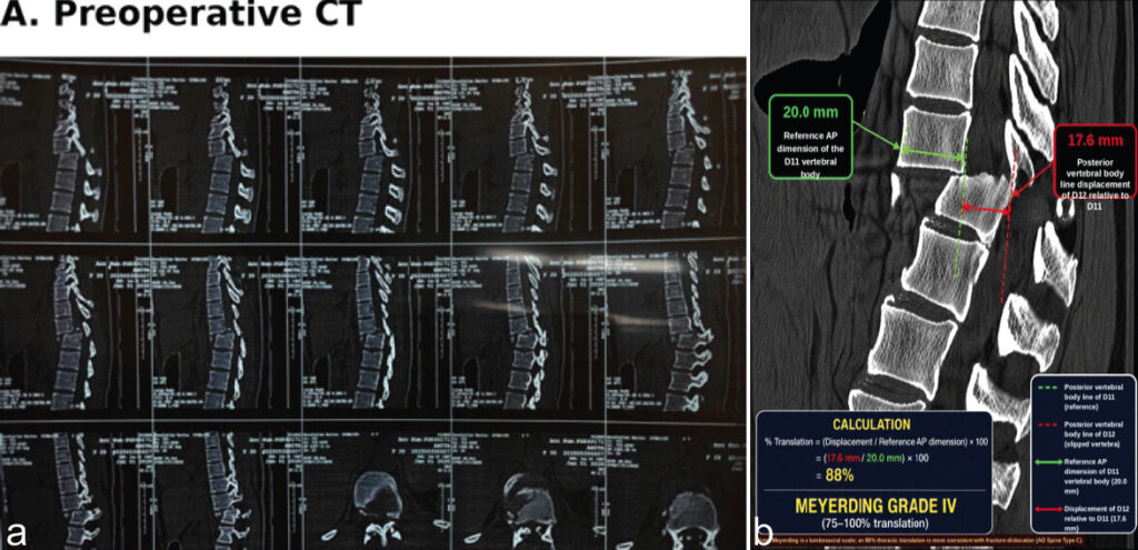
The initial neurological examination was consistent with a complete traumatic SCI, documented as AIS A, with an upper extremity motor score (UEMS) of 50/50, lower extremity motor score (LEMS) 0/50, total motor score 50/100, light-touch score 32/56, pin-prick score 25/56, and total sensory score 57/112; neurological function above the injury level was fully preserved. Pre-operative sagittal computed tomography (CT) demonstrated approximately 88% anterior translation of D11 relative to D12, measured using the Meyerding method, consistent with Meyerding Grade IV traumatic spondylolisthesis, with marked anterolisthesis and disruption of thoracolumbar alignment (Fig. 1a). Magnetic resonance imaging demonstrated severe D11–D12 translational injury with marked focal spinal cord compression and intramedullary T2/short tau inversion recovery hyperintensity consistent with traumatic cord edema/contusion. Associated disruption of the D11–D12 intervertebral disc and posterior ligamentous complex was present, with surrounding paraspinal soft-tissue edema. No definite complete cord transection was identified.
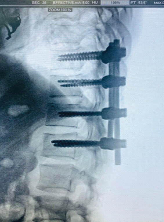
Posterior reduction and D10–L1 pedicle screw-rod fixation were performed on January 01, 2026, within approximately 24 h of injury, through a standard midline posterior approach under general anesthesia. Intraoperative fluoroscopy confirmed satisfactory implant position and restoration of spinal alignment (Fig. 2). The post-operative course was uneventful, without wound complications or early implant-related problems. A multidisciplinary SCI rehabilitation program was instituted postoperatively, individualized to neurological status and tolerance and delivered in five progressive phases from acute stabilization through community reintegration, with therapy intensity advancing from daily bedside sessions to five to six gait-focused sessions per week as motor recovery progressed (Table 1). Therapy progressed from prevention of secondary complications, joint range-of-motion work, bed mobility, sitting balance, and transfer training toward task-specific standing and gait training as voluntary lower-limb motor function emerged, with body-weight-supported or robot-assisted gait training used as an adjunct when clinically appropriate [11,12]. A representative locomotor training session during the intensive phase comprised approximately 90 min, combining warm-up and range-of-motion work, lower-limb strengthening, bed mobility and transfer practice, sitting and standing balance, supported standing and weight-shifting, body-weight-supported or robotic gait training, and overground functional walking (Table 2). Bladder and bowel management, pressure-injury prevention, and patient and family education were incorporated throughout.
Rehabilitation program by phase
| Phase | Approx. period | Main rehabilitation | Rough dosage |
|---|---|---|---|
| Phase 1: Acute stabilization | Post-operative days 1–7 | Positioning, pressure-injury prevention, respiratory care, passive ROM, prevention of contractures, bed mobility, bowel/bladder program | Daily, 30–60 min PT/OT divided into sessions |
| Phase 2: Early rehabilitation | Week 2–4 | Active-assisted ROM as movement emerges, strengthening preserved muscles, rolling, supines→sitting, sitting balance, transfer training, wheelchair skills, standing preparation | 5–6 days/week, ~60–90 min/day |
| Phase 3: Motor recovery | Weeks 4–8 | Progressive lower-limb strengthening, sit-to-stand, supported standing, weight shifting, stepping initiation transfer training, balance | 5 days/week, ~60–120 min/day |
| Phase 4: Locomotor training | Weeks 6–12 | BWSTT/robot-assisted gait where appropriate+overground gait+strengthening+balance | 3–5 gait sessions/week, ~30–60 min/session |
| Phase 5: Functional/community rehabilitation | 3–6 months | Overground walking, assistive device training, stairs, endurance, transfers, ADL training, community mobility | 3–5 days/week, progressively individualized |
| Home/community phase | After discharge | Home exercise, walking practice, strengthening, balance, ADLs, community participation | Individualized; progressive |
ROM: Range of motion, PT/OT: Physical therapy/occupational therapy, BWSTT: Body-weight supported treadmill training, ADL: Activities of daily living
Representative locomotor training session (intensive phase, ~90 min)
| Component | Approx. time |
|---|---|
| Warm-up/ROM | 10 min |
| Lower-limb strengthening | 15 min |
| Bed mobility/transfers | 15 min |
| Sitting/standing balance | 15 min |
| Supported standing/weight shifting | 10 min |
| BWSTT or robotic gait training | 20 min |
| Overground gait/functional walking | 5–10 min |
| Cool-down/education | 5 min |
| Total | ~90 min |
BWSTT: Body-weight supported treadmill training, ROM: Range of motion
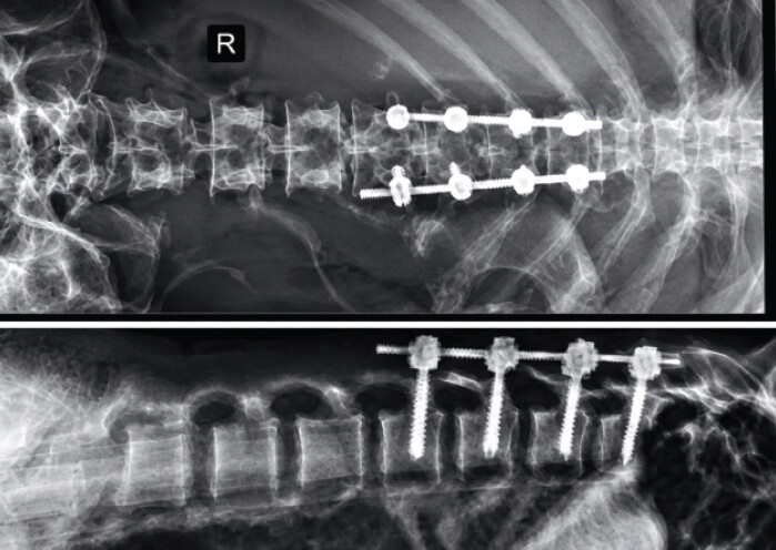
Neurological status improved to AIS C at discharge (LEMS 12/50, total motor score 62/100) and continued to improve at 2 weeks (LEMS 15/50), 6 weeks (LEMS 18/50), and 3 months (LEMS 22/50), reaching AIS D at 6 months (LEMS 27/50, total motor score 77/100) (Table 3). UEMS remained at the ceiling of 50/50 throughout, consistent with intact upper-extremity function above the injury level, while light-touch and pin-prick scores rose in parallel. SCIM III and WISCI II tracked this trajectory closely, improving from 15/100 and 0/20 at presentation to 29/100 and 1/20 at discharge, and to 56/100 and 9/20 at 6 months, corresponding to a transition from dependent, non-ambulatory status to short-distance assisted ambulation (Table 4). The 6-month radiograph confirmed maintained reduction and satisfactory implant position without evidence of construct failure (Fig. 3). Neurological function above the injury level remained clinically intact throughout follow-up, so the major change occurred below the level of injury. All serial AIS, ISNCSCI, SCIM III, and WISCI II assessments at every time point were performed by the same senior resident, minimizing inter-rater variability in the reported scores. Patient-reported outcomes, including pain, quality of life, satisfaction with recovery, and functional confidence, were also recorded serially and improved in parallel with the neurological and functional measures, with the patient progressing from being unable to resume usual activities to partial return by 6 months (Table 5); no follow-up data beyond 6 months were obtained.
Serial ISNCSCI neurological assessment
| Parameter | Admission | Discharge | 2 week | 6 week | 3 month | 6 month |
|---|---|---|---|---|---|---|
| AIS | A | C | C | C | C | D |
| NLI | T11† | T11† | T11† | T11† | T11† | T11† |
| UEMS /50 | 50 | 50 | 50 | 50 | 50 | 50 |
| LEMS/50 | 0 | 12 | 15 | 18 | 22 | 27 |
| Total motor/100 | 50 | 62 | 65 | 68 | 72 | 77 |
| LT/56 | 32 | 35 | 37 | 39 | 41 | 44 |
| PP/56 | 25 | 29 | 31 | 33 | 36 | 39 |
| Total sensory/112 | 57 | 64 | 68 | 72 | 77 | 83 |
| DAP | Absent | Present | Present | Present | Present | Present |
| VAC | Absent | Absent | Absent | Absent | Present | Present |
| Sacral sensory sparing | No | Yes | Yes | Yes | Yes | Yes |
| Sacral motor sparing | No | No | No | No | Yes | Yes |
| Motor ZPP | T11† | T12† | T12† | L1† | L2† | L2f |
| SCIM III/100 | 15 | 29 | 34 | 40 | 47 | 56 |
| WISCI II/20 | 0 | 1 | 2 | 4 | 6 | 9 |
ISNCSCI: International standards for neurological classification of spinal cord injury, AIS: American spinal injury association impairment scale, NLI: Neurological level of injury, UEMS: Upper extremity motor score, LEMS: Lower extremity motor score, LT: Light touch, PP: Pin prick, DAP: Deep anal pressure, VAC: Voluntary anal contraction, ZPP: Zone of partial preservation, SCIM III: Spinal cord independence measure, version III, WISCI II: Walking index for spinal cord injury II. †Motor level/ZPP reported by spinal segment. At admission, absent DAP, absent VAC, and absent sacral sensory and motor sparing confirmed a neurologically complete (AIS A) injury per ISNCSCI criteria; sacral sensory sparing returned at discharge, voluntary anal contraction returned at 3 months, and sacral motor sparing was regained by 3 months, tracking the AIS conversion to D at 6 months
Serial SCIM III and WISCI II functional assessment
| Time point | AIS | SCIM III/100 | WISCI II/20 | Functional picture |
|---|---|---|---|---|
| Admission | A | 15 | 0 | Dependent; non-ambulatory |
| Discharge | C | 29 | 1 | Basic sitting/transfer ability beginning; requires substantial assistance |
| 2 weeks | C | 34 | 2 | Improved transfers; assisted standing |
| 6 weeks | C | 40 | 4 | Standing and very limited assisted ambulation |
| 3 months | C | 47 | 6 | Short-distance assisted walking |
| 6 months (final) | D | 56 | 9 | Meaningful functional recovery; walking with assistance/device |
SCIM III: Spinal Cord Independence Measure III, WISCI II: Walking Index for spinal cord injury II, AIS: American spinal injury association impairment scale
Serial patient-reported outcomes
| Patient-reported outcome | Admission | Discharge | 3 months | 6 months |
|---|---|---|---|---|
| Pain NRS (0–10) | 8 | 5 | 3 | 2 |
| WHOQOL-BREF overall QoL (0–10) | 2 | 4 | 6 | 7 |
| Satisfaction with recovery (0–10) | 1 | 4 | 7 | 8 |
| Overall functional confidence (0–10) | 1 | 3 | 5 | 6 |
| Return to usual activities | Unable | Dependent | Partial | Partial |
NRS: Numeric rating scale, WHOQOL-BREF: World Health Organization Quality of Life-BREF. No follow-up beyond 6 months was obtained
Discussion
The defining feature of this case is the combination of severe structural disruption and substantial neurological and functional recovery, quantified longitudinally rather than at a single time point. Traumatic spondyloptosis, defined by greater than 100% vertebral displacement, is the extreme form of this injury spectrum. Yadla et al. reported that four of five patients with sagittal-plane traumatic thoracolumbar spondyloptosis presented with complete AIS A deficits, and only the patient with an incomplete injury recovered neurological function postoperatively [4]. Subsequent case reports have nonetheless described meaningful recovery to AIS D after traumatic lumbar spondyloptosis when reduction and stabilization were achieved [5,6,8]. Ver et al.’s systematic review and case series proposed a classification for traumatic lumbar spondylolisthesis based on disc integrity and recommended interbody fusion when disc injury is present, reporting an overall fusion rate of 74% and significant improvement in pain and neurological symptoms at final follow-up [3]. The Meyerding system, used here because it is well established for describing sagittal translation, is complementary to more comprehensive injury-classification schemes such as the AOSpine thoracolumbar spine injury classification system, which additionally grades fracture morphology and neurological status [13]. Applying this scheme to the present case, the injury is classified as morphology type C (translational injury, 8 points), neurological modifier N4 (complete SCI, 4 points), with no case-specific modifiers identified (M2, 0 points), for a composite AOSpine score of C + N4 (12 points). This classification corroborates the severity implied by the Meyerding Grade IV translation and situates the case for more direct comparison with other traumatic thoracolumbar SCI cohorts. Quantitative measurement of vertebral translation demonstrated 88% anterior displacement, confirming the injury as Meyerding Grade IV rather than grade V/spondyloptosis. We classify the present injury as Meyerding Grade IV rather than spondyloptosis, a distinction with direct bearing on how the injury compares with the published spondyloptosis literature.
The magnitude of recovery in this patient is notable against the wider evidence base. Khorasanizadeh et al., analyzing 114 studies and nearly 20,000 patients, reported a pooled AIS/Frankel conversion rate of only 19.3% among patients initially graded A, with recovery rates increasing significantly with longer follow-up [9]. This patient’s trajectory illustrates that principle directly: She improved to AIS C during the index admission but continued to recover to AIS D by 6 months, a change that assessment restricted to discharge would have missed. AIS is an overall neurological classification rather than a grade assigned separately above and below the lesion; in this patient, function above the injury level remained clinically intact throughout, while motor and sensory function below the level progressively recovered, as shown by rising LEMS, total motor score, and sensory scores.
The timing of surgery is relevant to interpreting this outcome. Definitive fixation was performed within approximately 24 h of injury. A 2024 systematic review update supports early decompression within this window when feasible: patients decompressed early were roughly twice as likely to improve by two or more AIS grades at 6 and 12 months [14]. The temporal association between early surgical stabilization and subsequent neurological recovery is notable; however, causality cannot be established in this single case. The observed recovery may reflect the combined effects of early mechanical stabilization, natural neurological recovery following traumatic SCI, resolution of secondary SCI processes, and intensive rehabilitation. The magnitude of recovery should therefore be interpreted as an important clinical observation rather than evidence that early surgery alone was responsible for the neurological outcome.
Reporting SCIM III and WISCI II alongside AIS and ISNCSCI scores adds clinically meaningful granularity that AIS grade alone cannot provide, since a categorical neurological grade does not capture graded change in self-care independence, mobility, or walking capacity [15, 16]. In this patient, both functional measures improved in close parallel with the neurological conversion, moving from dependent and non-ambulatory status at presentation to short-distance assisted walking at 6 months, consistent with the broader rehabilitation literature supporting body-weight-supported and robot-assisted gait training as effective adjuncts to conventional overground rehabilitation in motor-incomplete SCI [11,12].
The clinical implication is that prognostication after severe traumatic SCI should remain evidence-based and longitudinal rather than anchored to the earliest available examination. An initial AIS A examination is an important marker of injury severity but should not be treated as an immutable endpoint for counseling patients and families. Serial standardized neurological and functional assessment, extending well beyond hospital discharge, can capture clinically important recovery that would otherwise be missed.
This report has several important limitations. As a description of a single patient, the findings cannot be generalized to the broader population of patients with traumatic Meyerding Grade IV thoracolumbar spondylolisthesis or complete traumatic SCI; pooling this case with others through a multicenter case series, registry, or systematic review would allow the trajectory described here to be interpreted against a larger, more representative sample. There was no concurrent comparator managed non-operatively, with delayed surgery, or with an alternative surgical strategy, so it is not possible to determine whether the observed recovery is attributable to the treatment chosen; a prospective cohort or registry-based comparison matching patients by injury severity, level, and AIS grade would allow the effect of early surgical stabilization to be estimated more directly. Consequently, causality between early surgical stabilization and the neurological improvement cannot be established: spontaneous neurological recovery, resolution of spinal shock, resolution of cord edema, and the structured rehabilitation program may all have contributed independently or synergistically to the trajectory described.
The 6-month follow-up period, while longer than that of many published case reports, may still be insufficient to capture this patient’s ultimate neurological and functional trajectory, since meaningful recovery after traumatic SCI can continue well beyond this interval; extending follow-up to 12 and 24 months, as recommended for SCI recovery studies, would better establish whether the recovery trajectory plateaus or continues to improve. The relative contribution of individual rehabilitation components to the observed recovery cannot be isolated, since multiple interventions (positioning, strengthening, gait training, and assistive-device practice) were delivered concurrently as part of a single multidisciplinary program. Despite the AIS conversion from A to D, the patient remained substantially functionally impaired at 6 months, requiring assistance or a device for ambulation; the distinction between categorical neurological grade conversion and the achievement of clinically meaningful functional independence deserves emphasis and should not be conflated. Continued longitudinal tracking of SCIM III, WISCI II, and the patient-reported outcomes in Table 5 beyond 6 months would clarify whether functional independence continues to approach the neurological grade achieved.
Psychological status was not formally assessed with a validated instrument, although pain, quality of life, satisfaction, and functional confidence were captured serially (Table 5). Longer-term assessment of fusion status, implant integrity, adjacent-segment change, residual deformity, and late complications beyond 6 months was similarly not performed, even though early alignment was maintained and no implant failure was observed; scheduled follow-up with dynamic radiographs or CT at 12 and 24 months would confirm durable fusion and detect late hardware or adjacent-segment complications not yet apparent at 6 months. Finally, the magnitude of neurological recovery in a patient initially classified as AIS A is unusually favorable relative to published cohorts and may represent an exceptional outcome; case reports are inherently susceptible to selection and publication bias toward favorable results, and this trajectory should not be interpreted as evidence that similar recovery is expected in most patients with comparable injuries. Systematic registration of both favorable and unfavorable outcomes in SCI registries, and inclusion of this case in future pooled analyses or systematic reviews, would help contextualize how representative this recovery trajectory is and mitigate the bias inherent to isolated case reporting.
Conclusion
Severe traumatic D11–D12 Meyerding Grade IV spondylolisthesis may present with a complete neurological deficit but does not invariably preclude meaningful recovery. In this patient, neurological status improved from AIS A at presentation to AIS C at discharge and AIS D at 6 months, corroborated by serial ISNCSCI motor and sensory scores and by SCIM III and WISCI II functional data, while neurological function above the injury level remained intact throughout. The case reinforces the importance of precise radiological grading and of sufficiently long, standardized serial neurological and functional assessment before offering a final prognosis.
Clinical Message
In severe traumatic thoracolumbar translation injuries, an initial complete neurological deficit should prompt careful longitudinal assessment rather than definitive pessimistic prognostication. Early stabilization, structured rehabilitation, and serial standardized neurological and functional testing over at least 6 months can reveal clinically meaningful recovery, as illustrated by this patient’s improvement from AIS A to AIS D.
Conflict of Interest:
Source of Support:
Nil
Consent:
The authors confirm that informed consent was obtained from the patient for publication of this article
References
- Wang Y.Global, regional, and national burdens of traumatic brain injury, spinal cord injury, and skull fracture and their attributable risk factors from 1990 to 2021: A systematic analysis of the global burden of disease study 2021. Front Public Health 2025;13:1622693. Google Scholar | PubMed
- Meyerding HW.Spondylolisthesis: Surgical treatment and results. Surg Gynecol Obstet 1932;54:371-7. Google Scholar | PubMed
- Ver ML, Dimar JR2nd , Carreon LY.Traumatic lumbar spondylolisthesis: A systematic review and case series. Global Spine J 2019;9:767-82. Google Scholar | PubMed
- Yadla S, Lebude B, Tender GC, Sharan AD, Harrop JS, Hilibrand AS.Traumatic spondyloptosis of the thoracolumbar spine. J Neurosurg Spine 2008;9:145-51. Google Scholar | PubMed
- Amesiya R, Orwotho N, Nyati M, Mugarura R, Mwaka ES.Traumatic spondyloptosis of the lumbar spine: A case report. J Med Case Rep 2014;8:453. Google Scholar | PubMed
- Barwar N.Post-traumatic L4-L5 spondyloptosis with cauda equina syndrome: A case report. Trauma Case Rep 2021;33:100475. Google Scholar | PubMed
- Sharma GK, Salaria AK, Verma H, Singh S.Acute traumatic lumbar spondyloptosis with complete neurological deficit: A case report and literature review. J Orthop Case Rep 2025;15:180-5. Google Scholar | PubMed
- Gautam S, Tamang RS, Joshi K, Bhusal S, Shrestha R.Surgical management of traumatic spondyloptosis: A case series. J Med Case Rep 2024;18:468. Google Scholar | PubMed
- Khorasanizadeh M, Yousefifard M, Eskian M, Lu Y, Chalangari M, Harrop JS.Neurological recovery following traumatic spinal cord injury: A systematic review and meta-analysis. J Neurosurg Spine 2019;30:683-99. Google Scholar | PubMed
- American Spinal Injury Association, International Spinal Cord Society. International Standards for Neurological Classification of Spinal Cord Injury, Revised 2019. Richmond, VA: ASIA; 2019. Google Scholar | PubMed
- Yang FA, Chen SC, Chiu JF, Shih YC, Liou TH, Escorpizo R.Body weight-supported gait training for patients with spinal cord injury: A network meta-analysis of randomised controlled trials. Sci Rep 2022;12:19262. Google Scholar | PubMed
- Arroyo-Fernández R, Menchero-Sánchez R, Pozuelo-Carrascosa DP, Romay-Barrero H, Fernández-Maestra A, Martínez-Galán I.Effectiveness of body weight-supported gait training on gait and balance for motor-incomplete spinal cord injuries: A systematic review with meta-analysis. J Clin Med 2024;13:1105. Google Scholar | PubMed
- Vaccaro AR, Oner C, Kepler CK, Dvorak M, Schnake K, Bellabarba C.AOSpine thoracolumbar spine injury classification system: Fracture description, neurological status, and key modifiers. Spine 2013;38:2028-37. Google Scholar | PubMed
- Fehlings MG, Hachem LD, Tetreault LA, Skelly AC, Dettori JR, Brodt ED.Timing of decompressive surgery in patients with acute spinal cord injury: Systematic review update. Global Spine J 2024;14:38S-57S. Google Scholar | PubMed
- Furlan JC, Noonan V, Singh A, Fehlings MG.Assessment of disability in patients with acute traumatic spinal cord injury: A systematic review of the literature. J Neurotrauma 2011;28:1413-30. Google Scholar | PubMed
- Ditunno JF Jr, Ditunno PL, Scivoletto G, Patrick M, Dijkers M, Barbeau H.The walking index for spinal cord injury (WISCI/WISCI II): Nature, metric properties, use and misuse. Spinal Cord 2013;51:346-55. Google Scholar | PubMed
© 2026 Journal of Orthopaedic Case Reports - Published by Indian Orthopaedic Research Group
About the Authors
-

Dr. Sharath Raj
-

Dr. Saloni Rani Kumar
-

Dr. Deepak Kumar
-

Dr. Shrinivas VB
-

Dr. Vijay G Goni
Related Articles in Journal of Orthopaedic Case Reports
 September 1, 2026 Blunt Traumatic Posterior Cord Syndrome Following a Hangman Fracture: A Case Report
 February 1, 2026 Virtual reality rehabilitation to enhance functional recovery initiated from the acute phase following cervical cord injury: A case report
 November 1, 2025 Posterior Decompression and Stabilization in Thoracic and Lumbar Spinal Tuberculosis: A Prospective Observational Study
 September 1, 2025 Treatment of Bilateral Distal Femoral Shaft Fractures in a Paraplegic Patient with Intramedullary Nailing: A Case Report