


Introduction
Spinal cord injury (SCI) affects approximately 11,000 people annually in the United States [1,2]. The extent of cord involvement varies widely between patients, and the International Standards for Neurological Classification of SCI (ISNCSCI) formally recognizes five incomplete SCI syndromes: Central cord syndrome, Brown-Sequard syndrome, anterior cord syndrome, conus medullaris syndrome, and cauda equina syndrome [3]. Even syndromes with a well-established clinical picture can present atypically. A recent report described Brown-Sequard syndrome following only minor trauma in a patient with an underlying cervical segmentation anomaly, a reminder that these classification patterns describe a spectrum rather than fixed rules, and that careful modality-by-modality sensory and motor testing matters more than the mechanism of injury alone [4].
Posterior cord syndrome (PCS) is not separately classified within the ISNCSCI system, and its reported incidence is below 1%, a figure that probably reflects under-recognition as much as true rarity [1,5,6,7]. Clinically, PCS produces loss of proprioception, vibration sense, two-point discrimination, and deep pressure sensation below the level of injury, occasionally with pain or paresthesia, while motor power, light touch, and pinprick sensation are relatively preserved [2,3,8,9]. Reported causes include posterior spinal artery ischemia, tumor, disc herniation, Vitamin B12 deficiency, and trauma [6,10,11]; iatrogenic injury has also been described, including a case of PCS following electrode lead placement during spinal cord stimulation for chronic pain.[12] Traumatic PCS is the least common of these etiologies and is generally attributed to a hyperextension mechanism [11,13]. Recognizing PCS early also has practical consequences: The sensory ataxia it produces can be mistaken for a non-organic complaint or for a coexisting peripheral neuropathy, which delays rehabilitation planning that specifically targets proprioceptive retraining rather than motor strengthening alone. We report a case of blunt traumatic PCS following a hangman fracture, with the aim of sharpening clinical suspicion for this injury pattern and clarifying the role of delayed imaging in confirming the diagnosis when acute studies are inconclusive.
Case Report
A 24-year-old man with a history of depression and prior appendicectomy, and an otherwise unremarkable family and social history, presented after a road traffic accident. He reported paresthesia involving his entire body, neck pain, and subjective weakness of the right arm. Computed tomography of the cervical spine identified a right C1 transverse process fracture, a hangman fracture of C2 with associated lamina, spinous process, and teardrop fracture components, and a C3 spinous process fracture without subluxation (Fig. 1).
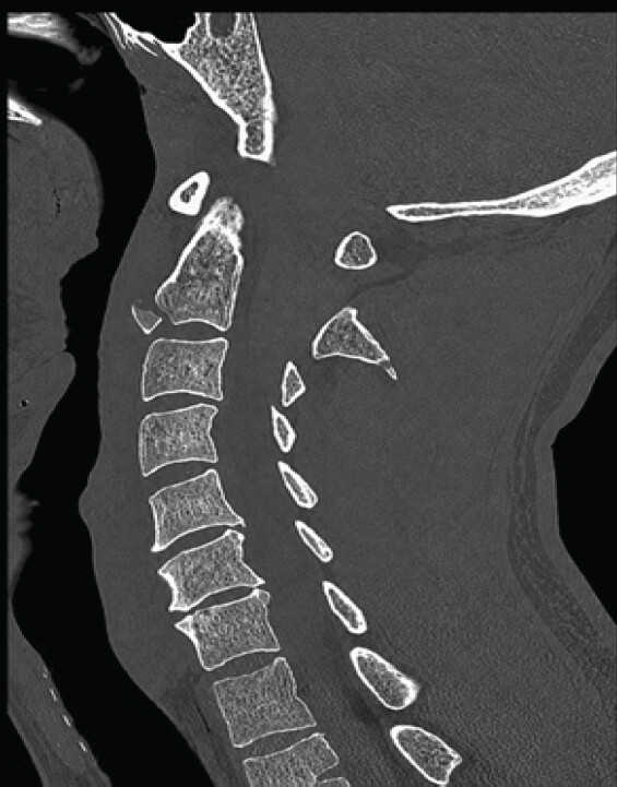
Neurological examination followed a standard ISNCSCI protocol, testing motor power, light touch, pinprick, joint position sense, and vibration in each limb. The findings were consistent with an incomplete SCI predominantly affecting the dorsal columns: paresthesia extended from the neck distally to involve joint position sense bilaterally in both upper and lower limbs. Strength testing graded his right arm at antigravity power, his left hand showed only minimal weakness, and both legs were full strength. Because arm weakness was present without corresponding leg weakness, central cord syndrome was also considered in the differential diagnosis at this stage. Cervical magnetic resonance imaging (MRI) showed faint, diffuse cord edema together with disruption of the anterior longitudinal ligament at C2-C3 (Fig. 2), but it did not clearly localize the injury within the substance of the cord itself.
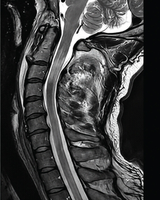
The risks and benefits of operative stabilization with halo immobilization were discussed at length and weighed against a trial of collar immobilization alone. The patient elected to proceed with surgery and gave informed consent for both the operation and the halo orthosis. He underwent anterior cervical interbody fusion with plating at C2-C3, supplemented with a halo orthosis, tolerated the procedure without swallowing difficulty, and began physical therapy and mobilization soon afterward. Because his deficit was primarily sensory rather than motor, his rehabilitation program was weighted toward balance training and proprioceptive re-education alongside standard mobilization, rather than the strength-focused protocol that would typically follow a motor-predominant cord injury.
At his first outpatient review, he had no measurable motor deficit but showed slight impairment of fine finger dexterity. By 3 months postoperatively, mild balance dysfunction persisted alongside improving dysesthesia, and he was ambulating independently without walking aids. At 1 year, his neurological examination was essentially normal apart from minor residual proprioceptive loss in the hands and feet; this residual numbness did not interfere with his daily activities, and nerve conduction studies performed at that visit were normal. He continued to describe some anxiety about the residual weakness, though objectively his function had returned close to baseline. A follow-up cervical MRI without contrast, obtained 10 months after the injury, clearly delineated the injury to the posterior columns (Fig. 3a and b), anatomically confirming a posterior SCI and establishing the diagnosis of PCS in retrospect.
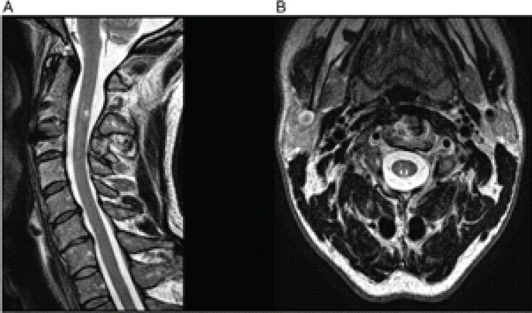
Discussion
Traumatic PCS remains uncommon in the published literature. Most reported cases of PCS are atraumatic in origin [5,8,11], and among the small number of traumatic cases, we identified, all but one involved the cervical spine [2,5,9,13,14,15]. Hyperextension is the mechanism most often implicated [1,13,15], and both hangman fractures and teardrop fractures of the axis, as seen in our patient, are recognized hyperextension injuries [1, 7]. Only two prior reports specifically discuss surgical management of traumatic PCS [13,15], which makes the operative course and 1-year outcome described here a useful addition to a thin evidence base.
Belen and Weingarden described a 41-year-old man injured in a motorcycle collision who presented with paresthesia and bilateral upper- and lower-limb weakness [15]. His examination differed from ours in that weakness was bilateral rather than confined to one side, pinprick sensation was lost below C5, and rectal tone was reduced, although proprioception was similarly absent distally, with milder loss proximally at the shoulders. A cervical myelogram in that patient showed stenosis, and he underwent C4-C7 laminectomy; by 3 months, strength had recovered to 4/5 in all limbs, though proprioception and vibration remained absent in the right leg and only partially returned on the left. The authors attributed the upper-limb weakness to a coexisting central cord component, a combination that appears to be common after hyperextension injury.
Steidl reported a construction worker struck on the head by a falling wooden block, who developed tingling in the neck and shoulders followed by severe bilateral hypersensitivity in the C3-C4 dermatomes, without any radiographic abnormality [9]. His symptoms resolved spontaneously within 3 weeks. Because imaging was normal, Steidl proposed that the posterior gray matter is particularly vulnerable to contusion, and that transient perivascular bleeding causing limb paralysis lasting up to 4 h could occur through excessive anteroposterior motion of the cord rather than direct bony injury. The same author suggested that PCS results from cord compression by inward buckling of the ligamentum flavum and ventral osteophytes, a proposed mechanism distinct from that of central cord syndrome [8].
Distinguishing PCS from central cord syndrome, and more broadly from other incomplete SCI patterns, is not always straightforward at initial assessment, since arm-predominant weakness can arise in either condition and both are common consequences of hyperextension injury. A recently published case from the Journal of Orthopedic Case Reports illustrates this overlap well: A 63-year-old man with C5-C6 bilateral facet dislocation presented with quadriparesis heavier in the upper than the lower limbs, a pattern classically ascribed to central cord syndrome, and was managed successfully with staged posterior and anterior stabilization after closed reduction [16]. Our patient’s asymmetric arm weakness raised the same differential early on, and it was only the specific pattern of sensory loss, together with the appearance of the follow-up MRI, that ultimately pointed to a posterior rather than a central lesion. The anatomical basis for this distinction is worth stating plainly: Central cord syndrome reflects damage concentrated in the central gray matter and medially situated corticospinal fibers serving the upper limb, whereas PCS reflects damage confined to the dorsal white matter columns carrying proprioceptive and vibratory input, with motor pathways relatively spared. Both patterns can coexist after a single hyperextension event, and where they overlap, as in the Belen and Weingarden case discussed above, the resulting picture may not fit cleanly into either textbook category. Incomplete SCI syndromes can also emerge from unexpected mechanisms, as in the Brown-Sequard case described above, which is a further reminder that the physical examination, and not the mechanism of injury on its own, should drive the diagnosis [4].
Other rare SCI patterns make the same point: Clinical findings and imaging findings do not always evolve at the same pace. White cord syndrome, a reperfusion injury first described after decompressive surgery for chronic cord compression, produces new post-operative deficits accompanied by delayed T2 hyperintensity on MRI that can be mistaken for a fresh structural injury rather than recognized as a consequence of sudden reperfusion to chronically ischemic tissue [17]. Anterior spinal cord syndrome from fibrocartilaginous embolism can similarly mimic transverse myelitis on early imaging, with a vascular pattern only becoming apparent once inflammatory and demyelinating causes have been excluded and the clinical course is reviewed in full [18]. In each of these entities, as in the case reported here, imaging obtained too early in the clinical course can under-represent, or simply fail to localize, the true anatomical extent of injury, and a period of watchful reassessment with repeat imaging is often what finally establishes the diagnosis. This is a practical argument for building a planned follow-up MRI into the management pathway of any patient with a clinically incomplete cord syndrome and an acute study that does not fully explain the examination findings, rather than treating a normal or non-specific early scan as reassurance that no structural injury is present.
PCS is a rare subset of SCI that can be missed entirely if vibration and proprioception are not specifically tested during a standard ISNCSCI examination [2,6,18]. In our patient, MRI performed in the acute phase failed to delineate the injury clearly within the substance of the cord, so PCS could easily have gone unrecognized at that stage. Patients with PCS frequently exhibit a positive Romberg sign, which is sometimes misattributed to neurosyphilis [2,3,5], and because PCS sits outside the formal ISNCSCI syndrome classification, it is arguably even more prone to being overlooked than the five recognized syndromes [1,3,6]. What sets our case apart is that we were able to confirm the site of injury anatomically to the posterior columns on a follow-up MRI obtained a year after the initial presentation, an anatomical correlation that most of the prior reports we reviewed did not achieve.
Conclusion
This report describes PCS diagnosed after blunt trauma to the axis, with near-complete resolution of symptoms following cervical fusion at C2-C3. A thorough neurological examination, with deliberate attention to proprioception and vibration rather than pinprick and light touch alone, is central to recognizing PCS, since routine screening will otherwise miss it. Where the acute MRI is inconclusive, as it was here, we suggest that a sub-acute or delayed study, obtained once the initial cord edema has settled, can help confirm the anatomical diagnosis and guide counseling about long-term recovery. For spine surgeons managing patients with predominantly sensory deficits after cervical trauma, this case supports setting expectations around a slower, proprioception-focused recovery trajectory rather than assuming that a normal early motor examination and an unremarkable acute scan rule out a clinically significant cord injury.
Clinical Message
Arm-predominant weakness after cervical hyperextension trauma is not always central cord syndrome. When proprioceptive and vibratory loss is disproportionate to motor findings, clinicians should specifically examine the dorsal columns and consider PCS, repeating MRI later in the clinical course if the initial study does not fully explain the findings.
Conflict of Interest:
Nil
Source of Support:
Nil
Consent:
The authors confirm that informed consent was obtained from the patient for publication of this article
References
- Marcon RM, Cristante AF, Teixeira WJ, Narasaki DK, Oliveira RP, De Barros Filho TE. Fractures of the cervical spine.. Clinics (Sao Paulo) 2013;68:1455-61. [Google Scholar] | [PubMed]
- McKinley W, Santos K, Meade M, Brooke K. Incidence and outcomes of spinal cord injury clinical syndromes. J Spinal Cord Med 2007;30:215-24. [Google Scholar] | [PubMed]
- Maynard FM, Bracken MB, Creasey G, Ditunno JFJr, Donovan WH, Ducker TB. International standards for neurological and functional classification of spinal cord injury. American Spinal Injury Association. Spinal Cord 1997;35:266-74. [Google Scholar] | [PubMed]
- Mukherjee S, Pandian KP, Senthil V. A rare case of C5-C6 bilateral facetal dislocation with central cord syndrome managed by staged stabilization. J Orthop Case Rep 2026;16:268-72. [Google Scholar] | [PubMed]
- Zhang S, Wang Z, Zhang S, Lu C, Liu Z, Kang C. Brown Sequard syndrome in a patient with Klippel-Feil syndrome following minor trauma: A case report and literature review.. BMC Musculoskelet Disord 2023;24:722. [Google Scholar] | [PubMed]
- Bosch A. Incomplete traumatic quadriplegia. A ten-year review.. JAMA 1971;216:473. [Google Scholar] | [PubMed]
- Cochrane M, Hess M, Sajkowicz N. Posterior cord syndrome associated with postoperative seroma: The case to perform a complete neurologic exam. J Spinal Cord Med 2020;43:892-4. [Google Scholar] | [PubMed]
- Kim HJ, Lee KY, Kim WC. Treatment outcome of cervical tear drop fracture.. Asian Spine J 2009;3:73-9. [Google Scholar] | [PubMed]
- Diaz E, Morales H. Spinal cord anatomy and clinical syndromes.. Semin Ultrasound CT MRI 2016;37:360-71. [Google Scholar] | [PubMed]
- Steidl L. Contusion injuries of the cervical spinal cord--contusio cervicalis posterior.. Acta Chir Orthop Traumatol Cech 1972;39:290-4. [Google Scholar] | [PubMed]
- Ferrando A, Bas P, Bas T. Late neurological complications due to laminar hook compression in idiopathic scoliosis surgery.. Spinal Cord Ser Cases 2017;3:17081. [Google Scholar] | [PubMed]
- Chanideh I, Khodaei A, Ghadiri M, Shakibaee Fard Z, Falahi Tabar MM, Yavari A. “White cord syndrome”: A reperfusion injury following laminectomy and spinal decompression surgery-a case report and literature review. Case Rep Med 2026;2026:5539165. [Google Scholar] | [PubMed]
- Hills AS, McKinley W. Poster 94: Posterior cord syndrome: Demographics and Rehabilitation Outcomes.. PM R. 2017;9:S166. [Google Scholar] | [PubMed]
- Kwon YM. Posterior cord syndrome after spinal cord stimulation electrode lead insertion: A case report.. Korean J Neurotrauma 2022;18:393-8. [Google Scholar] | [PubMed]
- Belen JG, Weingarden SI. Posterior central cord syndrome following a hyperextension injury: Case report.. Paraplegia 1988;26:209-11. [Google Scholar] | [PubMed]
- Sarkar B, Ahuja K, Choudhury AK, Jain R. Penetrating spine injury bisecting thoracic spinal canal with no significant neurological deficits-the midline cord syndrome.. Spinal Cord Ser Cases 2018;4:102. [Google Scholar] | [PubMed]
- Boda SA, Mandyam SR, Bhukya B, Joseph J, Sarkar C. When the disc strikes: rethinking fibrocartilaginous embolism.. Cureus 2026;18:e113143. [Google Scholar] | [PubMed]
- Itzoe M, Sciubba DM. Posterior cord syndrome.. In: Encyclopedia of Trauma Care.Springer: Berlin Heidelberg20151273. [Google Scholar] | [PubMed]
© 2026 Journal of Orthopaedic Case Reports - Published by Indian Orthopaedic Research Group
About the Authors
-

Dr. Sharath Raj
-

Dr. Deepak Kumar
-

Dr. Saloni Rani Kumar
-

Dr. Shrinivas VB
-

Ms. Urvi Goni
-

Dr. Vijay G Goni
Related Articles in Journal of Orthopaedic Case Reports
 March 1, 2025 Charcot Spinal Arthropathy Secondary to Spinal Cord Injury – A Case Report
 October 1, 2026 Brown-Séquard Syndrome after Self-administered Manual Manipulation of the Cervical Spine: A Case Report
 October 1, 2026 Bilateral Medial Medullary Syndrome Following Anterior Odontoid Screw Fixation for a Type II Fracture: A Case Report
 September 1, 2026 Three-Grade Neurological Recovery (ASIA Impairment Scale A to D) Following Surgical Stabilization of Traumatic D11–D12 Meyerding Grade IV Spondylolisthesis: A Case Report