


Introduction
Hoffa fractures are coronal-plane shear fractures involving one or both condyles of the distal femur. They are rare, accounting for approximately 0.65% of all femoral fractures. These fractures are further subclassified into lateral condylar, medial condylar and bicondylar fractures. Among them, lateral unicondylar fractures are predominant, representing roughly 75–85% of all Hoffa fractures [1, 2, 3]. In the pediatric (skeletally immature) population, Hoffa fractures are extremely rare [4, 5].
The mechanism of injury leading to Hoffa fractures is most commonly associated with high-energy trauma, such as motor vehicle accidents or sports-related injuries; however, it remains incompletely understood. Biomechanically, Hoffa fractures are believed to result from an axial load transmitted through the knee in deep flexion (≥90°), often combined with valgus or varus stress and rotational forces [6, 7].
This case reports presents a 14-year-old girl who sustained a lateral Hoffa fracture during a minor trauma which resulted in a locked knee in fixed flexion. Only limited literature describes the occurrence of mechanical knee locking as a presenting symptom of Hoffa fractures. This clinical manifestation is very rare and has mainly been reported in isolated case reports or small case series, typically in association with displaced osteochondral fragments or interposition of the fractured condylar fragment within the knee joint. In the available literature, mechanical knee locking related to Hoffa fractures has most often been described in delayed or initially missed diagnoses, where displaced intra-articular fragments impede normal knee motion [7, 8]. In this case report, the fracture was treated with closed reduction and followed by arthroscopic fixation.
Case Report
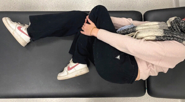
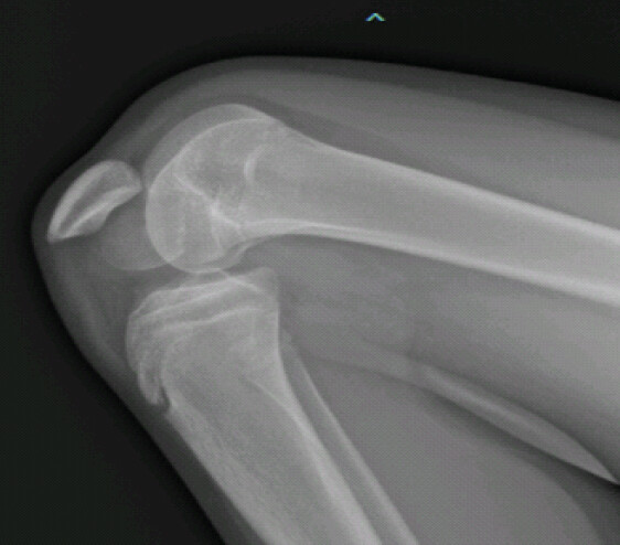
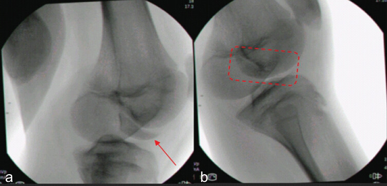
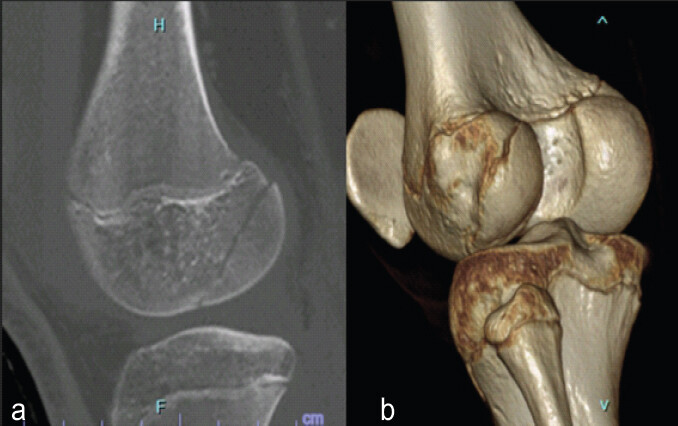
A 14-year-old girl presented to the emergency department with a mechanically locked left knee, fixed at approximately 130° of flexion (Fig. 1). She reported a seemingly trivial, low-energy injury mechanism consisting of a twisting motion while standing still, during which she heard an audible crack, followed by a fall. Following the injury, she experienced extreme pain and the inability to extend the knee. Physical examination revealed no neurovascular deficits in the affected limb. Conventional anteroposterior and lateral radiographs of the knee demonstrated a fracture of the lateral femoral condyle, accompanied by caudal displacement of the patella, locked distal to this fracture border (Fig. 2). Due to extreme pain and fixed flexion deformity, an urgent intervention under general anesthesia was performed. The patella was unlocked, and the Hoffa fracture could be reduced with manual posterolateral compression with fluoroscopic confirmation of reduction (Fig. 3). The patient was put in a long leg cast in full extension. An additional computed tomography (CT) scan confirmed the presence of the Hoffa Fracture, which was anatomically reduced (Fig. 4). Three days after trauma, the patient was surgically treated for this fracture. The patient was positioned supine on the operating table with the knee flexed to 90°. A tourniquet was applied. Standard anteromedial and anterolateral arthroscopic portals were established. Hemarthrosis was evacuated to improve visualization. Intra-articular inspection revealed Grade 1 chondral lesions in all three compartments, intact menisci, intact cruciate ligaments, and the already reduced lateral Hoffa fracture. The fracture fragment was temporarily reduced and stabilized using a large bone clamp, followed by definitive fixation using 2 headless compression screws through 2 additional lateral portals with fluoroscopic confirmation of reduction (Fig. 5).
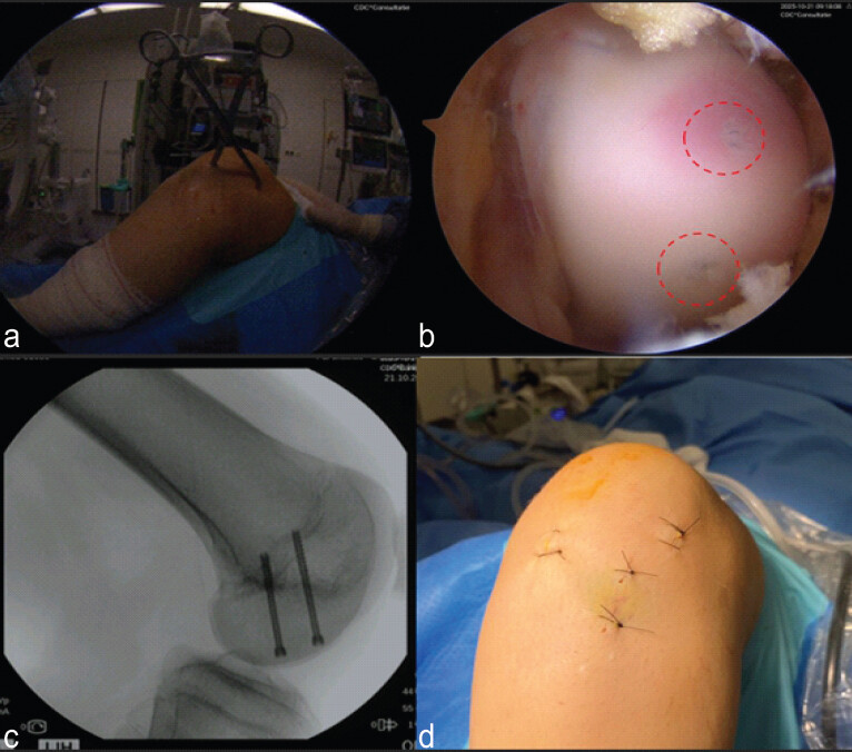
Postoperatively, a long leg cast in extension was applied for 2 weeks, followed by free mobilizations of the knee. The patient was not allowed to weight bear for 6 weeks.
Six weeks after trauma the patient had a full range of motion and was able to start partially weight bear. A rehabilitation program could be started, focusing on restoring mobility, flexibility, and quadriceps strength under the supervision of a physiotherapist. Radiographs at 0, 2, 6, and 16 weeks showed perfect position and healing of the Hoffa fracture (Fig. 6). Four months after trauma, there was a full range of motion, normal quadriceps muscle strength and waist, comparable to the contralateral right side. At that time, she had a Knee Injury and Osteoarthritis Outcome Score of 93, a Tegner score of 7, and she was able to return to normal activity and sport level. Due tothe excellent clinical and radiological examination, no additional control CT was performed to avoid additional irradiation in this young patient.
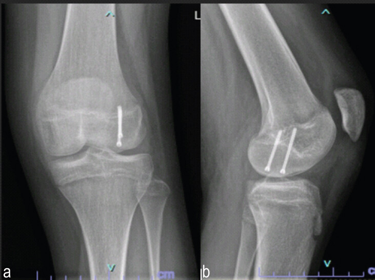
Discussion
Despite classically described after high-energy trauma in adults, in this case, a minor trauma caused the Hoffa fracture. Several other authors have reported this type of fracture occurring after low-energy trauma in skeletally immature patients [4, 9]. This susceptibility in children and adolescents has been attributed to incomplete bone mineralization, a higher cartilage-tobone ratio, and the relative mechanical weakness of the immature distal femur, predisposing it to shear-type intra-articular fractures even after minor trauma [4, 10]. This case report, however, represents a single pediatric case, which limits the ability to generalize our findings to all Hoffa fractures in skeletally immature patients.
Conservative (non-operative) management of Hoffa fractures has consistently been associated with poor clinical outcomes, including malunion, nonunion, fragment displacement, premature post-traumatic osteoarthritis, and avascular necrosis of the femoral condyle. Consequently, open reduction and internal fixation has long been regarded as the gold-standard treatment to achieve anatomical reduction and stable fixation of this intra-articular fracture. In recent years, minimally invasive and fully ar throscopic reduction and fi xation techniques have been described; however, their use remains limited to selected cases and is predominantly reported in small case series and case reports [7, 8, 10, 11, and 12].
Arthroscopic reduction and fixation of Hoffa fractures offer several potential advantages compared to open techniques, including preservation of soft tissues, reduced surgical exposure, and minimized disruption of periarticular structures. These benefits are associated with decreased intraoperative blood loss and shorter operative times, which may in turn reduce the risk of surgical site infection [8, 10]. Furthermore, arthroscopic management allows for early post-operative protocols, potentially facilitating faster recovery of knee range of motion and functional outcomes [10, 12]. An additional advantage of the arthroscopic approach is the ability to directly visualize the intra-articular surface and to diagnose and treat concomitant intra-articular lesions – such as chondral defects, meniscal tears, or ligamentous injuries – during the same surgical procedure [10, 11]. In this way, an additional magnetic resonance imaging scan could be avoided. In this case, penetration of the lateral femoral physis by the fixation screws could not be avoided. Given the patient’s age and near skeletal maturity, the risk of clinically significant growth disturbance is considered low. However, to fully assess this possible complication, yearly clinical and radiological follow-up until skeletal maturity is advised.
This case report has some limitations. Due to the rarity of Hoffa fractures with acute fixed flexion deformity, comparison with larger studies and standardized treatment protocols is limited. The short follow-up of 4 months is insufficient to assess long-term complications such as cartilage injuries, post-traumatic osteoarthritis, avascular necrosis, physeal disturbance, or implant-related issues.
In this case, the fracture was anatomically reduced before definitive fixation. It is difficult to determine whether similar outcomes would occur in irreducible or more comminuted Hoffa fractures. However, Souto et al. reported good clinical and radiological outcomes of a comminuted Hoffa fracture after arthroscopic reduction and fixation [12].
In the literature, so far, no complications were associated with arthroscopic fixation. Larger studies or case series are needed to determine possible complications such as infection, hardware irritation, or stiffness.
Being a case report from a single institution, the favorable outcome may reflect surgeon experience and careful patient selection, limiting broader applicability of the conclusions.
Conclusion
Closed reduction and arthroscopic anatomic fixation of certain Hoffa fractures lead to restoration of the anatomy and biomechanics and excellent clinical outcomes.
Clinical Message
Clinicians should consider the possibility of a Hoffa fracture in adolescents presenting with a locked knee after seemingly trivial trauma. Although rare in young patients, these fractures can be successfully managed with arthroscopic techniques. Nevertheless, urgent reduction may be required in selected cases to prevent complications and restore normal knee function.
Conflict of Interest:
Source of Support:
Nil
Consent:
The authors confirm that informed consent was obtained from the patient for publication of this article
References
- Patel PB, Tejwani NC.The Hoffa fracture: Coronal fracture of the femoral condyle a review of literature. J Orthop 2018;15:726-31. Google Scholar | PubMed
- Anwer A, Siddiqui YS, Abbas M, Chowdhry M.Conjoint bicondylar Hoffa's fracture with associated supracondylar fracture. J Bone Joint Dis 2023;38:115-9. Google Scholar | PubMed
- Pathak S, Salunke A, Karn S, Ratna HV, Thivari PS, Sharma S.Hoffa's fracture with associated injuries around the knee joint: An approach to a rare injury. Cureus 2020;12:e7865. Google Scholar | PubMed
- De Beer A, Brown MJ.Pediatric cartilaginous lateral femoral condyle Hoffa fracture: A case report and review of the literature. BMC Pediatr 2023;23:626. Google Scholar | PubMed
- Xiao Y, Arthur C, Liu X.Results of surgical treatment of Hoffa fractures in pediatric population: 8-case series. Front Pediatr 2025;13:1552420. Google Scholar | PubMed
- Letenneur J, Labour PE, Rogez JM, Lignon J, Bainvel JV.[Hoffa's fractures. Report of 20 cases (author's transl)]. Ann Chir 1978;32:213-9. Google Scholar | PubMed
- Lewis SL, Pozo JL, Muirhead-Allwood WF.Coronal fractures of the lateral femoral condyle. J Bone Joint Surg Br 1989;71-B:118-20. Google Scholar | PubMed
- Zhou Y, Pan Y, Wang Q, Hou Z, Chen W.Hoffa fracture of the femoral condyle: Injury mechanism, classification, diagnosis, and treatment. Medicine (Baltimore) 2019;98:e14633. Google Scholar | PubMed
- Flanagin BA, Cruz AI, Medvecky MJ.Hoffa fracture in a 14-year-old. Orthopedics 2011;34:138. Google Scholar | PubMed
- Chen ZH, Wang HF, Wang HY, Li F, Bai XF, Ni JL.Hoffa's fracture in a five-year-old child diagnosed and treated with the assistance of arthroscopy: A case report. World J Clin Cases 2022;10:13458-66. Google Scholar | PubMed
- Manfredini M, Gildone A, Ferrante R, Bernasconi S, Massari L.Unicondylar femoral fractures: Therapeutic strategy and long-term results. A review of 23 patients. Acta Orthop Belg 2001;67:132-8. Google Scholar | PubMed
- Souto CS, Machado DB, Rodrigues N, Tavares N.All arthroscopic management of an Hoffa fracture of the medial femoral condyle: Case report. J Orthop Rep 2024;4:100335. Google Scholar | PubMed
Related Articles in Journal of Orthopaedic Case Reports
 July 10, 2022 Localized Pigmented Villonodular Synovitis of the Bilateral Knee: A Case Report
 August 1, 2026 The Boba Tea Sign: A Novel Magnetic Resonance Imaging Finding in Synovial Chondromatosis of the Knee – A Case Report
 August 1, 2026 Intra-articular Ganglionic Cysts of Knee: A Cause for Anterior Knee Pain
 July 1, 2026 Angiofibroma of Knee Masquerading as Ganglion Cyst – A Rare Case Report