


Introduction
Diabetic foot infection (DFI) is a serious complication of diabetes mellitus and a major cause of non-traumatic lower-extremity amputation worldwide. It is associated with high morbidity, prolonged hospitalization, increased healthcare costs, and reduced quality of life [1]. Its pathogenesis is multifactorial, commonly involving peripheral neuropathy, peripheral arterial disease, impaired immunity, and poor glycemic control, all of which contribute to tissue breakdown and delayed wound healing [1,2]. Recurrent ulceration and infection further increase the risk of limb loss and mortality, making prevention and early intervention essential [3]. Current guidelines emphasize infection control, restoration of perfusion, pressure offloading, and metabolic optimization as cornerstones of management [4, 5].
Historically, patients with advanced infection and severe ischemia, especially after failed prior interventions, were frequently managed with major amputation. However, major amputation in diabetic patients is associated with substantial functional decline and high long-term mortality [6]. For this reason, multidisciplinary team (MDT) care has become central to the management of complex DFI. Combined input from vascular surgery, infectious disease, endocrinology, podiatric surgery, wound care, and rehabilitation has been associated with improved limb salvage and survival [4, 7]. Partial foot and midfoot amputations have also emerged as limb-preserving alternatives in selected patients, particularly when adequate perfusion and wound care can be achieved [8].
This case demonstrates the feasibility of proximal midfoot amputation as a limb-sparing alternative to major amputation in advanced DFI through sustained MDT care.
Case Report
A 62-year-old man with poorly controlled type 2 diabetes mellitus presented with a painful, chronic, non-healing right foot wound. His comorbidities included severe peripheral arterial disease and chronic heavy smoking. Admission laboratory findings were consistent with uncontrolled diabetes, active infection, and chronic inflammation: HbA1c 11.0%, white blood cell count 14.8 × 10⁹/L, hemoglobin 10.2 g/dL, platelet count 412 × 109/L, C-reactive protein 126 mg/L, erythrocyte sedimentation rate 92 mm/h, serum creatinine 1.1 mg/dL, blood urea nitrogen 24 mg/dL, serum albumin 2.8 g/dL, and random blood glucose 286 mg/dL. His history was notable for multiple failed limb-salvage attempts, including repeated debridements, first and second ray amputations, and multiple lower-extremity angioplasties. He reported severe ischemic pain requiring high-dose analgesics, with a pre-operative visual analog scale (VAS) pain score of 9/10. Despite repeated counseling, he consistently refused major amputation.
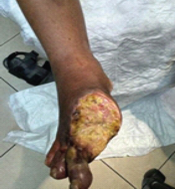
Examination revealed absent pedal pulses. The right foot showed an open ischemic wound with extensive slough, necrotic and fibrotic tissue, minimal pale granulation tissue, periwound maceration, and epidermal lysis. Gangrene involved the third and fourth toes (Fig. 1). Imaging confirmed advanced peripheral arterial disease with severe distal arterial insufficiency and poor pedal runoff. Given the persistent ischemia, progressive tissue loss, previous failed interventions, and refusal of major amputation, a limb-salvage strategy was pursued through coordinated MDT care. Pre-operative planning included repeat vascular assessment for revascularization, targeted antimicrobial therapy directed by the infectious disease team, and glycemic optimization by the endocrinology team.
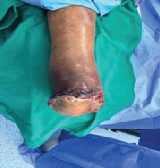
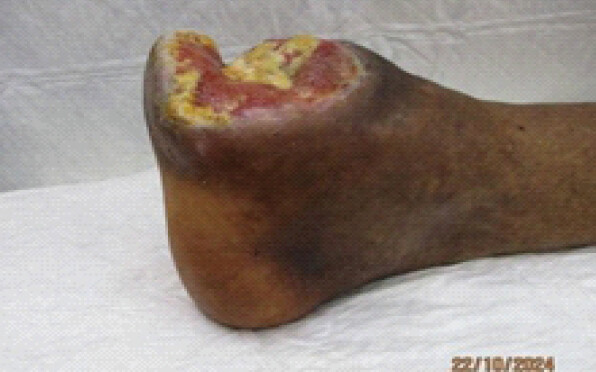
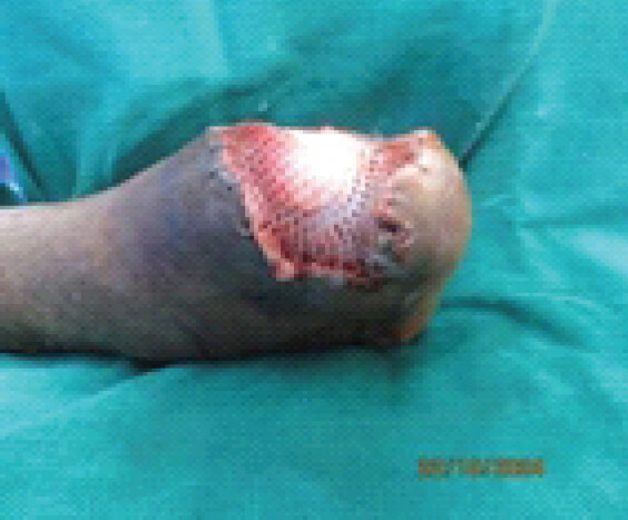
The patient initially underwent a right Lisfranc amputation. However, persistent ischemia and exposed bone necessitated further operative management. As part of the ongoing MDT plan, vascular surgery performed repeat revascularization, and the podiatric surgery and wound care teams proceeded with proximal midfoot revision and Achilles tendon lengthening to address equinus deformity. Immediate post-operative assessment showed local flap coverage with stable wound margins and preservation of a plantigrade foot (Fig. 2). During the post-operative course, wound dehiscence developed, with partial separation of the surgical incision and exposed soft tissue (Fig. 3). Serial debridements were then performed, followed by negative pressure wound therapy, acellular dermal matrix grafting (Fig. 4), and local flap coverage to promote healing and preserve function.
The patient remained under close MDT follow-up. Serial investigations showed progressive improvement: white blood cell count decreased to 8.9 × 10⁹/L, C-reactive protein to 18 mg/L, erythrocyte sedimentation rate to 38 mm/h, serum albumin improved to 3.5 g/dL, and random blood glucose decreased to 154 mg/dL. Renal function remained stable, with a serum creatinine level of 1.0 mg/dL. Clinically, the wound showed progressive granulation, resolution of necrotic tissue, and stabilization without evidence of systemic infection.
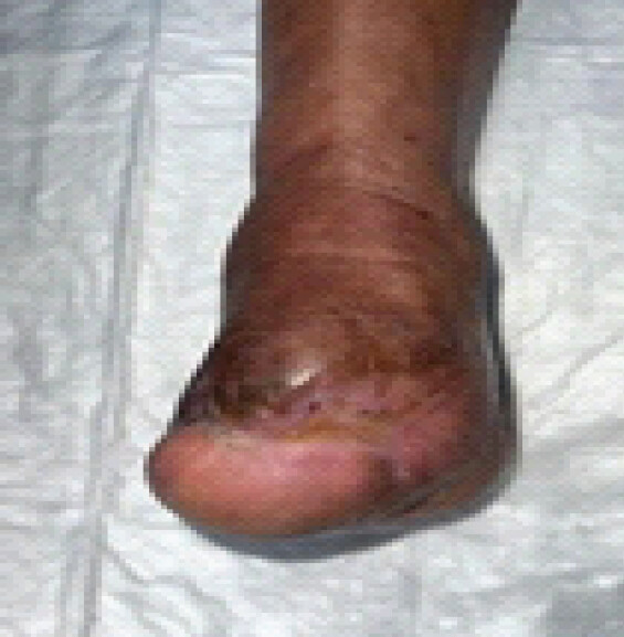
At 12-month follow-up, the patient was ambulating independently with custom footwear, reported minimal residual pain with a VAS score of 2/10, remained free of recurrent infection, and required no further amputation. Final assessment showed complete wound healing with a stable, plantigrade foot suitable for weight-bearing (Fig. 5).
Discussion
The major difficulties in this case were the coexistence of severe peripheral arterial disease, uncontrolled diabetes, recurrent infection, extensive tissue necrosis, multiple prior failed limb-salvage procedures, and the patient’s refusal of major amputation. Together, these factors created a very high risk of persistent wound failure, progressive infection, and eventual major limb loss. Previous studies have shown that DFI complicated by ischemia is strongly associated with poor healing, repeated interventions, and a higher risk of amputation, particularly in the presence of advanced peripheral arterial disease [1,2]. Poor glycemic control, nonviable tissue, and repeated prior surgical failure are also recognized predictors of adverse outcomes [2, 9].
A further challenge was attempting limb preservation after repeated unsuccessful debridements, ray amputations, and angioplasties. In such settings, major amputation is often considered the most definitive treatment, especially when ischemia and tissue loss coexist [4,6]. However, major amputation is associated with marked functional decline and substantial long-term mortality in diabetic patients [6]. In this case, the patient’s refusal of major amputation made functional limb salvage the only acceptable pathway.
This case suggests that proximal midfoot amputation can be a practical limb-preserving alternative in selected high-risk patients when supported by sustained MDT care. The favorable result was likely attributable not only to the amputation level itself but also to coordinated management of perfusion, infection, glycemic control, wound bed preparation, and soft-tissue reconstruction. This is consistent with reports showing improved limb salvage when diabetic foot care is delivered through multidisciplinary models [4,7]. Restoration of perfusion was especially important, as ischemia is a major determinant of poor healing in diabetic foot disease [2].
Our findings also align with the literature supporting partial foot and midfoot amputation as alternatives to major amputation when preservation of a plantigrade, functional foot is achievable [8]. In our patient, despite severe disease and post-operative wound complications, the final outcome was a stable plantigrade foot that allowed independent ambulation with custom footwear, supporting the functional value of this approach [8]. In addition, the use of negative pressure wound therapy is supported by prior evidence demonstrating its role in promoting granulation tissue formation and wound bed preparation in complex diabetic wounds [10].
This report has limitations. As a single-case study, its findings cannot be generalized broadly. Successful implementation required repeated interventions, prolonged follow-up, advanced wound care, vascular optimization, patient adherence, and access to an experienced MDT. The long-term durability of limb function and the risk of recurrent infection or ulceration after proximal midfoot amputation remain uncertain and require further study.
Conclusion
This case highlights that proximal midfoot amputation, when supported by coordinated MDT care, can preserve limb function and avoid major amputation in selected patients with advanced DFI and poor prognostic factors. Sustained multidisciplinary management may achieve functional limb salvage, maintain independence, and improve quality of life even when major amputation appears inevitable.
Clinical Message
Selected patients with advanced diabetic foot infection may achieve functional limb salvage with proximal midfoot amputation when perfusion, infection, glycemic control, wound reconstruction, and offloading are managed through a coordinated multidisciplinary team.
Conflict of Interest:
Source of Support:
Nil
Consent:
The authors confirm that informed consent was obtained from the patient for publication of this article
References
- Armstrong DG, Boulton AJ, Bus SA.Diabetic foot ulcers and their recurrence. N Engl J Med 2017;376:2367-75. Google Scholar | PubMed
- Prompers L, Huijberts M, Apelqvist J.High prevalence of ischemia and infection in diabetic foot disease. Diabetes Care 2007;30:1442-7. Google Scholar | PubMed
- Lavery LA, Peters EJ, Armstrong DG.What are the most effective interventions in preventing diabetic foot ulcers?. Int Wound J 2008;5:425-33. Google Scholar | PubMed
- Hingorani A, LaMuraglia GM, Henke P, Meissner MH, Loretz L, Zinszer KM.The management of diabetic foot: A clinical practice guideline by the society for vascular surgery in collaboration with the American Podiatric Medical association and the society for vascular medicine. J Vasc Surg 2016;63:3S-21. Google Scholar | PubMed
- Jakosz N.Book review - IWGDF guidelines on the prevention and management of diabetic foot disease. Wound Pract Res 2019;27:144. Google Scholar | PubMed
- Izumi Y, Satterfield K, Lee S, Lavery LA.Mortality of first-time amputees in diabetics: A 10-year observation. Diabetes Res Clin Pract 2009;83:126-31. Google Scholar | PubMed
- Prompers L, Schaper N, Apelqvist J.Delivery of care to diabetic patients with foot ulcers in daily practice: Results of the eurodiale study, a prospective cohort study. Diabet Med 2008;25:700-7. Google Scholar | PubMed
- Schade VL, Roukis TS.The role of partial foot amputation in limb salvage. Clin Podiatr Med Surg 2014;31:65-78. Google Scholar | PubMed
- Hicks CW, Mathioudakis N, Canner JK, Sherman RL, Hines KF, Lum YW.The Society for Vascular Surgery WIfI classification system predicts wound healing but not major amputation in patients with diabetic foot ulcers treated in a multidisciplinary setting. J Vasc Surg 2016;64:838. Google Scholar | PubMed
- Blume PA, Walters J, Payne W.Comparison of negative pressure wound therapy using vacuum-assisted closure with advanced moist wound therapy in the treatment of diabetic foot ulcers: A multicenter randomized controlled trial. Diabetes Care 2008;31:631-6. Google Scholar | PubMed
© 2026 Journal of Orthopaedic Case Reports - Published by Indian Orthopaedic Research Group
About the Authors
-

Dr. Abdulaziz F Aljehani
-

Dr. Magdi S Alzir
-

Dr. Abdulrahman Bin Haidarah
-

Dr. Mohammed O Alnadhairi
-
Dr. Abdulelah K Alqawlaq
-

Dr. Hani Mohammed Badahdah
Related Articles in Journal of Orthopaedic Case Reports
 September 1, 2026 Limb Salvage for Entire Tibial Ewing Sarcoma in a 4-year-Old Child Using Extracorporeal Irradiation and Free Flap Reconstruction: A Case Report and Surgical Technique
 September 1, 2026 Proximal Humerus Ewing Sarcoma: Technical Challenges and Strategies for Bone Defect Reconstruction
 September 1, 2026 Successful Limb Salvage Using Staged Applications of a Fish-Skin Xenograft (Kerecis) in a Heel Pad Degloving Injury: A Case Report
 August 1, 2026 Successful Salvage of Infected Tibial Shaft Non-union After Implant Removal with Ilizarov External Fixation: A Case Report