


Introduction
Septic arthritis is an orthopedic emergency that demands prompt diagnosis and intervention to prevent irreversible joint destruction and systemic sepsis. Staphylococcus aureus accounts for the majority of cases in immunocompetent adults; however, immunocompromised patients harbor a broader spectrum of pathogens, including Gram-negative bacilli [1].
Serratia marcescens is an opportunistic Gram-negative rod in the Enterobacteriaceae family, well recognized as a nosocomial pathogen in hemodialysis units and intensive care settings. Its occurrence as a cause of septic arthritis is infrequent, as Serratia orthopedic infections are typically associated with open trauma or direct nosocomial inoculation. Delayed hematogenous spread to a joint is exceedingly rare; the first documented case of S. marcescens joint infection dates to Atlas and Belding in 1968. Subsequent reports have been confined almost exclusively to immunocompromised hosts, including renal transplant recipients [2] and patients on hemodialysis [3]. Reported joint involvement has predominantly involved the sacroiliac joint [3] and sternoclavicular joint [4]; ankle involvement is rarely described [5]. In a series of seven orthopedic S. marcescens infections reported by Svensson et al., no ankle case was identified [6].
A targeted PubMed literature search was performed using the terms (“S. marcescens”) AND (“septic arthritis”) AND (“ankle”) for studies published between January 2017 and March 2026. No published case involving ankle septic arthritis due to S. marcescens in a patient with concomitant systemic lupus erythematosus (SLE) and end-stage renal disease (ESRD) was identified. The most recent comparable report was a 2022 case of S. marcescens hip septic arthritis and osteomyelitis following COVID-19 [7].
Patients with SLE on long-term corticosteroids carry a substantially elevated risk of opportunistic infection through complement deficiency, neutrophil dysfunction, and blunted acute-phase responses [8]. The skeletal consequences of prolonged corticosteroid use are well established: fracture risk increases within 3–6 months of initiation and correlates directly with cumulative dose [9]. When ESRD is superimposed, chronic kidney disease-mineral-bone disorder further impairs bone quality through secondary hyperparathyroidism, adynamic bone disease, and abnormal bone turnover [10]. These cumulative biological impairments have direct implications for implant selection in any surgical intervention in such a host.
We report a case of S. marcescens septic arthritis of the ankle in a patient with SLE and ESRD, managed with ring fixator-assisted ankle arthrodesis, and discuss the diagnostic strategy and surgical decision-making in the context of a biologically compromised host.
Case Report
A 45-year-old male with SLE (diagnosed 2011) on long-term corticosteroids developed ischemic stroke in 2014, resulting in the left hemiparesis with spastic ambulatory gait. By 2025, progressive lupus nephritis culminated in anuric ESRD; following arteriovenous fistula failure, a permacath was inserted for hemodialysis.
During a hemodialysis-related hospitalization (Day 0), he developed healthcare-associated pneumonia treated with 2 weeks of parenteral antibiotics, with apparent clinical resolution at discharge (Day 14). On Day 42, without intercurrent trauma or permacath manipulation, he presented with insidious left ankle pain, swelling, and complete inability to bear weight – an interval consistent with delayed hematogenous seeding in an immunosuppressed host.
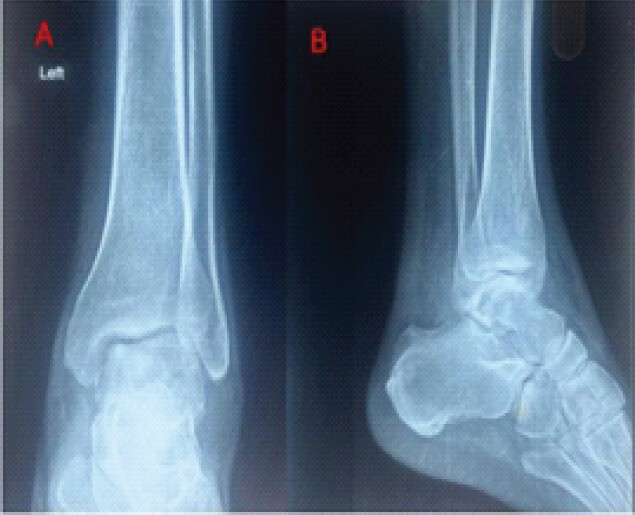
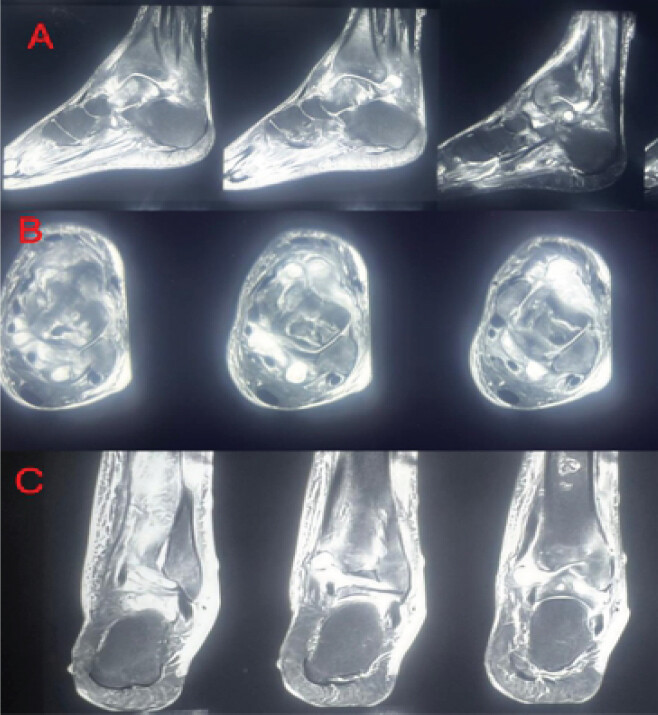
Examination revealed a diffusely swollen, erythematous, warm, tender left ankle in clear contrast to the normal contralateral limb (Fig. 1), with systemic low-grade pyrexia (38.1°C) and tachycardia (104/min). The permacath site was unremarkable. Investigations showed neutrophilic leukocytosis (white blood cell 14,800/mm3), elevated erythrocyte sedimentation rate (ESR) (92 mm/h), and C-reactive protein (CRP) (118 mg/L). Plain radiographs demonstrated periarticular soft-tissue swelling, periarticular osteopenia, and early tibiotalar joint space loss without cortical erosion (Fig. 1). Magnetic resonance imaging on T1, T2, and short tau inversion recovery sequences confirmed diffuse synovial thickening, complex effusion, and subchondral bone marrow edema of the tibial plafond and talar dome, consistent with septic arthritis (Fig. 2).
Diagnostic arthrocentesis yielded 16 mL of turbid seropurulent fluid. Concurrent blood and synovial aspirate cultures both grew S. marcescens supporting hematogenous dissemination. On antimicrobial susceptibility testing, the isolate was sensitive to amikacin, gentamicin, ciprofloxacin, imipenem, meropenem, and ertapenem, and resistant to ceftriaxone and cefoperazone-sulbactam. Synovial biopsy demonstrated acute-on-chronic inflammation without granulomata or fungal morphology, making tuberculosis (TB) and fungal arthritis unlikely, though paucibacillary TB could not be entirely excluded histologically (Fig. 3).
Intravenous meropenem (500 mg q12h, dose-adjusted for ESRD) was commenced in consultation with infectious disease and nephrology. A trial of conservative management was undertaken, with the ankle immobilized in a below-knee splint alongside culture-directed intravenous antibiotics. Despite 6 weeks of targeted therapy, inflammatory markers failed to normalize and repeat imaging confirmed progressive articular destruction. Intraoperative assessment revealed near-total loss of articular cartilage with exposed subchondral bone bilaterally, precluding joint salvage.
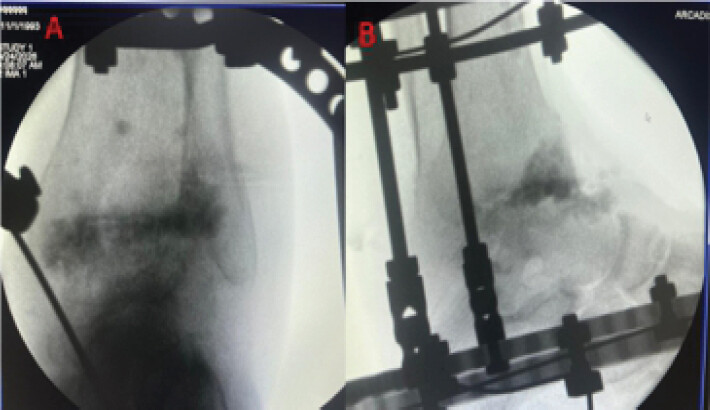
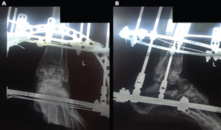
Following multidisciplinary discussion, ring fixator-assisted ankle arthrodesis was performed. Internal fixation was rejected given active infection, steroid-induced osteoporosis, and impaired wound healing. Through an anterior approach, thorough debridement, synovectomy, and lavage were performed with preparation of subchondral bone to bleeding surfaces. An Ilizarov ring fixator was applied with the ankle in neutral dorsiflexion and 5° of external rotation; antibiotic calcium sulfate beads were inserted. Intraoperative fluoroscopy confirmed satisfactory bone apposition and alignment; immediate post-operative radiographs demonstrated the construct (Fig. 4). Hemodialysis continued uninterrupted postoperatively.
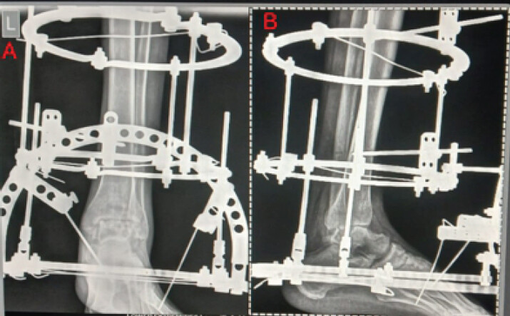
At 6-month follow-up, the visual analog scale (VAS) score improved from 8 out of 10 to 4 out of 10. Partial weight-bearing with aids was achieved at 10 weeks. Radiographs at 6 months demonstrated progressive trabecular bridging at the arthrodesis site, suggestive of ongoing union (Fig. 4); ESR and CRP trended to near-normal. The frame has been dynamized with removal planned at complete consolidation. No wound complications, pin-site infections, or sepsis recurrence were noted. At 9-month follow-up, radiographs demonstrated further consolidation across the arthrodesis site (Fig. 5), with further symptomatic improvement as the VAS pain score decreased from 4 out of 10 to 2 out of 10. The patient remains under continued clinical and radiological surveillance.
Discussion
The concurrent isolation of S. marcescens from both blood and joint aspirate cultures was the pivotal diagnostic finding, distinguishing hematogenous seeding from primary articular inoculation or percutaneous line infection. The temporal sequence of pneumonia on day 0, apparent clinical resolution, and ankle symptoms commencing on approximately day 28 post-discharge is consistent with the known pathophysiology of delayed hematogenous joint seeding in immunosuppressed hosts, in whom bacteremia may be low-grade and clinically occult before establishing in a susceptible joint [11]. Formal molecular strain comparison between pulmonary and articular isolates was not performed. Therefore, hematogenous dissemination was inferred from clinical presentation, temporal association, and concordant microbiological findings rather than confirmed by genotypic analysis.
A review of previously reported cases of S. marcescens septic arthritis in the literature provides context for the present case. Rogala and Cruess [2] described one of the earliest cases of hematogenous S. marcescens joint infection in a renal transplant recipient, with a fatal outcome, highlighting the severity of this infection in immunosuppressed hosts. Donovan et al. [12] subsequently reported seven cases predominantly involving large joints in a nosocomial setting, with variable outcomes. Svensson et al. [6] described seven orthopedic Serratia infections, none of which involved the ankle. More recent reports include a case of septic sacroiliitis by Simon et al. [3] and a case of sternoclavicular arthritis by Amao-Ruiz et al. [4], both in immunocompromised patients managed with antibiotics alone. Hadid et al. [5] described ankle and tibial involvement in an immunocompetent diabetic patient managed with surgical debridement. The most recent comparable case is a 2022 report of S. marcescens hip septic arthritis following COVID-19 [7]. Across these cases, treatment ranged from antibiotics alone to surgical debridement; none required arthrodesis. The present case is distinctive in its combination of ankle involvement, dual systemic immunocompromise from SLE and ESRD, concordant evidence of a hematogenous source, and the requirement for ring fixator-assisted arthrodesis as definitive management.
Healthcare-associated Serratia strains in hemodialysis populations frequently acquire extended-spectrum beta-lactamase or carbapenemase resistance [13], which would substantially restrict therapeutic options in a patient with ESRD and limited antibiotic dosing flexibility. Its absence made meropenem monotherapy with renal dose adjustment straightforward in practice, but not guaranteed in this infective context.
Synovial biopsy contributed decisively to the diagnostic pathway by demonstrating acute-on-chronic inflammation without granuloma or fungal morphology, making TB and fungal arthritis unlikely, both important differential diagnoses in an immunosuppressed patient from a TB-endemic region. The absence of acid-fast bacilli and the presence of culture-confirmed gram-negative growth collectively supported a single bacterial etiology, enabling targeted rather than empirical broad-spectrum management [11].
The rejection of internal fixation in favor of ring fixator arthrodesis was the central surgical decision in this case. Internal fixation was avoided because active infection, prolonged corticosteroid exposure, impaired bone quality, and ESRD-associated metabolic bone disease increased concerns regarding implant-related complications, wound problems, and fixation failure [9, 10,14]. External fixation, therefore, offered a biologically favorable alternative in this setting.
The Ilizarov ring fixator addresses these concerns through specific mechanisms. Tensioned transfixing wires exert compressive forces across the arthrodesis site without stress-shielding; the primary goal here is compression for fusion, not distraction, while stimulating osteogenic activity at the bone-contact surface. The percutaneous wire configuration places no implant bulk in the infected wound, eliminating the scaffold for biofilm. The frame remains external, allowing unobstructed inspection of the wound throughout the consolidation period. Axial dynamization applied to this patient allows controlled load sharing once the early callus is visible. Published series support this approach: Gessmann et al. reported 86.5% bony consolidation in 37 patients with infected ankle arthrodesis using the Ilizarov fixator at a mean follow-up of 46 months [15], and Wang et al. demonstrated 92% fusion in 54 patients with septic ankle arthritis at a minimum 6-year follow-up [16]. The decision to utilize an Ilizarov frame was dictated by the need to bypass the biologically compromised soft-tissue envelope and deficient bone stock characteristic of this patient’s dual SLE/ESRD pathology. The functional trajectory at 6 months, progressing radiological union, weight-bearing achieved, VAS reduction from eight to four, and no infective recurrence, is consistent with published benchmarks for this technique.
Limitations
Molecular strain typing was not performed; sputum cultures were unavailable, making pneumonia only a possible bacteremic source; follow-up duration remains limited, without computed tomography confirmation of fusion; functional assessment relied on VAS alone without validated scores (American Orthopedic Foot and Ankle Society/foot and ankle ability measure); and as a single case report, no generalizable conclusions can be drawn regarding optimal management.
Conclusion
S. marcescens septic arthritis of the ankle in a patient with SLE and ESRD is a rare but recognizable clinical presentation. Concurrent blood and synovial aspirate cultures are essential for establishing the mechanism of infection and guiding therapy. Synovial biopsy is a valuable adjunct to exclude granulomatous and fungal diagnoses. Ring fixator-assisted ankle arthrodesis may be a useful treatment option for immunocompromised patients with advanced joint destruction, providing stability and infection control while avoiding the need for internal fixation in an infected environment.
Clinical Message
Serratia marcescens septic arthritis of the ankle in a multiply immunocompromised host requires early joint aspiration, concurrent blood cultures, synovial biopsy, and ring-fixator arthrodesis when internal fixation is biologically untenable.
Conflict of Interest:
Source of Support:
Nil
Consent:
The authors confirm that informed consent was obtained from the patient for publication of this article
References
- Mathews CJ, Weston VC, Jones A, Field M, Coakley G.Bacterial septic arthritis in adults. Lancet 2010;375:846-55. [Google Scholar] | [PubMed]
- Rogala EJ, Cruess RL.Multiple pyogenic arthritis due to Serratia marcescens following renal homotransplantation. Report of a case. J Bone Joint Surg Am 1972;54:1283-7. [Google Scholar] | [PubMed]
- Simon EL, Wainblat EG, Krizo JA, Smalley CM, Fertel BS.Septic sacroiliitis caused by Serratia marcescens. Am J Emerg Med 2020;38:2758.e5-8. [Google Scholar] | [PubMed]
- Amao-Ruiz E, Correa-Fernandez AM, De La Fuente Galan L.Serratia marcescens septic sternoclavicular joint arthritis: A case report. Reumatol Clin 2016;12:238-9. [Google Scholar] | [PubMed]
- Hadid H, Usman M, Thapa S.Severe osteomyelitis and septic arthritis due to Serratia marcescens in an immunocompetent patient. Case Rep Infect Dis 2015;2015:347652. [Google Scholar] | [PubMed]
- Svensson O, Parment PA, Blomgren G.Orthopaedic infections by Serratia marcescens: A report of seven cases. Scand J Infect Dis 1987;19:69-75. [Google Scholar] | [PubMed]
- Tantikosol P, Sumanasrethakul C, Pakpianpairoj C.Serratia marcescens septic arthritis and osteomyelitis after COVID-19: A case report and literature review. J Southeast Asian Orthop 2022;47:51-5. [Google Scholar] | [PubMed]
- Huang JL, Hung JJ, Wu KC, Lee WI, Chan CK, Ou LS.Septic arthritis in patients with systemic lupus erythematosus: Salmonella and nonsalmonella infections compared. Semin Arthritis Rheum 2006;36:61-7. [Google Scholar] | [PubMed]
- Van Staa TP, Leufkens HG, Abenhaim L, Zhang B, Cooper C.Use of oral corticosteroids and risk of fractures. J Bone Miner Res 2000;15:993-1000. [Google Scholar] | [PubMed]
- Moe SM, Drüeke T, Lameire N, Eknoyan G.Chronic kidney disease-mineral-bone disorder: A new paradigm. Adv Chronic Kidney Dis 2008;15:3-12. [Google Scholar] | [PubMed]
- Mathews CJ, Coakley G.Septic arthritis: Current diagnostic and therapeutic algorithm. Curr Opin Rheumatol 2008;20:457-62. [Google Scholar] | [PubMed]
- Donovan TL, Chapman MW, Harrington KD, Nagel DA.Serratia arthritis. Report of seven cases. J Bone Joint Surg Am 1976;58:1009-11. [Google Scholar] | [PubMed]
- Hsieh MS, Lu CL, Lee CC.Bacteremia and septic arthritis in patients on hemodialysis: Increased susceptibility to gram-negative organisms. Nephrol Dial Transplant 2006;21:2240-6. [Google Scholar] | [PubMed]
- Katsenis D, Bhave A, Paley D, Herzenberg JE.Treatment of malunion and nonunion at the site of an ankle fusion with the Ilizarov apparatus. J Bone Joint Surg Am 2005;87:302-9. [Google Scholar] | [PubMed]
- Gessmann J, Özokyay L, Fehmer T, Muhr G, Seybold D.Arthrodesis of the infected ankle joint: Results with the Ilizarov external fixator. Z Orthop Unfall 2011;149:212-8. [Google Scholar] | [PubMed]
- Wang S, Li B, Yu X, Wu H, Liu L.Is ankle arthrodesis with an Ilizarov external fixator an effective treatment for septic ankle arthritis? A study with a minimum of 6 years of follow-up. Clin Orthop Relat Res 2023;481:717-25. [Google Scholar] | [PubMed]
© 2026 Journal of Orthopaedic Case Reports - Published by Indian Orthopaedic Research Group
About the Authors
-

Dr. Shubham Jaiswal
-

Dr. Sandeep Patel
-

Dr. Utkarsh Kumar Reddy Gopavaram
-

Dr. Harkaran Singh Khosa
Related Articles in Journal of Orthopaedic Case Reports
 September 1, 2025 A Rare Case of Septic Arthritis of the Knee Associated with Pathological Neck of Femur Fracture Caused by Serratia Marcescens – A Case Report
 September 1, 2026 Right Knee Ureaplasma Septic Arthritis in an Immunocompromised Host, Requiring Right Hip Disarticulation for Definitive Source Control
 July 1, 2026 Tuberculosis of the Elbow in a Chronic Kidney Disease Patient with Arteriovenous Fistula: A Case Report
 July 1, 2026 Concurrent Occurrence of Dysplasia Epiphysealis Hemimelica involving Lateral Malleolus and Sinus Tarsi: Rare Case with Review of Literature