


Introduction
Monteggia fracture-dislocations involve a fracture of the proximal ulna with dislocation of the radial head [1] and are commonly seen in pediatric patients but can also occur in adults [2]. While acute cases in adults are typically managed with open reduction and internal fixation, delayed diagnoses or non-anatomical reduction of the proximal ulna fracture may lead to chronic Monteggia lesions characterized by radial head dislocation, ulnar malunion, progressive joint dysfunction, and disabilities [2, 3].
Corrective osteotomy remains the cornerstone of treatment for chronic Monteggia injuries [4, 5]. In recent years, the application of three-dimensional (3D) technology encompassing virtual surgical planning, patient-specific cutting guides, and custom implants has gained popularity in complex deformity correction across various anatomical sites [6, 7].
Here, we report a case of chronic Monteggia fracture-dislocation in an adult patient successfully managed using 3D-assisted pre-operative planning and a patient-specific osteotomy guide, adding to the emerging evidence on the role of this technology in upper-limb malunions.
Case Report
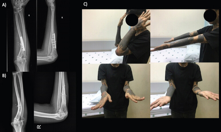
A 24-year-old male presented with a 1-year history of persistent right forearm pain. He had sustained a Monteggia fracture of the right forearm approximately 1 year prior, initially managed at another institution with open reduction and internal fixation with small dynamic compression plate fixation (Fig. 1a) and subsequently implant removal after fracture union (Fig. 1b). Despite these interventions, he continued to experience pain and dysfunction. Clinical evaluation (Fig. 1c) revealed limited elbow flexion and pronation. The patient, however, experienced pain and discomfort while performing daily activities. The radiographs revealed malunion of the proximal ulna with anterior dislocation of the radial head, consistent with a chronic Monteggia fracture-dislocation (Bado type I) of the right elbow [8].
Surgical technique and 3D workflow
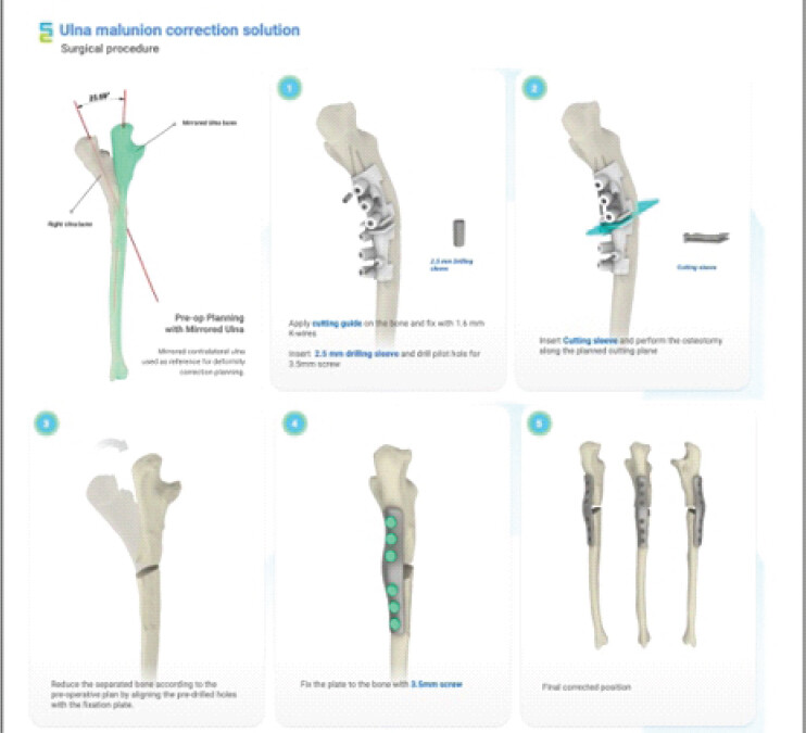
The surgical management was divided into a virtual planning phase and an execution phase using patient-specific instrumentation (Fig. 2). Under general anesthesia and in a supine position, a standard surgical approach was performed along the ulnar crest to expose the deformed bone.
-
Virtual Surgical Planning: Pre-operative computed tomography (CT) data of both the affected and contralateral forearms were utilized to create 3D virtual models. The deformity of the right ulna was analyzed by mirroring the healthy contralateral side to serve as an anatomical template, revealing a required correction angle of 25.69°
-
Patient-Specific Guide Design: A customized 3D-printed osteotomy guide was engineered based on pre-operative CT imaging to fit the unique cortical topography of the proximal ulnar shaft. This guide incorporated built-in drilling sleeves and a precise cutting slot to dictate the exact location and trajectory of the osteotomy
-
Intraoperative Execution: The customized surgical cutting guide was positioned to match the planned anatomical location and secured with 1.6 mm K-wires. Positioning was confirmed with fluoroscopy before pre-drilling the pilot holes for definitive 3.5 mm screws and performing the osteotomy through the guide’s designated cutting slot
-
Reduction and Internal Fixation: After completing the osteotomy, the separated bone segments were reduced. Precise anatomical alignment was achieved by matching the pre-drilled holes to the corresponding holes in a 3D-printed customized bone plate. Final alignment and implant position were checked to confirm proper correction before proceeding with any necessary radial head stabilization.
Following the ulnar correction, the radial head remained unstable, so an open reduction was performed. The annular ligament was reconstructed using a palmaris longus tendon graft to stabilize the radiocapitellar joint. A temporary transarticular pin was inserted to secure the joint. An autologous iliac crest bone graft was harvested and placed at the osteotomy site. After achieving hemostasis, the subcutaneous tissue and skin were closed. A posterior long arm slab was then applied.
Total operative time was 120 min, with a fluoroscopy exposure time of 49 s. Guide design and manufacturing required 14 days, with an estimated additional cost of 990 USD for the patient-specific guide and implant relative to conventional fixation.
Following the surgical procedure, the patient’s elbow was immobilized with a long arm slab for 3 weeks. At that point, the transarticular pin was removed. For the next 3 weeks, the patient was encouraged to perform passive range of motion exercises under supervision. After 6 weeks postoperatively, active range of motion and strengthening exercises were permitted.
Post-operative radiographic measurement demonstrated an achieved correction angle of 22.38°, compared with the planned correction of 25.69°. Post-operative recovery was initially favorable. At early follow-up, the patient achieved an elbow flexion-extension range of motion from 0° to 120°, with 80° of both supination and pronation. However, at 11 months postoperatively, radiographs revealed signs of oligotrophic non-union at the osteotomy site, without bridging callus formation. Notably, the patient had continued smoking throughout the post-operative period.
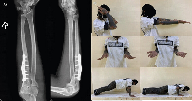
The patient subsequently underwent revision surgery with repeat bone grafting for the non-union 1 year after the initial osteotomy. Post-operative imaging confirmed progression toward union. At 9 months following the revision procedure, and 1.9 years after the initial corrective osteotomy, the patient had regained functional elbow range of motion, with only mild limitations in forearm rotation. Radiographs demonstrated solid union at the osteotomy site (Fig. 3).
Discussion
Chronic Monteggia fracture-dislocations remain a formidable challenge in orthopedic practice due to delayed diagnosis, malunion of the ulna, and persistent radial head dislocation. These lesions, particularly Bado type I injuries, often present with pain, limited range of motion, and joint instability, significantly impairing function [9]. In such cases, reconstructive strategies must address both osseous deformity and soft tissue imbalance to restore normal biomechanics and prevent long-term complications.
The cornerstone of surgical management in chronic Monteggia lesions is corrective ulnar osteotomy [5]. However, traditional techniques often rely on intraoperative judgment and fluoroscopic guidance, which may lead to suboptimal alignment or increased operative time. Historically, corrective osteotomy for chronic Monteggia lesions was performed freehand, using fluoroscopic landmarks to estimate the osteotomy plane and angulation [2,10]. Techniques such as closing-wedge, dome, or step-cut osteotomies have been described, but their success depends heavily on intraoperative experience and remains susceptible to residual malalignment or recurrent subluxation [2,11]. The overarching goal in managing these injuries is to restore ulnar length and alignment to allow stable reduction of the radial head with congruent radiocapitellar articulation [3]. The conventional treatment algorithm begins with ulnar osteotomy and fixation, followed by assessment of radial head stability. If reduction is unstable, adjunctive procedures such as annular ligament reconstruction or temporary transcapitellar pinning are employed [12]. In enduring or irreducible cases, salvage options such as radial head excision or prosthetic replacement may be considered, though they are reserved for refractory cases due to concerns about long-term joint function [3,13].
In this case, we utilized three-dimensional (3D) technology to overcome the limitations of traditional management. In recent years, 3D-assisted techniques have been increasingly applied in upper-limb deformity correction, particularly for distal radius, forearm, and humeral malunions [7]. Pre-operative CT data can be converted into virtual models, enabling accurate assessment of deformity and simulation of corrective osteotomies. Patient-specific guides allow precise transfer of the surgical plan to the operative field, reducing reliance on intraoperative estimation and fluoroscopy. Several series have demonstrated that 3D planning improves accuracy of correction, reduces operative time, and facilitates early functional recovery compared to conventional freehand osteotomies [14,15 16].
Although the application of 3D printing has been increasingly reported in the correction of upper extremity deformities, such as distal radius and forearm malunions, there is a relative paucity of literature focusing specifically on chronic Monteggia fracture-dislocations [11,12]. Only a small number of prior reports, most notably a two-case series describing custom-made surgical guides for malunited forearm fractures with associated radial head dislocation [16], have specifically addressed 3D-assisted correction in this injury pattern. Unlike these prior reports, the present case combines a patient-specific osteotomy guide with a custom-matched bone plate and concurrent annular ligament reconstruction within a single reconstructive strategy for a chronic Monteggia lesion, specifically, a combination not previously described for this injury. Our case supports its potential role in improving anatomical accuracy, joint congruency, and overall functional outcomes in this challenging subset of elbow pathology.
Despite the initial success, our patient developed oligotrophic non-union at the osteotomy site. This was likely multifactorial, with the patient’s continued smoking being a significant contributor. Smoking is well-established as a negative modulator of bone healing, and its impact in the context of osteotomy should not be underestimated [17]. Revision surgery with repeat autologous bone grafting ultimately led to union and satisfactory functional recovery. Thus, this case also underscores the importance of addressing modifiable risk factors, such as tobacco use, during pre-operative counseling and post-operative follow-up.
Furthermore, our case incorporated a comprehensive surgical approach combining osteotomy, annular ligament reconstruction, and temporary radiocapitellar pinning. This multimodal strategy likely contributed to early stability and radial head relocation. The use of radiocapitellar pinning, although debated in the literature, provided temporary stabilization, and facilitated ligament healing in our patient [18,19]. An additional aspect highlighted by this case is the importance of interdisciplinary cooperation between surgeons and biomedical engineers in the application of 3D technology. The accurate design of patient-specific guides and implants relies on meticulous communication of clinical requirements to engineers, while surgeons must interpret and validate the virtual plan for feasibility in the operative field. Prior studies in orthopedic oncology and complex limb reconstruction have emphasized that such collaborative workflows improve accuracy, reduce intraoperative uncertainty, and ultimately translate into better functional outcomes for patients [7,14,15].
Conclusion
The successful outcome in this case suggests that 3D-assisted surgical planning may be particularly beneficial in chronic Monteggia injuries where conventional alignment techniques may fall short due to prior surgical alterations or complex deformity, achieving anatomical ulnar alignment and successful radiocapitellar relocation through precise pre-operative simulation and a patient-specific guide. However, the subsequent development of a post-operative non-union in this patient, who continued smoking throughout treatment, underscores that personalized surgical guides must be coupled with strict adherence to biological optimization, particularly tobacco cessation, to achieve reliable union. As 3D printing becomes increasingly accessible and cost-effective, its role in upper-limb reconstruction, particularly in individualized deformity correction, is likely to expand, though larger studies and long-term follow-up are needed to validate its routine use and assess cost-effectiveness compared to standard methods.
Clinical Message
Patient-specific 3D surgical planning and osteotomy guides can improve the precision of corrective ulnar osteotomy and radial head reduction in chronic Monteggia fracture-dislocation. This technical precision must nonetheless be paired with rigorous biological optimization, particularly pre-operative and post-operative tobacco cessation, to achieve reliable bone union.
Conflict of Interest:
Source of Support:
Nil
Consent:
The authors confirm that informed consent was obtained from the patient for publication of this article
References
- Lattanza LL, Chen S.Monteggia fracture-dislocations. In: The Pediatric Upper Extremity New York: Springer; 2014 1-14. Google Scholar | PubMed
- Wintges K, Cramer C, Mader K.Missed monteggia injuries in children and adolescents: A treatment algorithm. Children (Basel) 2024;11:391. Google Scholar | PubMed
- Zhang R, Wang X, Xu J, Kang Q, Hamdy RC.Neglected Monteggia fracture: A review. EFORT Open Rev 2022;7:287-94. Google Scholar | PubMed
- Konrad GG, Kundel K, Kreuz PC, Oberst M, Sudkamp NP.Monteggia fractures in adults: Long-term results and prognostic factors. J Bone Joint Surg 2007;89-B:354-60. Google Scholar | PubMed
- Thirumavalavan J, Penvose IR, Prokuski-Lund V.Adult monteggia fracture-dislocations. Oper Tech Orthop 2023;33:101026. Google Scholar | PubMed
- Gao S, Stephens JD, Piatt C, Hockman T, Hills A, Thompson J .3D Printing in Orthopedics. Amsterdam: Elsevier eBooks; 2018 85-94. Google Scholar | PubMed
- Jarayabhand R, Jiamton C, Puncreobutr C, Lohwongwatana B, Theerachai A, Fang C.Invited review article: Patient-specific 3D-Printed metal plates for osteotomy and fracture fixation: An illustrative series with workflow and regulatory considerations. J Orthop Surg (Hong Kong) 2025;33:10225536251356724 [TPS1.1] Google Scholar | PubMed
- Bado JL.The Monteggia lesion. Clin Orthop Relat Res 1967;50:71-86. Google Scholar | PubMed
- Xu P, Zhang Z, Ning B, Wang D.Outcomes and experience after open reduction for chronic Monteggia fracture in children. Transl Pediatr 2022;11:1122-9. Google Scholar | PubMed
- Xu Z, Li Y, Wang Z, Cai H.Open reduction combined with CORA-based osteotomy of the ulna in the treatment of missed Bado type I Monteggia injury. Medicine 2017;96:e8609-9. Google Scholar | PubMed
- Garg RN, Gupta D, Kundu S, Vaje SS.Step-cut, biplanar, elongation and angulation ulnar osteotomy with annular ligament reconstruction in neglected Monteggia fracture dislocation in children: A retrospective study. Int J Res Orthop 2023;10:162-9. Google Scholar | PubMed
- Langenberg LC, Beumer AC, The B, Koenraadt KL, Eygendaal D.Surgical treatment of chronic anterior radial head dislocations in missed Monteggia lesions in children: A rationale for treatment and pearls and pitfalls of surgery. Shoulder Elbow 2019;12:422-31. Google Scholar | PubMed
- Rodgers WB, Waters PM, Hall JE.Chronic Monteggia lesions in children. Complications and results of reconstruction. J Bone Joint Surg 1996;78:1322-9. Google Scholar | PubMed
- Saravi B, Lang G, Steger R, Vollmer A, Zwingmann J.Corrective osteotomy of upper extremity malunions using three-dimensional planning and patient-specific surgical guides: Recent advances and perspectives. Front Surg 2021;8:615026. Google Scholar | PubMed
- Reyniers P, Verrewaere D, Es EV, Colaris J, Schweizer A, Verstreken F.The role of 3D technology in corrective osteotomy for forearm malunion. J Hand Surg Eur Vol 2025;50:771-80. Google Scholar | PubMed
- Oka K, Murase T, Moritomo H, Yoshikawa H.Corrective osteotomy for malunited both bones fractures of the forearm with radial head dislocations using a custom-made surgical guide: Two case reports. J Should Elbow Surg 2012;21:e1-8. Google Scholar | PubMed
- Scolaro JA, Schenker ML, Yannascoli S, Baldwin K, Mehta S, Ahn J.Cigarette smoking increases complications following fracture: A systematic review. J Bone Joint Surg 2014;96:674-81. Google Scholar | PubMed
- Gryson T, Van Tongel A, Plasschaert F.The management of chronic paediatric Monteggia fracture-dislocation. J Orthop 2021;24:65-76. Google Scholar | PubMed
- Demirel M, Sağlam Y, Tunalı O.Posterior interosseous nerve palsy associated with neglected pediatric Monteggia fracture-dislocation: A case report. Int J Surg Case Rep 2016;27:102-6. Google Scholar | PubMed
© 2026 Journal of Orthopaedic Case Reports - Published by Indian Orthopaedic Research Group
About the Authors
-

Dr. Abhishet Varama
-

Dr. Chittawee Jiamton
-

Dr. Natthaphat Viriya
-

Dr. Chedtha Puncreobutr
-

Dr. Boonrat Lohwongwatana
Related Articles in Journal of Orthopaedic Case Reports
 June 1, 2026 Oblique Close Wedge versus Modified Step Cut Osteotomy for Cubitus Varus Deformity in Children: A Prospective Non-Randomized Study
 January 1, 2026 Harnessing 3D Printing Technology for Complex Acetabular Reconstruction in Revision Total Hip Arthroplasty: From Childhood Hip Trauma to Customized Modern Solutions
 March 1, 2025 A Case Report of Bilateral Shepherd Crook Deformity of Proximal Femur in an Adolescent Managed by Corrective Osteotomy, Double Angle Blade Plate and Bone Graft
 July 6, 2017 Cerclage Wiring as an Adjunct for the Treatment of Femur Fractures: Series of 11 Cases