


Introduction
Paget’s disease of bone (PDB) is a localized disorder of bone remodeling, resulting in a disorganized mosaic of woven and lamellar bone, which is susceptible to deformity and fracture [1]. The PDB was traditionally reported to have a restricted geographical distribution and to be less common in Asians than Caucasians [2]. The diagnosis is often straightforward when skeletal involvement is classical. In a few situations, such as atypical skeletal involvement or comorbidities, for example, chronic kidney disease (CKD), establishing the diagnosis can be difficult [3]. The treatment options for PDB in CKD are also limited.
Case Report
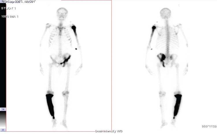
A 51-year-old male with CKD Stage 3 at the time of initial CKD diagnosis presented with progressive right leg pain, deformity, and a spontaneous fracture. He had a 5-year history of bilateral knee pain managed with analgesics. He was diagnosed with Stage 3 CKD 4 years before presentation, progressing to stage 5 at the time this case was evaluated. There was no history of swellings elsewhere in the body. Physical examination revealed a non-tender bony swelling with crepitus over the right leg without features of local inflammation. Laboratory evaluation showed a serum creatinine of 5.74 mg/dL, an estimated glomerular filtration rate of 10 mL/min/1.73 m2, and elevated total alkaline phosphatase (ALP) levels as tabulated in Table 1. A skeletal survey demonstrated the classical radiological features of PDB. Bone scintigraphy revealed increased tracer uptake involving the right tibia, left hemipelvis, and left humerus (Supplementary Fig. 1).
Investigations at initial presentation
| Parameter | Patient’s value | Reference range |
|---|---|---|
| Hemoglobin | 12.7 g/dL | 13.5–17.5 g/dL |
| Creatinine | 5.74 mg/dL | 0.5–1.2 mg/dL |
| Alkaline phosphatase | 257 IU/L | 44–147 IU/L |
| Corrected calcium | 8.7 mg/dL | 8.5–10.5 mg/dL |
| Phosphate | 4.7 mg/dL | 2.5–4.5 mg/dL |
| Intact PTH | 266.7 pg/mL | 10–65 pg/mL |
| 25(OH) vitamin D | 51.93 ng/mL | 30–100 ng/mL |
| pH | 7.245 | 7.35–7.45 |
| HCO₃⁻ | 15.6 mmol/L | 22–28 mmol/L |
Treatment course
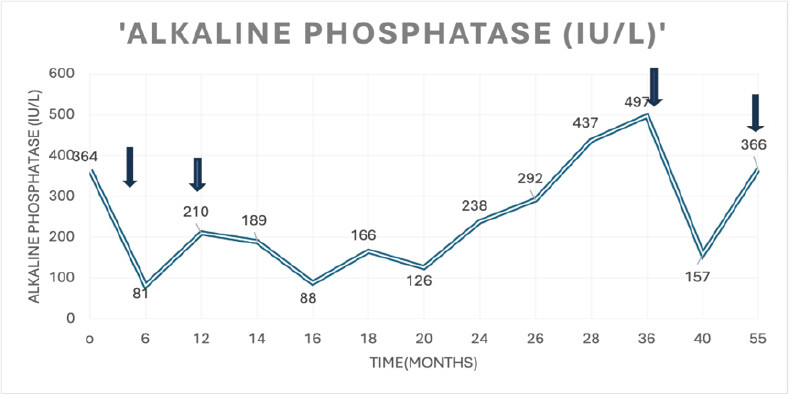
In view of end-stage renal disease, denosumab (60 mg subcutaneously) was initiated, with the understanding that this represented an off-label use in this population. Initial response was noted, with symptomatic improvement and a reduction in total ALP, as shown in Supplementary Fig. 2. Serum calcium and phosphate levels were monitored at baseline, 2 weeks, 1 month, and 3 months after each injection, with no severe hypocalcemia (defined as serum calcium <6.5 mg/dL) observed; however, calcium supplementation was administered when levels approached the lower limit of normal, and vitamin D levels were maintained. Recurrence of bone pain and a rise in ALP occurred after 4 months, prompting a second denosumab dose at 6 months after the first. He underwent surgery for right tibia fracture correction a month after the second denosumab injection. The fracture fixation itself contributed to symptom relief and functional improvement in the immediate post-operative period. Subsequent dosing (third dose after 2 years, fourth dose after 18 months following the third) was based on biochemical and clinical recurrence. Over the approximately 4-year follow-up period, the patient remained on dialysis with stable renal function.
Discussion
Diagnostic considerations
The index case presented with bone pain in the context of CKD. Importantly, not all bone pain in CKD can be attributed to CKD-mineral bone disorder (CKD-MBD), though the distinction can be challenging. Any persistent, localized pain with deformity or fracture should raise the possibility of an alternate diagnosis such as Paget’s disease.
Measurement of bone-specific ALP would be ideal for distinguishing PDB from CKD-MBD, but when unavailable in resource-limited settings, imaging plays a crucial role. In our patient, bone scintigraphy demonstrated focal, intense tracer uptake in a characteristic distribution (tibia, pelvis, humerus), which helped differentiate PDB from CKD-MBD, in which uptake is typically diffuse. Plain radiography showed cortical thickening and trabecular coarsening, supporting the diagnosis. However, histological confirmation by bone biopsy was not performed, limiting definitive diagnostic certainty, although the clinical and radiological findings were highly consistent with PDB.
Literature context
We reviewed 14 previously published case reports of PDB in CKD to contextualize our findings (Table 2). These cases were heterogeneous in CKD stage, skeletal involvement sites, treatment modalities, and follow-up duration, limiting direct comparison. Most patients were elderly males with advanced CKD (stages 4–5). The pelvis was the most commonly affected site. ALP levels were markedly elevated in all cases and appeared to reflect disease activity and treatment response.
Studies assessing Paget’s disease of bone in chronic kidney disease patients
| S. No. | Author, year | Age and gender | Baseline ALP (IU/L) | CKD stage | Skeletal involvement | Therapeutic agent used | Follow-up period |
|---|---|---|---|---|---|---|---|
| 1 | Schwarz et al. 2012 [8] | 86/M | 147 | 5 | Pelvic | Denosumab, 60 mg, SC, at 0, 6, 9, 12, 15 months | 15 months |
| 2 | Ringe and Delling 1985 [12] | 48/F | 496 | – | NA | NA | NA |
| 3 | Etemadi et al. 2008 [13] | 77/F | 6336 | On HD-5D | Skull | Alendronate 70 mg/week- 6 months | 6 months ALP-600 |
| 4 | Wu et al. 2009 [14] | 77/F | 108 | On PD | Lumbar L3 | Calcitonin, sc and nasal Surgical decompression | – |
| 5 | Cianciolo et al. 2010 [15] | 69/F | 5D | Skull | Clodronate | 12 months | |
| 6 | De Sousa-Amorim et al. 2012 [16] | 72/M | – | PD | Polyostotic cervical spine, first finger of the right hand, left ulna, right sacroiliac joint, left malleolus. | – | Asymptomatic |
| 7 | Kostine et al. 2016 [9] | 79/M | 379 | 4 | Pelvis and lumbar spine | Denosumab, 60 mg, SC single dose | 18 months |
| 8 | Kuthiah and Er 2019 [10] | 63/F | 312 | 4 | Left iliac, pubic bones, tibia | Denosumab, 60 mg, SC, six-monthly | 3 months ALP-118 |
| 9 | Chan et al. 2019 [17] | 80/M | 168 | 5 | Bilateral pelvic bones – asymptomatic | Nil | Nil |
| 10 | Panuccio and Tripepi 2020 [18] | 60/M | 6000 | 4 | Skull and long bones | Clodronate 800 mg twice (3 m) → calcitonin nasal (6 m) | 9 months ALP-1000 |
| 11 | Muthu et al. 2020 [19] | 60/M | 319 | Right femur | Pamidronate 30 mg/kg after 6 months, every 6 months | 18 months | |
| 12 | Elbüken et al. 2022 [20] | 47/M | 1,693 | 3 | Left iliac bone, right femur, and tibia | Denosumab, 60 mg, SC, three monthly | – |
| 13 | Guillermo et al. 2021 [21] | 80/M | 4500 | PD | Left iliac bone | Denosumab | – |
| 14 | Lorho et al. 1998 [22] | 81/M | – | 5D | Pelvis | None | – |
M: Male, F: Female, ALP: Alkaline phosphatase, CKD: Chronic kidney disease, PD: Paget’s disease
Treatment options in CKD-PDB
Bisphosphonates are the standard of care for active PDB. Zoledronate achieves superior biochemical and clinical remission compared with oral agents, with sustained suppression of bone turnover and conversion of woven to lamellar bone [4, 5]. It is a relatively safer drug, with minor flu-like adverse events seen in around 25% of patients [4]. However, due to nephrotoxicity, current guidelines advise against their use in patients with an eGFR below 35 mL/min [6]. Calcitonin may be used in CKD, but its efficacy is modest and often incomplete [7].
Denosumab rationale and limitations
Denosumab, a monoclonal antibody that targets RANKL, represents a potential alternative in advanced CKD because of its non-renal clearance. Several case reports and small case series have demonstrated biochemical and scintigraphic responses in patients with CKD and PDB. However, significant limitations exist: (i) evidence is limited to individual case reports and small heterogeneous series; (ii) variable efficacy has been observed, with some cases showing recurrence after treatment cessation; (iii) optimal dosing intervals remain unclear, with the need for repeated dosing in several cases; (iv) the risk of severe hypocalcemia – particularly in advanced CKD – remains a major concern, requiring careful monitoring; and (v) long-term safety data in this population are sparse. Our case, while showing initial improvement, also demonstrated recurrence after 4 months, illustrating the challenge of sustained disease control [8, 9, 10, 11].
Confounding factors in this case
It is important to acknowledge inherent confounders that limit attribution of improvement solely to denosumab. The spontaneous tibial fracture and subsequent surgical intervention for fracture correction (with internal fixation) likely contributed significantly to pain relief and functional recovery independent of denosumab therapy. In addition, the initial improvement in ALP and symptoms after the first denosumab dose may have been influenced by the natural history of post-fracture bone healing. These confounders make it difficult to attribute the observed biochemical and clinical improvements solely to denosumab, a limitation inherent in single-case reports.
Limitations
This case report has several important limitations:
-
Single case: Findings cannot be generalised to a population; individual responses vary markedly.
-
Lack of control group: No comparison with other treatment modalities (e.g., zoledronate in a matched CKD stage 5 patient).
-
Confounding variables: Surgical fracture correction and natural bone healing post-fracture may have contributed to symptom improvement independent of denosumab.
-
Disease recurrence: Recurrence after 4 months and need for repeated dosing raise questions about sustained efficacy.
-
Unclear optimal dosing: The ideal dosing interval in CKD remains uncertain; this case required doses every 6–18 months.
-
Limited biochemical markers: Bone-specific ALP was unavailable in our institution; total ALP is less specific for bone disease. Bone turnover markers (P1NP, CTX) were not measured.
-
Hypocalcemia monitoring: While severe hypocalcemia did not occur, the monitoring protocol may not have captured all asymptomatic episodes or long-term cumulative effects.
-
No histological confirmation: Diagnosis was based on biochemical and imaging findings; bone biopsy was not performed.
-
Medium-term follow-up: Approximately 4 years; long-term safety and efficacy beyond this period remain unknown.
-
Literature heterogeneity: The 14 cases reviewed differed substantially in CKD stage, disease sites, treatments, and follow-up duration.
Conclusion
PDB in the setting of CKD represents a rare diagnostic and therapeutic challenge. This case illustrates that denosumab may serve as a potential therapeutic option in patients with advanced CKD where bisphosphonates are contraindicated due to renal impairment. However, the clinical evidence supporting its use remains limited to individual case reports and small case series. Important considerations include the need for repeated dosing (as demonstrated in this case by recurrence after 4 months), the risk of hypocalcemia, necessitating careful monitoring, and the unclear optimal dosing intervals. Larger, controlled studies and longer-term follow-up data are needed to establish the role of denosumab in this population. Until such evidence accumulates, denosumab should be considered a reasonable alternative in selected patients with advanced CKD and PDB for whom bisphosphonates are contraindicated, with the caveat that careful biochemical monitoring and informed consent regarding the limited evidence are essential.
Clinical Message
Patients with CKD presenting with focal bone pain and deformity should be evaluated for alternative diagnoses beyond CKD-MBD, including PDB. Imaging modalities such as bone scintigraphy can aid differentiation. Denosumab may serve as a therapeutic option in advanced CKD where bisphosphonates are contraindicated, which can help clinicians feel more confident in managing complex cases, though treatment decisions should account for the need for repeated dosing and hypocalcemia monitoring.
Conflict of Interest:
Nil
Source of Support:
Nil
Consent:
The authors confirm that informed consent was obtained from the patient for publication of this article
References
- Gennari L, Rendina D, Falchetti A, Merlotti D.Paget's disease of bone. Calcif Tissue Int 2019;104:483-500. Google Scholar | PubMed
- Corral-Gudino L, Borao-Cengotita-Bengoa M, Del Pino-Montes J, Ralston S.Epidemiology of Paget's disease of bone: A systematic review and meta-analysis of secular changes. Bone 2013;55:347-52. Google Scholar | PubMed
- Cundy T.Paget's disease of bone. Metabolism 2018;80:5-14. Google Scholar | PubMed
- Reid IR, Miller P, Lyles K, Fraser W, Brown JP, Saidi Y.Comparison of a single infusion of zoledronic acid with risedronate for Paget's disease. N Engl J Med 2005;353:898-908. Google Scholar | PubMed
- Reid IR, Lyles K, Su G, Brown JP, Walsh JP, del Pino-Montes J.A single infusion of zoledronic acid produces sustained remissions in Paget disease: Data to 6.5 years. J Bone Miner Res 2011;26:2261-70. Google Scholar | PubMed
- Singer FR, Bone HG, Hosking DJ, Lyles KW, Murad MH, Reid IR.Paget's disease of bone: An endocrine society clinical practice guideline. J Clin Endocrinol Metab 2014;99:4408-22. Google Scholar | PubMed
- Doyle FH, Pennock J, Greenberg PB, Joplin GF, MacIntyre I.Radiological evidence of a dose-related response to long-term treatment of Paget's disease with human calcitonin. Br J Radiol 1974;47:1-8. Google Scholar | PubMed
- Schwarz P, Rasmussen AQ, Kvist TM, Andersen UB, Jørgensen NR.Paget's disease of the bone after treatment with Denosumab: A case report. Bone 2012;50:1023-5. Google Scholar | PubMed
- Kostine M, Mehsen-Cetre N, Bannwarth B.Denosumab-induced severe hypocalcemia in a patient with Paget's disease of bone and impaired renal function. Therapies 2017;72:383-5. Google Scholar | PubMed
- Kuthiah N, Er C.Effective Treatment of Paget's disease of the bone in a Chinese woman. Ann Acad Med Singap 2018;47:528-30. Google Scholar | PubMed
- Reid IR, Sharma S, Kalluru R, Eagleton C.Treatment of Paget's disease of bone with denosumab: Case report and literature review. Calcif Tissue Int 2016;99:322-5. Google Scholar | PubMed
- Ringe JD, Delling G.[Unusual course of renal osteopathy with Paget's disease and aluminium deposits. Dtsch Med Wochenschr 1985;110:411-5 German]. Google Scholar | PubMed
- Etemadi J, Bagheri N, Falaknazi K, Ardalan M, Rahbar K, Nobakht Haghighi A.Paget's disease of bone in a patient on hemodialysis. Iran J Kidney Dis 2008;2:105-7. Google Scholar | PubMed
- Wu LC, Tseng CH, Chiang YF, Tsuang YH.Monostotic Vertebral Paget's disease of the lumbar spine. J Chin Med Assoc 2009;72:52-5. Google Scholar | PubMed
- Cianciolo G, Manna GL, Capelli I, Donati G, Persici E, Cuna V.A case of Paget's disease in hemodialysis. Clin Nephrol 2010;73:403-7. Google Scholar | PubMed
- De Sousa-Amorima E, Del Peso-Gilsanza G, Bajo-Rubioa MA, Selgas-Gutiérreza R.Enfermedad de paget poliostótica asintomática asociada a hiperparatiroidismo secundario en un paciente en diálisis peritoneal. Nefrología 2012;32:553. Google Scholar | PubMed
- Chan PK, Lyu SY, Lu CC.Paget disease of bone in an elderly patient with chronic renal disease and weight loss: A case report. Medicine (Baltimore) 2019;98:e17458. Google Scholar | PubMed
- Panuccio VA, Tripepi R.Paget's disease and secondary hyperparathyroidism: Is Healing Possible?. Front Cell Dev Biol 2020. May 29;8:399. Google Scholar | PubMed
- Muthu S, Ramakrishnan E, Chellamuthu G.'Bone within bone appearance' in a pathological fracture. BMJ Case Rep 2020;13:e238567. Google Scholar | PubMed
- Elbüken G, BiLen Oİ, Ağgül H.Denosumab experience in a patient with end-stage renal disease and Paget's disease of the bone. Turkiye Klinikleri J Case Rep 2022;30:110-3. Google Scholar | PubMed
- Guillermo FP, Gabriel SJ, Fernando ZJ, Daniel JV, Arturo OL.Paget´s disease of bone in a patient on peritoneal dialysis. Int J Case Rep 2021;5:196. Google Scholar | PubMed
- Lorho R, Mourad G, Couret I, Argiles A.The dialysis patient with persisting elevation of bone alkaline phosphatase after parathyroidectomy. Nephrol Dial Transplant 1998;13:2394-6. Google Scholar | PubMed
Related Articles in Journal of Orthopaedic Case Reports
 July 1, 2026 Tuberculosis of the Elbow in a Chronic Kidney Disease Patient with Arteriovenous Fistula: A Case Report
 December 1, 2025 Correlation between Biochemical Parameters and Radiographic Healing Score in Tibial Fractures
 March 1, 2025 Simultaneous Bilateral Knee Extensor Mechanism Injury in a Young Male: A Clinical Diagnosis and Literature Review
 July 10, 2024 Infective Spondylodiscitis of Unknown Origin in a Patient with Chronic Kidney Disease – Case report