


Introduction
Hip fracture among the geriatric population is a significant health burden in the world, and morbidity, mortality rates, and disability are high. Older patients who have hip fractures usually have a number of comorbidities, frailty, and low physiological reserves that can greatly affect the post-operative outcomes and survival. The mortality rates in patients after hip fracture surgery are still high, and studies indicate that short-term and 1-year mortality is high, which is why the reliability of prognostic indicators to make a clinical decision and manage the patient optimally must be considered [1]. “Sarcopenia, or progressive and generalized skeletal muscle mass and activity loss, has become a key factor that predetermines negative outcomes in older adults among other risk factors. It is very common in patients with hip fractures and closely linked to higher post-operative complications, slow recovery, low functional outcomes, and higher mortality [2].
Historically, the diagnosis of sarcopenia is based on a set of muscle mass measurements, muscle strength assessment, and physical performance evaluation as suggested by the European Working Group on Sarcopenia in Older People (EWGSOP2). Nevertheless, these functional tests are in most cases impractical or unreliable in the acute environment of hip fractures because of pain, immobility, and limitations imposed by the trauma. Consequently, the desire to find alternative ways of sarcopenia assessment, especially imaging-based ones, which can be conducted with the utilization of regularly obtained radiological data, has increased [3]. Computed tomography (CT)-based measurement has become one of the essential tools to measure body composition, such as skeletal muscle mass and muscle quality, in terms of muscle cross-sectional area, skeletal muscle index (SMI), and muscle attenuation [4].
Opportunistic assessment of sarcopenia can be carried out with no extra cost or patient burden in pre-operative CT scans that are often done in elderly patients to evaluate their diagnosis or surgical planning intentions [5]. Research has shown that muscle measurements obtained using CT, especially psoas muscle area and density, are good predictors of muscle mass and quality, which is indicative of both sarcopenia and myosteatosis. Notably, these CT-based indices have been demonstrated to be related to clinical outcomes, and therefore, they are promising biomarkers of risk stratification in surgical patients [6].
There is also emerging evidence that CT-based sarcopenia is an important predictor of death in elderly patients who have undergone hip fracture surgery [7]. Less muscle mass and low muscle density have been found to be independently related to elevated short-term and long-term mortality rates even after considering the confounding variables of age, sex, and comorbidity. Moreover, patients with a low muscle quantity alongside poor muscle quality have the worst prognosis, which underlines the significance of extensive muscle examination [8].
Besides mortality, sarcopenia has also been observed to be associated with compromised functional recovery after hip fracture surgery. Sarcopenic patients have increased chances of having long hospital stays, delayed mobilization, reduced activity of daily living independence, and increased chances of institutionalization. These results are especially important among geriatric populations, where one of the most important determinants of quality of life and healthcare use is functional independence [9].
Although the scientific evidence of the prognostic value of sarcopenia continues to mount, the standardized, practical, and universally acceptable ways of measuring the condition in the acute clinical setting are, nonetheless, lacking. Evaluation through CT can offer an answer to this question because there are objective, reproducible, and readily obtainable measurements of muscle parameters possible. However, methodological inaccuracy, inconsistency in cut-off values, and inconsistent anatomical landmarks have limited the widespread application of this diagnostic technique [10]. The purpose of the current scientific paper lies in the pre-operative detection of sarcopenia, which occurs in the form of CT prediction of mortality and functional outcomes in geriatric patients undergoing hip fracture surgery. This investigation will help develop several robust prognostic models that could be used for risk stratification and perioperative care of these vulnerable patients. Therefore, the present study was conducted to evaluate the role of pre-operative CT-based sarcopenia assessment in predicting mortality and functional outcomes following hip fracture surgery in elderly patients.
Materials and Methods
Study design
This prospective observational cohort study was conducted in the Departments of Orthopaedics and Radiology of a tertiary care teaching hospital over a period of 18 months to evaluate the utility of CT-derived assessment of sarcopenia in predicting mortality and functional outcomes among elderly patients undergoing hip fracture surgery. The study protocol was approved by the Institutional Ethics Committee, and written informed consent was obtained from all participants or their legally authorized representatives before enrollment in the study.
Participants
Inclusion criteria
-
Patients aged ≥60 years
-
Diagnosed with hip fracture (intertrochanteric or femoral neck fracture)
-
Undergoing surgical management
-
Availability of pre-operative CT scan including abdominal or pelvic region
-
Provided informed consent
Exclusion criteria
-
Pathological fractures (e.g., malignancy-related)
-
Previous hip surgery on the affected side
-
Patients with neuromuscular disorders affecting muscle mass
-
Polytrauma patients or those with severe head injury
-
Incomplete clinical or imaging data
-
Refusal to participate in the study
Study sampling and sample size
A consecutive sampling method was employed, wherein all eligible patients presenting during the study period were enrolled until the target sample size of 50 participants was achieved. The sample size was determined based on study feasibility, expected patient availability, and evidence from previous studies evaluating sarcopenia in orthopedic populations.
Study groups
Participants were classified into two groups based on pre-operative CT-based sarcopenia assessment: The sarcopenic group, comprising patients with reduced skeletal muscle mass and/or low muscle attenuation, and the non-sarcopenic group, comprising patients with normal muscle mass and quality. This classification enabled comparison of clinical and functional outcomes between the two groups.
Study parameters
Data collected included demographic variables (age, sex, and body mass index), clinical variables (comorbidities, American Society of Anesthesiologists [ASA] grade, and fracture type), and radiological parameters obtained from CT imaging, including psoas muscle area, SMI, and muscle attenuation. The primary outcome measure was post-operative mortality. Muscle strength and physical performance tests, including handgrip strength and gait speed recommended by EWGSOP2, were not performed because acute hip fractures prevented reliable functional assessment before surgery. Secondary outcomes included post-operative complications, length of hospital stay, and functional recovery assessed using the Harris Hip Score.
Study procedure
Clinical and radiological examination was carried out on admitted patients who qualified for the study. Pre-operative CT images were studied while muscle measurements were done at specific anatomic locations such as the third lumbar vertebra or psoas muscle site. Surgery was carried out using established orthopedic methods. After surgery, treatment such as pain relief and physical therapy were offered. Follow-ups were carried out at regular intervals to check survival rates and functional improvement.
Data analysis
Data were analyzed using IBM Statistical Package for the Social Sciences software. Continuous variables were expressed as mean ± standard deviation, while categorical variables were presented as frequencies and percentages. Comparisons between sarcopenic and non-sarcopenic groups were performed using the independent t-test and Chi-square test, as appropriate. A P < 0.05 was considered statistically significant; because of the relatively small sample size, multivariable regression analysis was not performed, and the findings should therefore be interpreted as unadjusted associations rather than independent predictors.
Results
A total of 50 elderly patients undergoing hip fracture surgery were included in the study. Based on pre-operative CT assessment, 24 patients (48%) were classified as sarcopenic and 26 (52%) as non-sarcopenic. The majority of patients were aged 70–79 years, and a higher proportion of sarcopenic patients were aged ≥80 years. Sarcopenic patients were more frequently underweight and had higher ASA grades (III–IV), indicating poorer pre-operative physiological status. The distribution of fracture types was comparable between the groups, with intertrochanteric fractures accounting for approximately 62% of cases (Table 1).
Baseline characteristics of the study population
| Variable | Sarcopenic (n=24) (%) | Non-sarcopenic (n=26) (%) |
|---|---|---|
| Age ≥80 years | 8 (33.3) | 7 (26.9) |
| Male gender | 14 (58.3) | 12 (46.2) |
| Underweight BMI | 8 (33.3) | 3 (11.5) |
| ASA grade III–IV | 16 (66.7) | 10 (38.5) |
| Intertrochanteric fracture | 15 (62.5) | 16 (61.5) |
ASA: American Society of Anesthesiologists, BMI: Body mass index
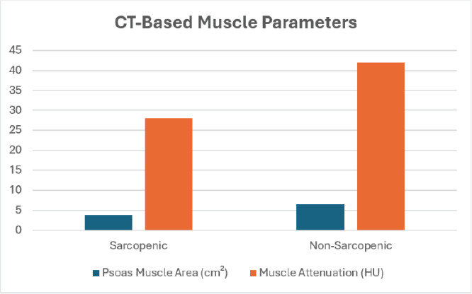
CT-based muscle assessment demonstrated significantly lower psoas muscle area (3.8 ± 0.9 cm2 vs. 6.5 ± 1.2 cm2; P < 0.001) and muscle attenuation (28 ± 5 Hounsfield Units [HU vs. 42 ± 6 HU; P < 0.001) among sarcopenic patients, confirming reduced muscle quantity and quality (Fig. 1). Post-operative complications occurred more frequently in the sarcopenic group (58.3% vs. 23.1%), and these patients experienced longer hospital stays (12.5 ± 3.2 days vs. 8.4 ± 2.1 days).
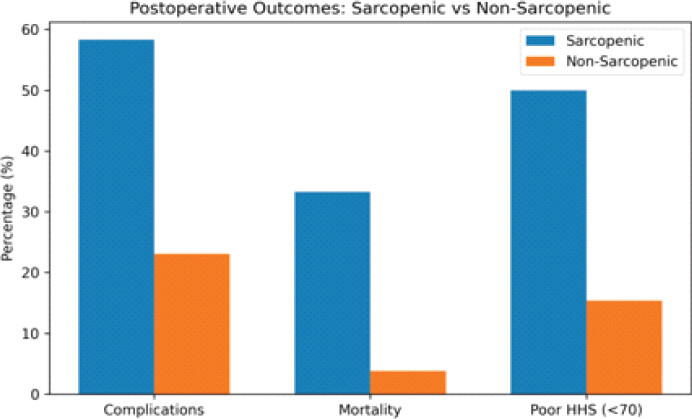
Mortality was substantially higher among sarcopenic patients, with 33.3% mortality compared to 3.8% in the non-sarcopenic group. Functional recovery was also inferior, as 50% of sarcopenic patients demonstrated poor Harris Hip Scores (<70), whereas more than half of non-sarcopenic patients (53.8%) achieved good-to-excellent outcomes (>80) (Table 2, Fig. 2). These findings indicate that pre-operative CT-defined sarcopenia is strongly associated with increased post-operative morbidity, prolonged hospitalization, higher mortality, and poorer functional recovery following hip fracture surgery.
Computed tomography-based muscle parameters and post-operative outcomes
| Parameter | Sarcopenic | Non-sarcopenic | P-value |
|---|---|---|---|
| Psoas muscle area (cm2) | 3.8±0.9 | 6.5±1.2 | <0.001 |
| Muscle attenuation (Hounsfield Units) | 28±5 | 42±6 | <0.001 |
| Hospital stay (days) | 12.5±3.2 | 8.4±2.1 | <0.001 |
| Post-operative complications | 58.30% | 23.1% | — |
| Mortality | 33.30% | 3.8% | — |
| Poor Harris Hip score (<70) | 50.00% | 15.4% | — |
Discussion
The present study demonstrated that pre-operative CT-based sarcopenia is a significant predictor of mortality, post-operative complications, and functional outcomes in geriatric patients undergoing hip fracture surgery. In this cohort of 50 patients, 24 (48%) were identified as sarcopenic, which is comparable to the prevalence reported by Jiang et al., [11] where 35.6% of elderly hip fracture patients were diagnosed with sarcopenia using the Asian Working Group for Sarcopenia 2019 criteria. The slightly higher prevalence observed in the present study may be attributed to differences in diagnostic criteria and imaging parameters. Age-wise distribution in the present study showed that 33.3% of sarcopenic patients were aged ≥80 years, supporting the findings of Jiang et al., [11], who reported that age ≥80 years significantly contributed to adverse outcomes such as early post-operative cognitive dysfunction (POCD) (odds ratio [OR] = 3.364). This highlights that advanced age and sarcopenia often coexist and synergistically worsen prognosis.
The CT-based muscle assessment in the present study revealed significantly reduced psoas muscle area (3.8 ± 0.9 cm2) and muscle attenuation (28 ± 5 HU) in the sarcopenic group compared to 6.5 ± 1.2 cm2 and 42 ± 6 HU in the non-sarcopenic group (P < 0.001). These findings are consistent with Boutin et al., [12], who demonstrated that decreased muscle size and attenuation on CT were significantly associated with reduced survival, with thoracic muscle size showing an OR of 0.66 (95% confidence interval [CI]: 0.49–0.87) and attenuation ranging between OR 0.67–0.72 for mortality. This agreement confirms that both muscle quantity and quality are critical determinants of survival and reinforces the validity of CT-based sarcopenia assessment as a prognostic biomarker. Although psoas muscle measurements are widely used as practical imaging biomarkers of sarcopenia, they may not fully reflect total skeletal muscle mass, and future studies should incorporate whole-body or lumbar skeletal muscle indices together with standardized CT diagnostic thresholds.
Post-operative complications were markedly higher in the sarcopenic group (58.3%) compared to the non-sarcopenic group (23.1%) in the present study, indicating a strong association between sarcopenia and increased morbidity. Although Chang et al. [13] did not directly quantify complication rates, their findings showed that low SMI and paraspinal muscle density were independently associated with adverse surgical outcomes, including prolonged hospitalization (P = 0.008 and P = 0.032, respectively). These results are in concordance with the present study, where sarcopenic patients had a significantly longer hospital stay (12.5 ± 3.2 days) compared to non-sarcopenic patients (8.4 ± 2.1 days), suggesting delayed recovery and increased healthcare burden.
Mortality analysis in the present study revealed a substantial difference between groups, with 33.3% mortality in the sarcopenic group compared to only 3.8% in the non-sarcopenic group. This finding strongly aligns with Veizi et al., [14], who reported that sarcopenia was independently associated with increased 1-year mortality (Hazard Ratio [HR]: 1.80, 95% CI: 1.04–3.13, P = 0.034), and sarcopenic obesity further increased mortality risk (HR: 2.68). Similarly, Boutin et al. [12] reported increasing mortality rates up to 79.5% at 5 years, with reduced muscle mass and attenuation significantly associated with decreased survival. These comparisons clearly validate the present study findings and emphasize that sarcopenia is a powerful independent predictor of mortality in elderly hip fracture patients.
Functional outcome assessment in the present study using the Harris Hip Score showed that 50% of sarcopenic patients had poor outcomes (<70), while only 20.8% achieved good to excellent recovery (>80), compared to 53.8% in the non-sarcopenic group. These findings are indirectly supported by Jung et al., [15], who demonstrated that significant post-operative reductions in muscle mass of the iliopsoas and rectus femoris were associated with impaired recovery and emphasized the need for targeted rehabilitation strategies. The decline in muscle mass observed postoperatively further supports the notion that pre-existing sarcopenia can worsen functional outcomes if not addressed early.
In addition, the association between sarcopenia and broader post-operative complications is reinforced by Jiang et al., [11], who reported that sarcopenia was independently associated with early POCD with an odds ratio of 3.716 (95% CI: 1.618–8.814, P = 0.005).” Although cognitive outcomes were not directly assessed in the present study, the significantly higher complication rate (58.3%) in sarcopenic patients suggests that sarcopenia contributes to both physical and neurological post-operative decline.
Interestingly, while the present study demonstrated prolonged hospital stay in sarcopenic patients, Veizi et al. [14] reported that sarcopenia alone was not significantly associated with prolonged hospitalization after adjustment for confounders. This discrepancy may be due to differences in study design, sample size (311 vs. 50), or adjustment factors. However, the trend toward increased resource utilization in sarcopenic patients remains clinically relevant.
Overall, the findings of the present study are in strong agreement with existing literature, confirming that sarcopenia assessed through CT imaging is a robust and reliable predictor of adverse outcomes. Noteworthy differences in muscle parameters (P < 0.001), mortality rate (33.3% vs. 3.8%), incidence of complications (58.3% vs. 23.1%), and length of hospitalization (12.5 vs. 8.4 days) serve as evidence to prove that sarcopenia plays a crucial role as a prognostic factor. Noteworthy, a CT-assessed diagnosis of this pathological condition serves as an objective tool for identifying high-risk patients. As shown by this study and confirmed by previous findings, it can be concluded that the condition in question is not only an indicator of aging but also a serious medical condition with a direct impact on surgical outcome. Inclusion of CT-assessed sarcopenia screening into pre-surgical protocol would help to identify risks in time, apply appropriate interventions, and thereby ensure better survival and recovery.
The strengths of this study include its prospective design and objective CT-based assessment of muscle quantity and quality using routinely available pre-operative imaging. However, several limitations should be acknowledged. The relatively small sample size (n = 50), single-center design, and short follow-up period may have limited statistical power, generalizability, and assessment of long-term outcomes. As an observational study, it demonstrates associations rather than causality, and selection bias may have been introduced because only patients with available pre-operative CT scans were included. Although baseline demographic and clinical variables were assessed, important confounders such as nutritional status, frailty, cognitive impairment, osteoporosis, and socioeconomic factors were not fully adjusted for, and multivariable regression analysis was not performed.
Furthermore, CT assessment was primarily based on psoas muscle measurements, which may not accurately represent whole-body skeletal muscle mass, and the lack of standardized CT cut-off values limits reproducibility. Muscle strength and physical performance measures recommended by EWGSOP2, including handgrip strength and gait speed, could not be assessed because of the acute clinical condition of patients with hip fractures. Interobserver and intraobserver reliability of CT measurements was not evaluated, while heterogeneity in fracture patterns, surgical procedures, rehabilitation protocols, and perioperative care may have influenced outcomes. Mortality analysis did not distinguish specific causes of death. Therefore, larger multicenter studies with standardized imaging protocols, comprehensive geriatric assessment, multivariable analyses, and external validation are required to confirm the prognostic value of CT-based sarcopenia assessment.
Conclusion
Although pre-operative CT-based sarcopenia assessment appears to be a promising prognostic imaging biomarker, further multicenter studies with larger sample sizes and standardized diagnostic criteria are required before routine clinical implementation.
Clinical Message
CT-derived sarcopenia assessment provides an objective and readily available method for identifying high-risk elderly hip fracture patients and may aid perioperative decision-making.
Conflict of Interest:
Nil
Source of Support:
Nil
Consent:
The authors confirm that informed consent was obtained from the patient for publication of this article
References
- Cruz-Jentoft AJ, Bahat G, Bauer J, Boirie Y, Bruyère O, Cederholm T. Sarcopenia: Revised European consensus on definition and diagnosis. Age Ageing 2019;48:16-31. Google Scholar | PubMed
- Gustafsson T, Ulfhake B. Sarcopenia: What is the origin of this aging-induced disorder? Front Genet 2021;12:688526. Google Scholar | PubMed
- Fielding RA, Vellas B, Evans WJ, Bhasin S, Morley JE, Newman AB. Sarcopenia: an undiagnosed condition in older adults. Current consensus definition: Prevalence, etiology, and consequences. International working group on sarcopenia. J Am Med Dir Assoc 2011;12:249-56. Google Scholar | PubMed
- Donini LM, Busetto L, Bischoff SC, Cederholm T, Ballesteros-Pomar MD, Batsis JA. Definition and diagnostic criteria for sarcopenic obesity: ESPEN and EASO consensus statement. Obes Facts 2022;15:321-35. Google Scholar | PubMed
- Min D, Lee HS, Shin M. Consequences of fall-induced hip fractures on cognitive function, physical activity, and mortality: Korean longitudinal study of aging 2006-2016. Injury 2021;52:933-40. Google Scholar | PubMed
- Alpantaki K, Papadaki C, Raptis K, Dretakis K, Samonis G, Koutserimpas C. Gender and age differences in hip fracture types among elderly: A retrospective cohort study. Maedica (Bucur) 2020;15:185-90. Google Scholar | PubMed
- Benichou O, Lord SR. Rationale for strengthening muscle to prevent falls and fractures: A review of the evidence. Calcif Tissue Int 2016;98:531-45. Google Scholar | PubMed
- Cruz-Jentoft AJ, Landi F, Schneider SM, Zúñiga C, Arai H, Boirie Y. Prevalence of and interventions for sarcopenia in ageing adults: A systematic review. Report of the international sarcopenia initiative (EWGSOP and IWGS). Age Ageing 2014;43:748-59. Google Scholar | PubMed
- Gandham A, Mesinovic J, Jansons P, Zengin A, Bonham MP, Ebeling PR. Falls, fractures, and areal bone mineral density in older adults with sarcopenic obesity: A systematic review and meta-analysis. Obes Rev 2021;22:e13187. Google Scholar | PubMed
- Rubin PJ, Leyvraz PF, Aubaniac JM, Argenson JN, Estève P, De Roguin B. The morphology of the proximal femur. A three-dimensional radiographic analysis. J Bone Joint Surg Br 1992;74:28-32. Google Scholar | PubMed
- Jiang T, Wang P, Wu J, Liang X, Wang Y, Hu X. Sarcopenia is associated with early postoperative cognitive decline in older adults following hip fracture surgery: A prospective cohort study. BMC Geriatr 2025;26:62. Google Scholar | PubMed
- Boutin RD, Bamrungchart S, Bateni CP, Beavers DP, Beavers KM, Meehan JP. CT of patients with hip fracture: Muscle size and attenuation help predict mortality. AJR Am J Roentgenol 2017;208:W208-15. Google Scholar | PubMed
- Chang CD, Wu JS, Mhuircheartaigh JN, Hochman MG, Rodriguez EK, Appleton PT. Effect of sarcopenia on clinical and surgical outcome in elderly patients with proximal femur fractures. Skeletal Radiol 2018;47:771-7. Google Scholar | PubMed
- Veizi BG, Imeri V, Naldöven ÖF, Güven Ş. Sarcopenia and sarcopenic obesity: Their association with postoperative outcomes in patients with hip fractures. J Hosp Med 2025;20:816-23. Google Scholar | PubMed
- Jung SY, Kim HJ, Oh KT. Comparative analysis of preoperative and postoperative muscle mass around hip joint by computed tomography in patients with hip fracture. Hip Pelvis 2022;34:10-7. Google Scholar | PubMed
© 2026 Journal of Orthopaedic Case Reports - Published by Indian Orthopaedic Research Group
About the Authors
-

Dr. Mohammed Saleh AlSaifi
-

Dr. Rajani Thakur
-

Dr. Siroya Meet Manishbhai
-

Dr. Ketan Rupala
-

Dr. Mohanakrishnan B
-

Dr. Ashirwad Pasumarthy
Related Articles in Journal of Orthopaedic Case Reports
 April 1, 2026 Functional Outcome of Unstable Intertrochanteric Femur Fracture Treated Surgically With Long Proximal Femoral Nail
 September 1, 2026 The Evolution of CT-Based Robotic Systems in Total Knee Arthroplasty: From Precision Tools to Intelligent Surgical Partners
 September 1, 2026 Comparative Study of Post-operative Pain and Functional Outcomes in Robotic-assisted Total Knee Replacement versus Cruciate-retaining Manual Total Knee Replacement
 September 1, 2026 Serial Magnetic Resonance Imaging Graft Signal Intensity and its Correlation with Functional Recovery Following Primary Anterior Cruciate Ligament Reconstruction