


Introduction
The ankle joint is a highly specialized synovial hinge joint essential for locomotion and distributing body weight [1,2]. It functions as a complex mechanical structure that must simultaneously provide mobility and withstand significant forces generated during gait [3]. Anatomically, the ankle mortise is formed by the articulation between the distal tibia, the distal fibula, and the talar dome [1,4].
The medial malleolus forms the medial border of this mortise and anchors the deltoid ligament complex, playing a vital role in ankle stability [5,6]. It actively resists eversion, external rotation, and lateral displacement of the talus [7]. Because the medial malleolus constitutes a part of the weight-bearing articular surface, even minor displacement can lead to incongruity, uneven cartilage loading, and eventual post-traumatic osteoarthritis [8].
Fractures in this region typically result from excessive supination-external rotation or pronation-eversion forces [9]. Consequently, surgical treatment is frequently required to achieve anatomical reduction and rigid internal fixation [10].
Tension band wiring (TBW) is a well-established surgical option that converts tensile forces generated during ankle movement into compressive forces at the fracture site [11]. This dynamic compression enhances stability and promotes osteosynthesis [12]. TBW is particularly effective for transverse fractures, small bony fragments, and osteoporotic bone where other implants may fail to achieve adequate purchase [6]. Furthermore, it is a simple, cost-effective technique that requires minimal instrumentation, making it highly practical in high-volume trauma settings [13]. This study aimed to evaluate the functional and radiological outcomes of medial malleolar fractures treated exclusively with TBW and to assess its clinical efficacy.
Materials and Methods
Study design and patient selection
A prospective observational study was conducted at the Department of Orthopaedics, Gandhi Medical College and Hamidia Hospital, Bhopal, over an 18 months following approval from the Institutional Ethics Committee. The study cohort consisted of 20 skeletally mature patients (aged >18 years) who presented with isolated, closed medial malleolar fractures classified as Herscovici Types A, B, and C. We deliberately excluded patients with Herscovici Type D fracture pattern (as it requires an antiglide plate), open or pathological fractures, associated ipsilateral limb fractures, and those with a history of prior ankle surgeries. Standard pre-operative clinical evaluations were performed, accompanied by anteroposterior, lateral, and mortise radiographic views of the affected ankle to accurately classify the fracture geometry and plan the surgical approach, and the surgery was performed when the swelling subsided. Surgical technique – the procedure was performed under spinal anesthesia with tourniquet control. A slightly curved medial incision was made over the medial malleolus, the fracture site was cleared of periosteum and debris, the reduction was performed using bone-holding forceps, and anatomical alignment of the fracture was confirmed with fluoroscopic guidance. Two parallel Kirschner wires (K-wires) were inserted from the tip of the medial malleolus proximally into the tibial metaphysis; a unicortical screw, fitted with a washer, was inserted into the tibial metaphysis, leaving a space for the wire of 2–3 mm between the bone and the washer. A stainless-steel wire was looped in a figure-of-eight configuration around the K-wires and the screw beneath the washer, and the wire was tightened to achieve compression across the fracture site; the ends of the K-wires were cut and bent over to prevent migration.
A below-knee plaster splint was given with post-operative non-weight-bearing maintained for 2–3 weeks, followed by splint removal and progressive mobilization; full weight bearing was allowed at 6 weeks.
Patients were scheduled for regular clinical and radiographic follow-up evaluations at 6 weeks, 3 months, and 6 months post-surgery. At each interval, functional recovery was quantified using the American Orthopaedic Foot and Ankle Society (AOFAS) Ankle-Hindfoot scale. Range of motion was measured using a goniometer, whereas subjective pain was measured via the Visual Analog Scale (VAS). Fracture union was assessed on serial radiographs, defined by the appearance of bridging callus across the fracture lines without hardware failure. The IEC registration number ECR/1055/Inst/MP/2018, IEC protocol no – 40/IEC/2024, date of approval March 27, 2024.
Results
A total of 20 skeletally mature patients with isolated, closed medial malleolar fractures were treated using the TBW technique and all completed the 6-month follow-up. The cohort had a mean age of 33.05 ± 11.2 years, reflecting a young, active demographic. There was a clear male predominance, with 14 men (70%) and 6 women (30%) included in the study.
Right-sided ankle injuries were more frequent, accounting for 13 cases (65%), whereas the left side was involved in the remaining 7 cases (35%). Regarding the mechanism of injury, high-energy trauma was the leading cause; road traffic accidents accounted for exactly half of the cases (10 patients). Domestic or self-falls were responsible for 8 injuries (40%), and sports-related incidents caused the remaining 2 cases (10%).
Based on pre-operative radiographic assessment using the Herscovici classification, 4 cases (20%) were Type A, 9 cases (45%) were Type B, and the remaining 7 cases (35%) presented with Type C fracture patterns. On average, the surgical intervention was performed 3.20 ± 1.96 days following the initial injury, allowing adequate time for the initial soft-tissue swelling to subside.
Radiological and functional outcomes
All 20 patients successfully achieved clinical and radiological fracture union, with no instances of delayed union or non-union. The mean time to evident radiographic union – defined by the appearance of bridging callus across the fracture interface in both anteroposterior and lateral views – was 10.8 ± 0.7 weeks.
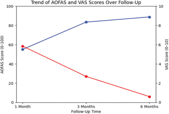
Post-operative pain, quantified using the VAS, demonstrated a consistent and significant downward trajectory throughout the rehabilitation period. At the 1-month follow-up, patients reported moderate discomfort with a mean VAS score of 5.85 ± 0.67. This pain subsided notably by the 3rd month, dropping to an average of 2.70 ± 0.66, and eventually settled at a minimal 0.60 ± 0.50 by the final 6-month evaluation.
Functional recovery was objectively tracked using the AOFAS Ankle-Hindfoot scale. Early in the recovery phase at 1 month, the mean AOFAS score stood at 55.00 ± 1.49. As weight-bearing and physiotherapy progressed, functional capacity improved substantially. By the 3-month mark, the average score reached 83.40 ± 1.67, culminating in an excellent final mean score of 88.80 ± 1.51 at 6 months (Table 1). Table 2 depicts the mean range of motion at final follow-up.
AOFAS ankle-Hindfoot scale over the 6-month post-operative period
| Follow-up interval | Mean AOFAS score | Standard deviation (±) | Clinical interpretation |
|---|---|---|---|
| 1 Month | 55 | 1.49 | Fair/Poor (Early mobilization phase) |
| 3 Months | 83.4 | 1.67 | Good |
| 6 Months | 88.8 | 1.51 | Excellent/Good (Final follow-up) |
AOFAS: American Orthopaedic Foot and Ankle Society
Range of motion at final follow-up
| Affected side | Unaffected side | |
|---|---|---|
| Dorsiflexion | 22.8±4.67 | 27.5±1.82 |
| Plantarflexion | 40.59±5.15 | 46.76±2.55 |
| Inversion | 23.52±4.62 | 29.43±1.43 |
| Eversion | 19.86±3.21 | 24.55±24 |
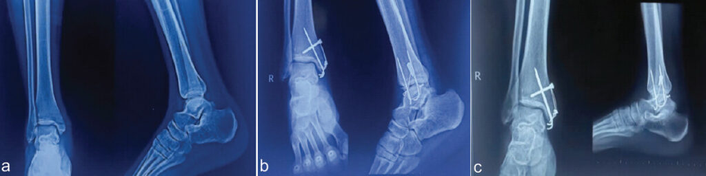
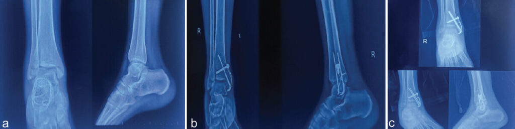
A comparative trend of the AOFAS scale versus the VAS scale is depicted in Fig. 1. Fig. 2 and 3 depict X-rays of clinical cases.
Complications
The overall complication profile for the tension band construct was highly favorable. An uneventful post-operative recovery was observed in 85% of the cohort (17 patients). Minor complications were limited to three individuals. Two patients (10%) experienced localized soft-tissue irritation due to the subcutaneous prominence of the K-wires, though neither required early hardware removal before fracture consolidation. In addition, a single patient (5%) developed a superficial surgical site infection, which was successfully managed and fully resolved with local wound care and a standard course of oral antibiotics (Table 3). Importantly, the cohort experienced zero instances of deep-seated infection, hardware failure, K-wire migration, or loss of reduction.
Complications
| Complication type | Number of patients (n=20) | Percentage | Management/Outcome |
|---|---|---|---|
| No complications | 17 | 85 | Uneventful recovery |
| Implant irritation | 2 | 10 | Conservative; hardware retained until union |
| Superficial infection | 1 | 5 | Resolved with oral antibiotics and local wound care |
| Non-union/Hardware failure | 0 | 0 | N/A |
Discussion
The primary objective of surgical intervention in medial malleolar fractures is the precise anatomical restoration of the articular surface to ensure the congruent mechanical alignment of the ankle mortise. This prospective study was specifically designed to evaluate the efficacy of TBW in achieving these goals. Our findings demonstrate that TBW provides excellent functional restoration, reliable and early fracture consolidation, and a highly acceptable safety profile.
Achieving rigid internal fixation that can withstand the dynamic forces of early mobilization is a cornerstone of modern orthopedic trauma care. The biomechanical advantage of TBW lies in its ability to convert the tensile forces generated across the convex cortex during ankle dorsiflexion into active compressive forces at the fracture interface. This dynamic compression is reflected in our cohort’s excellent radiographic union rate. We observed a 100% union rate with a mean consolidation time of 10.8 weeks. This aligns closely with the existing literature. For instance, in a comparative analysis, Aher et al. reported a mean union time of 12 weeks for their TBW cohort [2]. Conversely, Singh observed an even faster mean union time of 9.2 weeks [14]. Our 10.8-week average sits comfortably within this established spectrum, reinforcing that the figure-of-eight construct provides adequate stability to support reliable biological healing, particularly in the transverse and avulsion-type fracture patterns (Herscovici Type B) that comprised the majority of our cases.
Functional recovery is the most critical metric for patient satisfaction. Our cohort demonstrated a progressive and significant improvement in weight-bearing capacity and joint mobility, culminating in a mean AOFAS score of 88.8 at the 6-month final follow-up. This outcome is highly consistent with recent prospective evaluations. Kan et al. documented a mean AOFAS score of 89.2 in their TBW cohort [15], whereas Aher et al. reported a slightly higher mean of 91 at final evaluation [2]. Interestingly, in a large retrospective series evaluating predominantly complex Herscovici Type C fractures, Batar et al. noted a mean AOFAS score of 84.2 for patients treated with TBW [16]. The marginally higher functional scores in our study can likely be attributed to our strict patient selection and the high proportion of younger, active individuals (mean age 33.05 years) who were highly compliant with aggressive post-operative physiotherapy.
This functional recovery was inversely mirrored by a sharp decline in subjective pain. The mean VAS score in our cohort fell from 5.85 at 1 month to a negligible 0.60 by 6 months. These pain reduction trajectories parallel the findings of Mohan and Prasad, who noted that while immediate post-operative discomfort may vary, mid- and long-term pain resolution is excellent and highly comparable across established fixation techniques [17].
Despite its biomechanical and economic advantages, TBW is not without its specific limitations. The most frequently cited drawback is the subcutaneous prominence of the implant construct, which can lead to soft-tissue irritation. In our study, minor complications were restricted to three cases: Two instances of implant irritation (10%) and one superficial surgical site infection (5%). This complication profile is highly comparable to that reported by Choudhary et al., who observed a 6.7% infection rate in their TBW cohort alongside similar complaints of hardware prominence [18]. Similarly, Singh noted that while implant-related discomfort is slightly more frequent with TBW than with buried screw constructs, it rarely compromises the final functional outcome or necessitates premature hardware removal before complete fracture consolidation [14]. In our series, the instances of hardware irritation were managed conservatively, and no patients required early reoperation, confirming the overall safety of the technique.
Limitations of the study
Several limitations of this study warrant consideration. First, the research was conducted at a single tertiary care center with a relatively small sample size of 20 patients, which may limit the broader generalizability of the findings. Second, the follow-up period was restricted to 6 months. While this duration is sufficient for evaluating primary fracture consolidation and early- to mid-term functional recovery, it restricts the ability to assess late-stage complications. Specifically, the long-term incidence of post-traumatic ankle arthritis and the ultimate rate of hardware removal secondary to delayed implant irritation could not be fully captured within this timeframe. Future multicenter studies featuring larger cohorts and extended longitudinal follow-up are necessary to comprehensively evaluate the long-term survivorship and late complication profile of the tension band construct.
Conclusion
TBW remains a highly effective, reliable, and cost-efficient surgical strategy for the management of displaced medial malleolar fractures. By harnessing the principles of dynamic compression, the technique ensures excellent anatomical reduction, promotes reliable radiographic union at an average of 10.8 weeks, and facilitates robust functional recovery. While minor hardware irritation remains a known, manageable drawback, the absence of major complications combined with a near-complete resolution of long-term pain makes TBW an indispensable technique, particularly for small fracture fragments and in resource-limited or high-volume trauma settings.
Clinical Message
Tension band wiring (TBW) is a highly reliable, cost-effective, and biomechanically robust technique for managing displaced medial malleolar fractures. It is exceptionally well-suited for transverse fracture patterns and small avulsion fragments where screw purchase may be inadequate. By facilitating excellent dynamic compression, TBW promotes high union rates and facilitates early, aggressive rehabilitation. To maximize patient comfort and minimize the need for subsequent hardware removal, surgeons must prioritize meticulous soft-tissue handling and precise bending and impaction of the K-wire ends to reduce subcutaneous prominence.
Conflict of Interest:
Nil
Source of Support:
Nil
Consent:
The authors confirm that informed consent was obtained from the patient for publication of this article
References
- Standring S. Gray's Anatomy: The Anatomical Basis of Clinical Practice. 41st ed. London: Elsevier; 2016. Google Scholar | PubMed
- Aher D, Upasani T, Mehta S, Shah M, Mishra R. Bimalleolar ankle fracture: Comparative study between tension band wiring and cannulated cancellous screw fixation for medial malleolus. Int J Orthop Res 2019;1:4-6. Google Scholar | PubMed
- Mohammed AA, Abbas KA, Mawlood AS. A comparative study in fixation methods of medial malleolus fractures between tension bands wiring and screw fixation. Springerplus 2016;5:530. Google Scholar | PubMed
- Moore KL, Dalley AF, Agur AM. Clinically Oriented Anatomy. 7th ed. Philadelphia, PA: Lippincott Williams and Wilkins; 2014. Google Scholar | PubMed
- Datta S, Bandyopadhyay B, Khadilkar S, Tahir M, Subbaraman K, Hasan GM. Isolated medial malleolar fractures: Current concepts in management. Cureus 2024;16:e75934. Google Scholar | PubMed
- Pankovich AM, Shivaram MS. Anatomical basis of variability in injuries of the medial malleolus and the deltoid ligament. II. Clinical studies. Acta Orthop Scand 1979;50:225-36. Google Scholar | PubMed
- Michelson JD. Ankle fractures resulting from rotational injuries. J Am Acad Orthop Surg 2003;11:403-12. Google Scholar | PubMed
- Ramsey PL, Hamilton W. Changes in tibiotalar area of contact caused by lateral talar shift. J Bone Joint Surg Am 1976;58:356-7. Google Scholar | PubMed
- Lauge-Hansen N. Fractures of the ankle. II. Combined experimental-surgical and experimental-roentgenologic investigations. Arch Surg (1920) 1950;60:957-85. Google Scholar | PubMed
- Rockwood CA Jr, Green DP, Bucholz RW, Heckman JD. Rockwood and Green's Fractures in Adults. 8th ed. Philadelphia, PA: Lippincott Williams and Wilkins; 2015. Google Scholar | PubMed
- Georgiadis GM, White DB. Modified tension band wiring of medial malleolar ankle fractures. Foot Ankle Int 1995;16:64-8. Google Scholar | PubMed
- Ostrum RF, Litsky AS. Biomechanical comparison of medial malleolar fixation techniques. Orthopedics 1992;15:947-51. Google Scholar | PubMed
- Tailor D, Kapale J, Patil VH. Comparative study between CC screw and tension band wiring used in medial malleolus fractures. Int J Orthop Sci 2019;5:866-72. Google Scholar | PubMed
- Singh VK. A comparative study of tension band wiring and cannulated screw fixation for medial malleolar fractures. Int J Orthop Sci 2019;5:805-9. Google Scholar | PubMed
- Karthik Aiyanna NC, Deekshith Shetty K, Swamy M, Kailath KJ. Comparative analysis of tension band wiring and cannulated cancellous screws for displaced medial malleolus fractures: A prospective study. Int J Res Orthop 2025;11:314-8. Google Scholar | PubMed
- Batar S, Karataş ME, Oruç MM, Kemah B, Başak F. Comparison of fixation methods in medial malleolus fractures. Acta Orthop Traumatol Turc 2024;35:281-7. Google Scholar | PubMed
- Mohan DR, Prasad SR, Prabhakar P. Comparative study of tension band wiring and malleolar screw fixation for the treatment of medial malleolus fracture. Int J Orthop Sci 2021;7:455-9. Google Scholar | PubMed
- Choudhary V, Jangir R, Singh Y, Gomes N. A prospective comparative study of tension band wiring and malleolar screw fixation for medial malleolar fractures. Int J Life Sci. Biotechnol Pharma Res 2024;13:432-7. Google Scholar | PubMed
© 2026 Journal of Orthopaedic Case Reports - Published by Indian Orthopaedic Research Group
About the Authors
-

Dr. Vishal Champawat
-

Dr. Deepak Shah
-

Dr. Manish Patil
-

Dr. Suresh Uikey
-

Dr. Deepak Singh Patel
Related Articles in Journal of Orthopaedic Case Reports
 June 1, 2026 Malleolar Stress Fracture as the First Manifestation of Undiagnosed Crohn’s Disease: A Case Report
 January 1, 2026 Incidental Osteopoikilosis in an Ankle Fracture Patient: Case Report and Literature Review
 August 10, 2023 Open Displaced Fracture of Acromion with Associated Complete Supraspinatus Tear: A Rare Case Report
 February 10, 2021 Symptomatic Os Subtibiale Following Injury: A Case Report of Failed Conservative Treatment, Leading to Differential Diagnosis Made Intraoperatively