


Introduction
Shoulder joint is vulnerable for dislocation due to its shallow glenoid and thin, lax capsule. At any given time only one-fourth of the humeral head articulates with the glenoid. Shoulder instability and its treatment were described in ancient times approximately 4500–5000 years ago. The shoulder instability is first described by the Greek and Egyptian physicians in humankind’s oldest book. The most detailed description came from the Hippocratic texts of 500 BC [1].
The shoulder instability is well described by H.F. Moseley, who has a good section on the historical aspects of management of shoulder instability, and says that in a certain percentage of cases the middle glenohumeral ligament is attached to the neck of the scapula some distance from the fibrous labrum and that an anterior pouch exists of sufficient size to permit the anterior dislocation of the head [2]. The shoulder instability must have been experienced as post-traumatic pathology throughout human history, and evidence of shoulder dislocations is found in archaeological and paleopathological examinations of the shoulder for several 1000 of years. The shoulder instability is one of the most common disorders of the glenohumeral joint. It has gained extra mobility at the expense of its stability. The instability can be experienced in many ways; the patient experiences apprehension, recurrent subluxation, and dislocations.
The stabilizers are based on either of the two basics active (dynamic) or passive (static) mechanisms or stabilizers. The active mechanism includes the rotator cuff and biceps tendon, while the passive mechanism includes the capsulo-ligamentous structures that are the glenoid labrum, glenohumeral ligaments and fibrous capsule [3]. In an anterior dislocation, the arm is an abducted and externally rotated position. In the externally rotated position, the posterosuperior aspect of the humeral head abuts and drives through the anteroinferior aspect of the glenoid rim. This can damage the humeral head, glenoid labrum, or both. Due to the first episode of dislocation, there is stretching of capsule, or defect, glenohumeral ligaments causing the chances of recurrence. Due to this, there is an associated humeral head compression fracture or bone defect is described as a Hill-Sach’s lesion. If large enough, it can lead to locked dislocations that may require open reduction. The glenoid labrum is a fibrocartilaginous structure that rings the circumference of the glenoid fossa. Bankart lesions are injuries to the anteroinferior glenoid labrum complex and the most common capsulo-labral injury. A “bony Bankart” lesion refers to an associated fracture of the glenoid rim. These capsulo-labral lesions are risk factors for recurrent dislocation [4,5].
The strength or integrity of the anterior capsule is so important, as it gets damaged gradually during the dislocation episodes, makes painful and tensed on abduction and external rotation, causing a fear of getting a dislocation, which gives the anterior apprehension test positive.
Hill-Sach’s lesions are again described based on off-tracking or on- tracking based on the glenoid track and Hill-Sach’s interval. The engaging Hill-Sach’s lesion is more difficult to treat.
There are several methods of closed reduction described for acute dislocation. There are also several surgical methods described based on open and arthroscopic repairs. Open repairs and glenoid reconstruction surgeries have described with are based on the bony defects of both glenoid and posterior humeral head loss, and multiple surgeries are described based on bone defect or capsular defect. Capsular laxity is addressed either repairing the static stabilizers or strengthening static stabilizers using dynamic stabilizers.
Several of the open procedures have become obsolete after the minimal invasive arthroscopic repairs. However, this old method has still got it importance and good results in stabilizing the shoulder joint and preventing recurrences of dislocation, which is called the Boytchev Procedure, which many of them have not been heard of Boytchev is a Bulgarian orthopaedician who uses conjoined tendons of the coracobrachialis and short head of the biceps, together with the tendon of the pectoralis minor and the detached tip of the coracoid process is rerouted underneath the subscapularis muscle and reattached to the coracoid process at its anatomical location with a screw. This procedure is modified, and only the conjoined tendon is used for rerouting. The subscapularis tendon is functionally shortened due to this rerouting of the conjoined tendons beneath it, holding the humeral head posteriorly in the position of abduction and external rotation (ER). Subscapularis tightly adheres to the conjoined muscle tendons and is solidly scarred. The core concept of this procedure is that a muscular sling anterior to the joint is used to tighten the structures and give stability without disturbing the joint and maintain the proprioception at the same time.
Materials and Methods
Study design
This is a Cross-sectional Observational Study with a Study Period of 18 months (August 2022–February 2024) conducted at our institute, ESIC Medical College and Hospital, Sanatnagar (IEC Approval No – ESICMC/SNR/IEC/S0170/07–2022). The sample size is 30 patients. It is calculated by using n = Z2 * p(1-p)/d2 (Z = 1.96, p-prevalence=50%, d-0.05, with 10% dropout). The inclusion criteria include patients with recurrent anterior shoulder dislocations (>3 episodes) and who belong to the age group 18–50 years. Patients with 1st-time dislocation (acute), coracoid fractures, chronic unreduced dislocations, previous failed shoulder surgeries, glenohumeral arthritis, epilepsy, neuromuscular disorders, psychiatric illness and bony Bankart’s lesions are excluded.
Preoperative evaluation
-
Patient history, pain, and functional status were documented
-
Subscapularis muscle tone was palpated through the anterior axillary fold
-
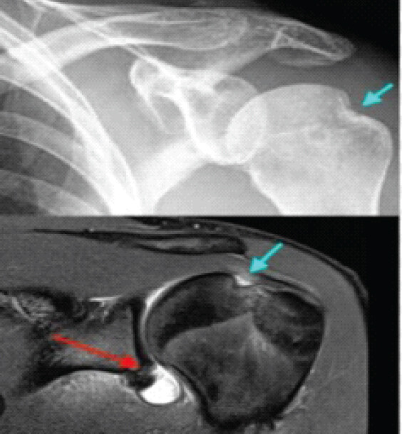
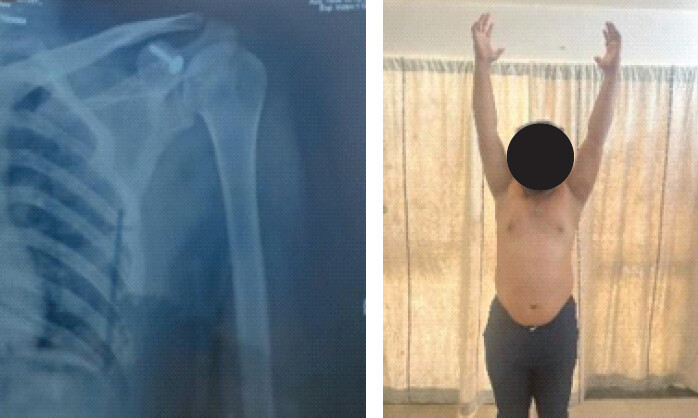
Shoulder instability/Ligamentous laxity was assessed by Oxford shoulder instability score (OSIS), Apprehension test and radiological evaluation (X-ray, magnetic resonance imaging and computed tomography evaluation) (Fig. 1).
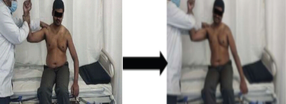
The apprehension test was done by the shoulder was abducted to 90° and externally rotated; the feeling of apprehension indicated a positive result (Fig. 2).
Surgical procedure
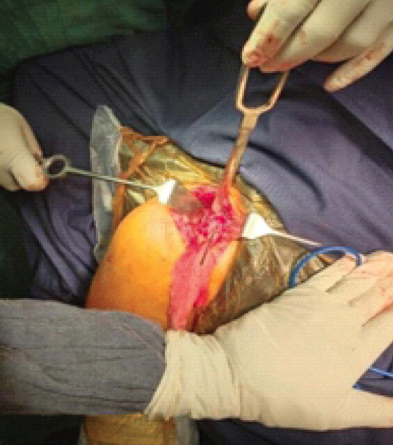
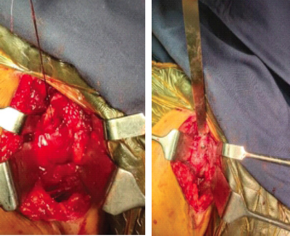
The position used was the beach-chair position under general anaesthesia. Patients were placed in supine position on the operating table with a sandbag placed under the operative shoulder. The deltopectoral approach was used, where there is a standard 10 cm incision made starting proximal to the tip of the coracoid process extending distally along deltopectoral groove. Cephalic vein was identified and retracted along with deltoid muscle laterally, and pectoralis major was retracted medially, exposing the tip of the coracoid process with its attached conjoint tendon. Pectoralis minor muscle was separated from the coracoid process medially, leaving behind coracobrachialis and the short head of biceps attached to the coracoid process. A hole was predrilled and tapped from the anterior part of the coracoid process along the axis of the coracoid. The coracoid process was osteotomized along with the attached coracobrachialis and short head of biceps muscle at a point 2 cm from the tip of the coracoid process (Fig. 3). The conjoint tendon was elevated till the lower border of the subscapularis muscle, taking care to protect the musculocutaneous nerve and anterior circumflex vessels. The shoulder was rotated externally, and the subscapularis muscle was identified. The lower border of subscapularis was identified by the leash of blood vessels. A tunnel was created between the subscapularis and the capsule by divided the subscapularis at the upper 2/3rd and lower 1/3rd junction, as the subscapular vessels lie in the lower 1/3rd of the muscle. The detached tip of the coracoid process with the conjoined tendons was passed through the tunnel underneath the subscapularis (Fig. 4). The tip of the coracoid process was reattached to its original site with a 4 mm cannulated cancellous screw with watcher (Fig. 5). The wound was closed in layers and arms were strapped to the chest in full internal rotation. All patients were given Velpeau strapping in the immediate post-operative period. At 2 weeks of the post-operative period, pendulum shoulder exercises were started after removal of the sutures. Follow-up was done postoperatively at 4 weeks, 12 weeks and 24 weeks. Clinical and Functional Outcome was evaluated using the Burkhead and Rockwood criteria and OSIS, scale (At 6 months). Radiologically standard shoulder X-ray to look for union, screw backout and breakage was done at regular follow-up (Fig. 5a and b).
Statistical analysis
Data were entered into an Excel sheet, and statistical analysis of data was done using the Statistical Package for the Social Sciences version 21. Continuous data were reported as mean ± SD) and percentages.
Results
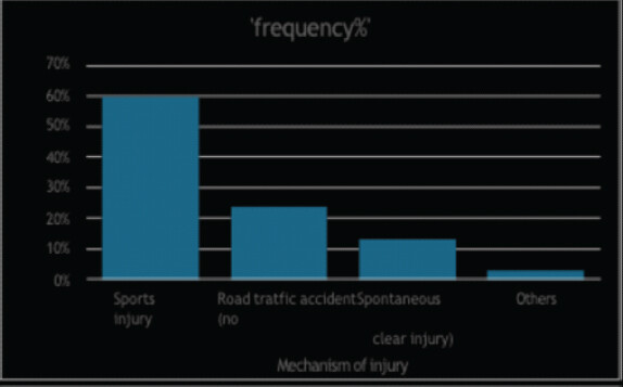
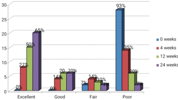
Among 30 patients, 50% of them belong to the age group <25 years, 33% belonged to the 26–40 years of age group, and 5% belonged to the more than 41 years of age group, with male predominance (87%). The age group at the initial episode of dislocation was studies and observed that 30% of initial dislocations occurred at <15 years of age, 67% at the age of 16–30 years and 3% at more than 31 years of age. The number of dislocations that occurred in 27% of the study subjects was 6–10 dislocations, and 11–15 dislocations occurred in 73% of the study subjects (Table 1). The most common mode of injury was found to be sports injury (60%) followed by road traffic accident (24%) with all of them being Right upper limb dominant (67%) than non-dominant (33%) (Fig. 6). The pre-operative OSIS score was observed to be having severe instability in 70% of them and 30% of them having moderate instability which were followed postoperatively and after 24 weeks, 86% of them improved based on OSIS scale (Tables 2 and 3). The functional outcome was measured using Burkhead and Rockwood criteria which was 93% of the patients who had poor outcome immediate postoperatively, which gradually reduced to 7% at 4 weeks, 12 weeks and 24 weeks. About 7% of the patients had fair functional outcome by 24 weeks. About 20% of the patients had good functional outcome by 24 weeks and 65% of the patients had excellent functional outcome by 24 weeks (Table 4, Fig. 7). There were no incidences of re-dislocation during the 18-month follow-up, one case showed screw loosening, two cases had hypertrophic scar and none of them had infection, coracoid fracture or musculocutaneous nerve injury (Table 5).
6–10 dislocations occurred in 27% of the study subjects and 11–15 dislocations occurred in 73% of the study subjects
| Number of dislocations | Frequency (n%) |
|---|---|
| 3–5 | 0 (0) |
| 6–10 | 8 (27) |
| 11–15 | 22 (73) |
Preoperatively, hypermobility was measured using the OSIS scale, revealing that a moderate level of instability was present in 30% of the study subjects and a severe level of instability was present in 70% of the study subjects
| OSIS | Frequency (n%) at pre-operative |
|---|---|
| Moderate | 9 (30) |
| Mild | 0 (0) |
| Severe | 21 (70) |
OSIS: Oxford shoulder instability score
Postoperatively, hypermobility was measured using the OSIS scale reveals that at 24 weeks, 86% of the patients have improved shoulder stability
| OSIS | Frequency (n%) at 24 weeks post-operative |
|---|---|
| Moderate | 4 (14) |
| Mild | 26 (86) |
| Severe | 0 (0) |
OSIS: Oxford shoulder instability score
Post-operative functional outcome using Burkhead and Rockwood criteria showing 93% of the patients had a poor outcome postoperatively, which gradually reduced to 7% at 4 weeks, 12 weeks, and 24 weeks. About 7% of the patients had a fair functional outcome by 24 weeks. About 20% of the patients had a good functional outcome by 24 weeks, and 65% of the patients had an excellent functional outcome by 24 weeks
| Functional outcome | 0 weeks (%) | 4 weeks (%) | 12 weeks (%) | 24 weeks (%) |
|---|---|---|---|---|
| Excellent | 0 (0) | 8 (27) | 15 (50) | 20 (65) |
| Good | 0 (0) | 4 (14) | 6 (20) | 6 (20) |
| Fair | 2 (7) | 4 (14) | 3 (10) | 2 (7) |
| Poor | 28 (93) | 14 (35) | 6 (20) | 2 (7) |
Post-operative complications – screw loosening/breakage was 3% and hypertrophic scar was 7%
| Complications | Frequency (n%) |
|---|---|
| Recurrence | 0 |
| Infection | 0 |
| Musculocutaneous nerve injury | 0 |
| Coracoid fracture | 0 |
| Screw loosening/breakage | 1 (3) |
| Hypertrophic scar | 2 (7) |
Discussion
Shoulder instability significantly affects the young and middle-aged population, impairing quality of life. Strengthening dynamic and static stabilizers enhances shoulder stability. About half a century ago, Boytchev, a Bulgarian orthopaedic surgeon, described a surgical technique for recurrent anterior dislocation of the shoulder. This procedure involves rerouting of the detached tip of the coracoid process with its attached conjoined tendon (short head of biceps and coracobrachialis) along with the pectoralis minor muscle deep to subscapularis and reattaching to its anatomical location [6]. Later, Conforty modified this procedure by rerouting the conjoined tendon of the short head of biceps and coracobrachialis only. The modified Boytchev procedure is biomechanically sound and acts by two possible mechanisms:
-
Dynamic muscular sling effect
-
Improved shoulder joint proprioception.
Treatment for recurrent anterior dislocation of the shoulder should provide an active corrective force that pushes the humeral head into the glenoid fossa, especially in the position of 90° abduction and ER.
The Boytchev procedure achieves these objectives in 3 ways:
1. Increased muscle bulk of subscapularis, coracobrachialis, and short head of biceps augments the bracing effect over the anteroinferior aspect of the shoulder joint and thus prevents anterior dislocation
2. Muscle bellies of two strong muscles are rerouted to lie more directly in contact with the joint capsule and humeral head so that their active contractions exert a strong control on the anterior exertion of the humeral head. Thereby, balancing the deforming forces of dislocation
3. Increases the pressure between the humeral head and subscapularis tendon and thus, increases proprioceptive stimuli in the subscapularis tendon which accelerate the protective reflex needed to prevent shoulder dislocation [7].
Biomechanical study of Halder et al. and cadaveric study of Jiang et al. provide valid scientific reason for the prevention of recurrence after the modified Boytchev procedure. In their study, the anterior, lateral and posterior deltoid, supraspinatus, short head of biceps, coracobrachialis and long head of triceps from ten cadaveric shoulders were tested in 0°, 30°, 60°, and 90° of glenohumeral abduction. A constant inferior force of 15 N was applied to the humerus. The tendons were loaded sequentially in proportion to their respective muscle’s cross-sectional area. Translations of the humeral head on the glenoid were recorded with a 3-Space™ tracking device. The results were found that the lateral deltoid (8.2 mm and SD 4.8 mm) was potentially most effective in superior translation of the humeral head, followed by the posterior deltoid (7.7 mm and SD 4.8 mm). The coracobrachialis and short head of biceps had considerable capability to translate the humeral head superiorly (2.8 mm and SD 1.3 mm), while the supraspinatus showed the weakest effects (1.3 mm, SD 0.5 mm) [8,9]. A study by Shibata et al. revealed that the efficacy of the modified Boytchev procedure was this increased pressure increases proprioceptive stimuli in the subscapularis tendon and thus accelerates the protective reflex needed to prevent shoulder dislocation [10].
In our study, the majority of the recurrent shoulder dislocations were due to injuries in sports (60%), which was in accordance with the study done by Zamora-Navas et al. and his team, where the original cause of dislocation was a sports injury in 11, a traffic accident in 5, an accidental fall in 9, and a minor injury in two. The level of physical activity before the first dislocation was high in 16 patients (regular sports activity in 13, manual work in 3) and medium in 9 (3 housewives, 6 sedentary professions). All patients were able to return to their normal activities [11].
Garg et al., and his team studied, and they observed that in terms of recurrence, range of motion, Visual Analog Scale Score, modified American shoulder and elbow surgeons score, and single assessment numeric evaluation score, none of the patients out of 38 of them had recurrence, nor anyone showed a positive load and shift test, sulcus test or any signs of hyperlaxity [12]. Similarly, in our research, pre-operative OSIS score was observed to be having severe instability in 70% of them, which was improved over 24 weeks to mild instability in 86% with no recurrence.
A study done by Dr. Sundar et al. out of 30 patients treated by the modified Boytchev procedure, 93% of patients had excellent/good outcome, and two patients had fair outcome due to traumatic dislocation after 1 year of post-operative period, which led to periarthritis of the affected shoulder and restricting abduction and ER movements [13]. In our analysis, the functional outcome was studied using Burkhead and Rockwood criteria; 65% (20 patients) of them showed excellent outcome, 20% (6 patients) of them showed good outcome, fair outcome was seen in 7% (2 patients) of them, and poor outcomes were seen in another 7% (2 patients).
Kumar et al. and his team demonstrated that there was no evidence of any post-operative infection or any radiological evidence of loosening, migration of the coracoid screw or any glenohumeral arthritis on subsequent follow-up in any of his patients [3]. There were no recurrences of dislocation, infection, musculocutaneous nerve injury and coracoid fracture in our research. Chandra et al. and his team observed that two patients had fragmentation of the coracoid process, which was fixed with non-absorbable suture [14]. There was screw loosening in one case and a hypertrophic scar in two cases.
Even though it is an old open technique, it has its own importance, being an easy, quick procedure and also easy to learn. This procedure does not need too much of instrumentation, which is costly, especially in arthroscopic procedures, can be done with basic general instrumentation, with only one cannulated cancellous screw, which is cheap and affordable. This has a good learning curve; even any orthopaedician can perform this surgery without arthroscopic experience, which has less post-operative pain, early rehabilitation, and good functional outcome.
Conclusion
This study has demonstrated with excellent functional outcomes with no recurrence, no infection, no nerve injury and good union of the coracoid process. This procedure gives confidence and stability in an abnormal or uncomfortable position of the shoulder (90° abduction and ER), which is vulnerable for re-dislocation. Modified Boytchev procedure fulfills most of the criteria of an ideal procedure with excellent functional outcome for recurrent anterior dislocation of shoulder, which is an effective procedure, technically simple, and does not require expert skills or any sophisticated instruments and can be performed by all orthopaedic surgeons where basic operative facility is present. It has an easy learning curve.
Clinical Message
Not only evolving into new skills and techniques (which are successful and great) but digging the past techniques can also give us great insights about the anatomy and the technique, which helps and guides us in less infrastructure and remote settings and be helpful for a basic orthopaedic surgeon who may be less skilled and not used to the new techniques.
Conflict of Interest:
Nil
Source of Support:
Nil
Consent:
The authors confirm that informed consent was obtained from the patient for publication of this article
References
- Te Slaa RL. The Acute First-Time Anterior Shoulder Dislocation (AFASD) [Thesis, Universiteit van Amsterdam] 2003 Google Scholar | PubMed
- Moseley HF. The basic lesions of recurrent anterior dislocation Surg Clin N Am 1963;43:1631-4. Google Scholar | PubMed
- Kumar S, Khan NA, Tandan BS. Management of recurrent anterior shoulder dislocation by modified boytchev procedure- our experience J Bone Joint Dis 2021;36:23-6. Google Scholar | PubMed
- Pak T, Kim AM. Anterior glenohumeral joint dislocation In: StatPearls Treasure Island, FL StatPearls Publishing 2025. Available from: https://www.ncbi.nlm.nih.gov/books/nbk557862. [Last accessed on 15 Marc h2026]. Google Scholar | PubMed
- Symeonides PP. The significance of the subscapularis muscle in the pathogenesis of recurrent anterior dislocation of the shoulder J Bone Joint Surg Br 1972;54:476-83. Google Scholar | PubMed
- Boytchev B. Treatment of recurrent shoulder instability Minerva Orthop 1951;2:377-9. Google Scholar | PubMed
- Conforty B. The results of the Boytchev procedure for treatment of recurrent dislocation of the shoulder Int Orthop 1980;4:127-32. Google Scholar | PubMed
- Halder AM, Halder CG, Zhao KD, O'Driscoll SW, Morrey BF, An KN. Dynamic inferior stabilizers of the shoulder joint Clin Biomech (Bristol) 2001;16:138-43. Google Scholar | PubMed
- Jiang LS, Cui YM, Zhou ZD, Dai LY. Stabilizing effect of the transferred conjoined tendon on shoulder stability Knee Surg Sports Traumatol Arthrosc 2007;15:800-5. Google Scholar | PubMed
- Shibata Y, Midorikawa K, Ogata K, Izaki T. Modified Boytchev and its combined operation for traumatic unstable shoulder Shoulder Joint 1999;23:327-1. Google Scholar | PubMed
- Zamora-Navas P, Borras Verdera A, Porras Garcia J, Padilla Marquez A, Linares P. Long-term results of the Boytchev procedure for the treatment of recurrent dislocation of the shoulder Acta Orthop Belg 2001;67:233-5. Google Scholar | PubMed
- Garg AK, Ayan S, Keshari V, Kundu D, Mukhopadhyay KK, Acharyya B. Modified Boytchev procedure for treatment of recurrent anterior dislocation of shoulder Indian J Orthop 2011;45:336-40. Google Scholar | PubMed
- Suryakumar S, Ram GG. Functional outcome of modified Boytchev procedure in recurrent shoulder dislocation Int J Orthop Sci 2018;4:225-7. Google Scholar | PubMed
- Chandra R, Sharma VK, Mahajan S, Bansal M, Arora S. Modified Boytchev procedure for recurrent anterior dislocation of shoulder J Orthop Surg (Hong Kong) 2014;22:204-8. Google Scholar | PubMed
© 2026 Journal of Orthopaedic Case Reports - Published by Indian Orthopaedic Research Group
About the Authors
-

Dr. Banoth Devandra Naik
-

Dr. Neerudi Sharon Rose
-

Dr. Sri Ravindranath Vutukuru
-

Dr. Velugu Prashanth
-

Dr. Venu Madhav Vuthpala
-

Dr. Mettu Anil Kumar
Related Articles in Journal of Orthopaedic Case Reports
 July 1, 2025 Fourteen Years of Living with Instability: A Case of Chronic Recurrent Anterior Shoulder Dislocation in a Middle-Aged Athlete
 November 10, 2022 Mallet Finger Caused by a Villonodular Synovitis: A Case Report and Literature Review
 April 1, 2026 Distal Femoral Replacement in Young Adults with Traumatic Comminuted Distal Femur Fractures: A Report of Two Cases
 July 10, 2024 The Curious Case of the Flying Femoral Head – A Neck of Femur Fracture-Dislocation with Intrapelvic Lie of the Fractured Femoral Head