


Introduction
Large talar bone cysts can cause pathologic fracture and damage to the articular cartilage that results in persistent swelling and pain of the subtalar joint and ankle joint [1]. When conservative treatment fails, surgery can be considered. There are many surgical approaches to treat symptomatic bone cysts [1,2,3,4,5,6,7]. Since majority of the talar surface is covered with articular cartilage, open surgical approaches to the talar bone cysts frequently require extensive soft tissue dissection, malleolar osteotomy, and sometimes damage to the talar articular cartilage [1,2]. Arthroscopic treatment of talar bone cyst is a good alternative, minimally invasive approach to reduce the surgical trauma and eliminate the need of osteotomy [1,2,3,4,5].
Subchondral cysts develop due to the valve mechanism of the damaged cartilage which allows unidirectional intrusion of fluid from the joint space into the subchondral bone [8]. The stress shielding by pressurized fluid and osteocyte death may cause cyst growth [9]. As a result of a small defect in the subchondral plate, huge subchondral bone cysts can form [8]. In some cases, normal cartilage adjacent to the osteochondral lesion may need to be removed to achieve adequate visualization and debridement of the cyst wall during the arthroscopic procedures [3]. Sometimes, after cyst decompression and bone grafting, the cyst might become uncontained and the bone graft of the cyst can drop into the ankle joint becoming an intra-articular loose body which can lead to persistent pain and arthritis [3]. If the cyst is decompressed from articular surface, restoring the cartilage surface is almost impossible. The posterior talar cysts can also be decompressed by arthroscopy [10]. The present technical note describes the arthroscopic approach of debridement and bone grafting of talar bone cysts without any disruption of the normal cartilage surface.
Case Report
Case details
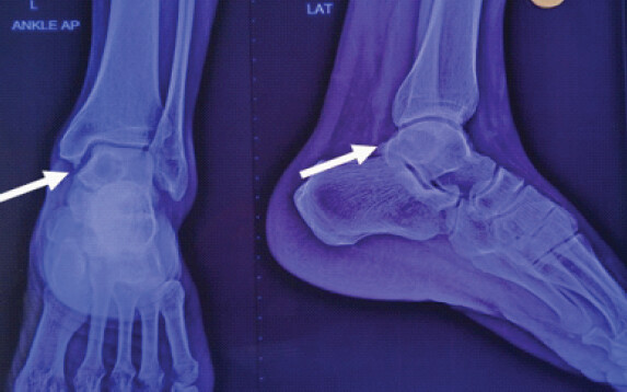
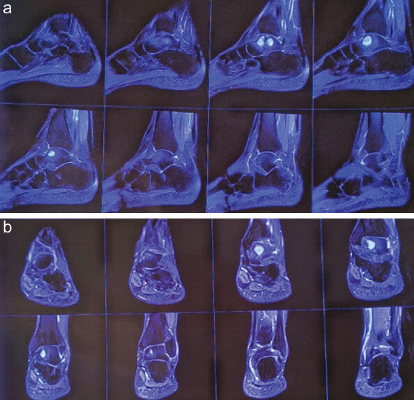
A 34-year-old male complaining of pain in the ankle while walking since 3 months. There is no history of trauma. Patient has no rest pain. X-ray ankle anteroposterior and lateral shows a cystic lesion in the talus, as shown in Fig. 1. An magnetic resonance imaging (MRI) was done which shows a cyst around 35 × 18 mm × 14 mm in the anteromedial talus, as shown in Fig. 2a and b. We preferred MRI over computed tomography scan as MRI gives the status of cartilage of tibiotalar joint along with cyst location and type of cyst also. Our initial plan was to do a diagnostic arthroscopy to see the cartilage status. If the cyst is accessible through arthroscopic portals, cyst decompression, curettage and bone grafting under arthroscopy was planned. If it is not accessible, an open decompression and bone grafting was the stendy procedure.
Procedure
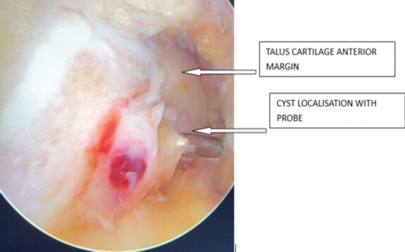
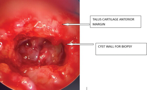
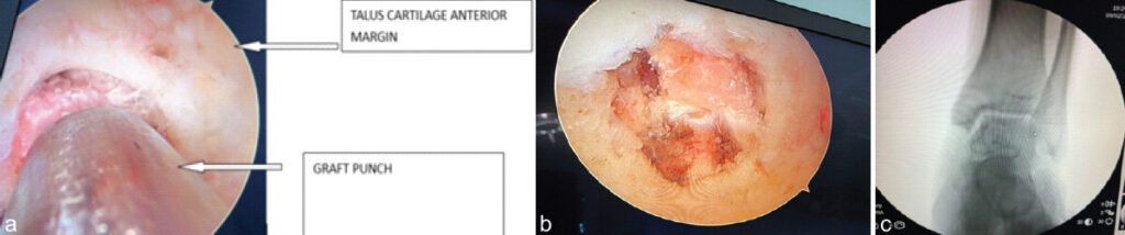
Patient under spinal anesthesia, in supine position with ankle overhanging the table as in standard ankle arthroscopy procedure. Bony landmarks are marked, dorsalis pedis artery is marked, and then, tourniquet was inflated. A 4.0-mm 30 arthroscope or a 2.7-mm 30 arthroscope can be used for the anterior talar bone cyst endoscopy. Here, we used a 4 mm 30 arthroscope. A thigh tourniquet is applied to provide a bloodless operative field. Joint inflation is done with a needle in anteromedial portal area. After joint is sufficiently inflated, anteromedial portal is made at the soft spot located between tibialis anterior and medial malleolus by nick and spread method. Then anterolateral portal is made in the soft spot between peroneus tertius and lateral malleoli under vision. A quick diagnostic arthroscopy was done with scope in AM portal, cartilage was found to be normal. Then, anteromedial talus distal to the cartilage border was visualized. An abnormal spot was seen, as shown in Fig. 3, which was corresponding with location of cyst. With a probe in AL portal, this spot was poked, a light green jelly liquid came out which confirmed the cyst. We switched to a dry arthroscopy with carbon dioxide to get good tissue for biopsy. The wall of the cyst was scraped and all the tissue was sent for biopsy, as shown in Fig. 4. After biopsy, we switched back to saline arthroscopy. A thorough curettage was done. Ipsilateral iliac crest bone graft was harvested. An accessory portal was made right opposite the cyst making sure no neurovascular structure which is injured and the cyst was packed with this graft carefully through accessory portal using small graft punch to neutralize the dead space, as shown in Fig. 5a and b. Cyst filled with graft was confirmed under C-arm, as shown in Fig. 5c. Ankle dorsiflexion and plantar flexion was done to rule out any impingement of graft with anterior tibial plateau. Portals were closed and below knee plaster of Paris was applied.
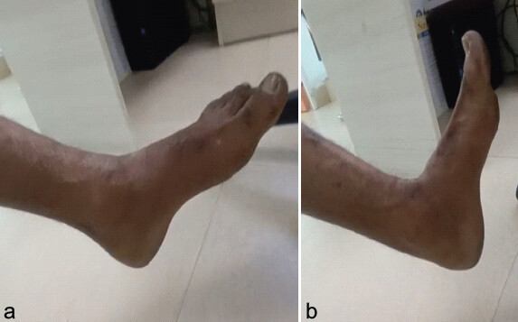
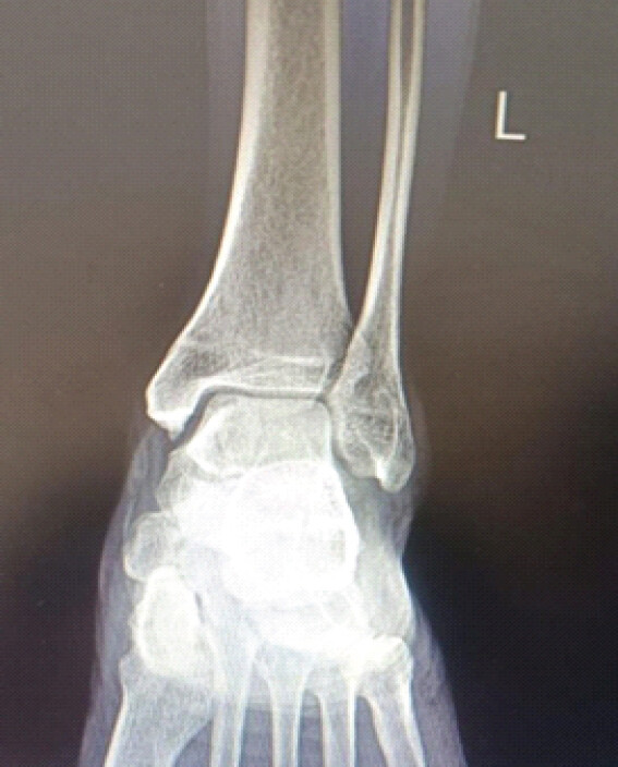
The biopsy reported as chronic inflammation. At 1 year follow-up, patient is walking pain-free with full range of motion, as shown in Fig 6a and b. X-ray shows good consolidation, as shown in Fig. 7.
Discussion
As compared with fluoroscopic-guided open curettage and bone grafting of the talar cyst, the arthroscopic approach can have better assessment of completeness of debridement and can manage the associated osteochondral lesion [3,6]. The advantages of this minimally invasive technique include better cosmetic result, minimal soft-tissue dissection, fewer wound complications, clear visualization of the cyst, and preservation of the articular cartilage. The potential risk of this technique includes iatrogenic fracture of the talus, injuries to the superficial peroneal nerve, recurrence of the cyst, and dropping of the graft into the ankle joint. If required, accessory portals can be made as per requirement for easy access. The portals should be spaced out to avoid crowding of instruments and incomplete visualization and curettage.
It is not suitable for bone cysts located just underneath a large osteochondral lesion, as the cyst can be approached through the osteochondral lesion. It is not suitable in cases of significant osteoarthrosis of the ankle.
Conclusion
Arthroscopic cyst decompression is a good alternative to open decompression with less morbidity and speedy recovery. Patient selection is the key factor. This procedure is indicated for talar bone cysts associated with small osteochondral lesions or intact articular surface but contraindicated if the cyst is adjacent to a large osteochondral lesion or there is significant osteoarthritis of the ankle joint.
Conflict of Interest:
Nil
Source of Support:
Nil
Consent:
The authors confirm that informed consent was obtained from the patient for publication of this article
Clinical Message
Arthroscopic cyst decompression is less invasive and has minimal chances of stiffness. Wherever possible choose arthroscopic decompression over open surgery.
How to Cite this Article
Yadoji HK, Gottemukkala AR, Juvvadi V, Yadoji GK. All Arthroscopic Decompression and Bone Grafting of Cyst in the Talus. Journal of Orthopaedic Case Reports 2026 October;16(10): 243-246.
References
- Zhu X, Yang L, Duan X. Arthroscopically assisted anterior treatment of symptomatic large talar bone cyst. J Foot Ankle Surg 2019;58:151-5. [Google Scholar] | [PubMed]
- Lui TH. Arthroscopic bone grafting of talar bone cyst using posterior ankle arthroscopy. J Foot Ankle Surg 2013;52:529-32. [Google Scholar] | [PubMed]
- Lui TH. Endoscopic curettage and bone grafting of huge talar bone cyst with preservation of cartilaginous surfaces: Surgical planning. Foot Ankle Surg 2014;20:248-52. [Google Scholar] | [PubMed]
- Lui TH. Arthroscopic curettage and bone grafting of bone cysts of the talar body. Arthrosc Tech 2017;6:e7-13. [Google Scholar] | [PubMed]
- Li CC, Lui TH. Management of bone cyst of talar body by endoscopic curettage, nanofracture, and bone graft substitute. Arthrosc Tech 2021;10:e1985-93. [Google Scholar] | [PubMed]
- Cebesoy O. Intraosseous ganglion of the talus treated with the talonavicular joint approach without exposing the ankle joint. J Am Podiatr Med Assoc 2007;97:424-7. [Google Scholar] | [PubMed]
- Li S, Lu R, Zhang J, Tao H, Hua Y. Outcomes of arthroscopic bone graft transplantation for Hepple stage V osteochondral lesions of the talus. Ann Transl Med 2021;9:884. [Google Scholar] | [PubMed]
- Van Dijk CN, Reilingh ML, Zengerink M, Van Bergen CJ. Osteochondral defects in the ankle: Why painful?. Knee Surg Sports Traumatol Arthrosc 2010;18:570-80. [Google Scholar] | [PubMed]
- Cox LG, Lagemaat MW, Van Donkelaar CC, Van Rietbergen B, Reilingh ML, Blankevoort L . The role of pressurized fluid in subchondral bone cyst growth. Bone 2011;49:762-8. [Google Scholar] | [PubMed]
- van Dijk CN, Scholten PE, Krips R. A 2-portal endoscopic approach for diagnosis and treatment of posterior ankle pathology. Arthroscopy 2000;16:871-876. [Google Scholar] | [PubMed]
© 2026 Journal of Orthopaedic Case Reports - Published by Indian Orthopaedic Research Group
About the Authors
-

Hari Krishna Yadoji
-

Ashok Raju Gottemukkala
-

Vasudeva Juvvadi
-

Guru Krishna Yadoji
Related Articles in Journal of Orthopaedic Case Reports
 October 1, 2026 Arthroscopic Piecemeal Excision of a Large Intra-articular Infrapatellar Osteochondroma of the Knee: A Case Report
 October 1, 2026 Arthroscopic Posterior Cruciate Ligament Tibial Spine Avulsion Fixation Using a Suture Pull-Through Single-Tunnel Technique
 September 1, 2026 Fixed Flexion Deformity due to Displaced Hoffa Fracture: A Case Report
 September 1, 2026 Arthroscopic ACL Tibial Spine Avulsion Fixation Using Suture Pull-Through Double-Tunnel Technique