


Introduction
Posterior malleolar fractures represent a distinct subset of ankle injuries that have gained increasing clinical attention due to their biomechanical importance and potential impact on long-term outcomes. Traditionally, posterior malleolar involvement was considered clinically insignificant unless associated with large fracture fragments or obvious ankle instability. However, recent studies have demonstrated that the posterior malleolus plays a critical role in ankle and syndesmotic stability [1], even when the fracture fragment is small or minimally displaced.
Most posterior malleolar fractures occur in conjunction with bi- or trimalleolar ankle fractures [2] and are commonly associated with rotational mechanisms of injury. Isolated posterior malleolar fractures without concomitant medial or lateral malleolar involvement are relatively uncommon and may be underrecognized. These injuries can present with mild or rapidly improving symptoms, an intact ankle mortise, and subtle radiographic findings, which may lead to potential underrecognition or delayed diagnosis [3].
Radiographic detection of posterior malleolar fractures on plain films can be challenging due to overlapping anatomy and minimal cortical disruption. As a result, scrutiny of lateral ankle radiographs is essential, particularly in patients with persistent swelling or a history of rotational trauma [4]. Computed tomography may be considered when plain radiographs are inconclusive or when fracture morphology requires further characterization [5]. While operative fixation is indicated in select cases to restore joint congruency and syndesmotic stability [6], stable non-displaced fractures are generally managed with conservative treatment. Recognition of these injuries is critical to prevent delayed diagnosis and potential long-term sequelae such as chronic pain, instability, or post-traumatic arthritis [7].
This case report describes an isolated, non-displaced posterior malleolus fracture sustained during recreational ice skating, notable for early complete symptom resolution and an intact ankle mortise, that was successfully managed with conservative treatment. The case highlights a clinical management pitfall and underscores the importance of careful radiographic evaluation and clinical judgment in the management of posterior malleolar fractures.
Case Report
A 29-year-old female presented to the emergency department with left ankle pain and swelling following a fall while recreational ice skating on December 28, 2025. The patient reported immediate pain and swelling after the injury and was unable to bear weight on the affected ankle. She denied any additional injuries.
On initial evaluation in the emergency department, physical examination demonstrated tenderness and swelling over the lateral aspect of the left ankle with inability to bear weight. Radiographs obtained the same day demonstrated a suspected non-displaced fracture of the posterior malleolus with associated soft-tissue swelling over the lateral malleolus. No fracture of the medial or lateral malleolus was identified. The ankle mortise appeared intact. The patient was treated with immobilization in a splint, was provided with crutches, and was discharged with instructions for orthopedic follow-up.
The patient was evaluated in a sports medicine clinic on January 05, 2026, approximately 1 week after the initial injury. At that time, she reported complete resolution of pain, with a pain score of 0/10, with improvement through rest, elevation, naproxen, and adherence to immobilization and weight-bearing restrictions. She denied instability or mechanical symptoms but reported persistent swelling and functional limitations related to her delivery service job, which required driving and loading items.
Physical examination demonstrated visible swelling over the lateral aspect of the left ankle without tenderness to palpation over the posterior, medial, or lateral malleoli. Range of motion testing revealed painless dorsiflexion and plantarflexion, as well as painless inversion and eversion against resistance.
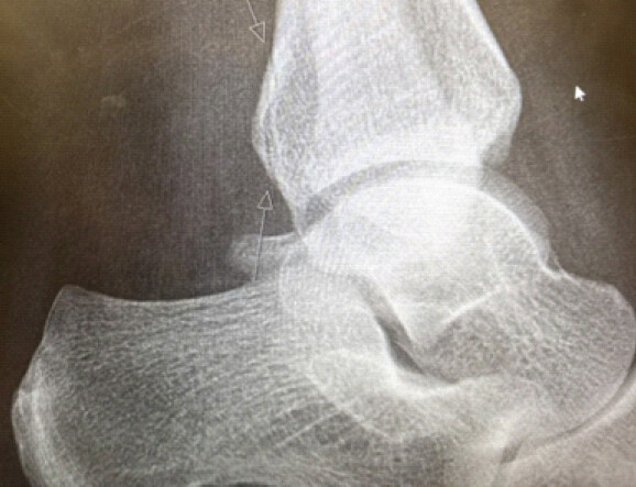
Repeat three-view radiographs of the left ankle demonstrated minimal cortical irregularity of the posterior malleolus, consistent with a non-displaced posterior malleolus fracture. No cortical irregularities of the medial or lateral malleoli were identified. The talar dome was intact, and the ankle mortise remained congruent without evidence of dislocation or talar subluxation (Fig. 1). The patient was transitioned from a splint to a walking boot and advised to continue immobilization, activity modification, and work restrictions.
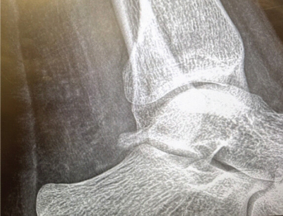
At a subsequent follow-up visit on January 28, 2026, the patient remained pain-free with a reported pain score of 0/10. Physical examination findings were unchanged, with persistent swelling but no tenderness or pain with range of motion. Repeat radiographs demonstrated persistent non-displaced cortical irregularity of the posterior malleolus without interval displacement or significant callus formation (Fig. 2). Given stable imaging findings and preserved ankle alignment, conservative management with continued immobilization in a walking boot and activity restrictions was maintained.
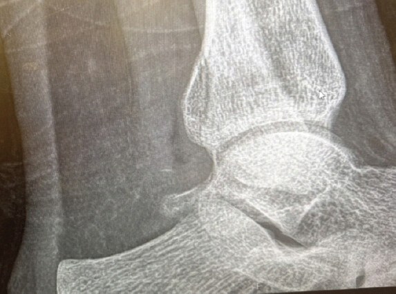
One month later, on February 25, 2026, follow-up radiographs demonstrated interval bony union of the posterior malleolus fracture with increased calcification and maintained alignment. The ankle mortise remained congruent with minimal residual fracture line visible (Fig. 3). The patient remained pain-free and was transitioned from a walking boot to an ankle brace with referral to formal physical therapy for gradual restoration of range of motion and progressive weight-bearing. Work restrictions were continued temporarily with planned reassessment for return to full duties.
Discussion
In this case, continued immobilization despite early symptom resolution resulted in maintained alignment and radiographic union without displacement, supporting the appropriateness of conservative management in carefully selected patients with non-displaced isolated posterior malleolus fractures.
Posterior malleolar fractures are most commonly encountered as part of bi- or trimalleolar ankle fracture patterns, typically resulting from rotational mechanisms of injury. The posterior malleolus serves as an important stabilizer of the ankle joint through its contribution to the distal tibiofibular syndesmosis [1], primarily via the posterior inferior tibiofibular ligament. Disruption of this structure can affect ankle stability and joint congruency even when fracture fragments are small or minimally displaced [8].
Isolated posterior malleolus fractures without associated medial or lateral malleolar injury are relatively uncommon. When they do occur, they may present diagnostic and management challenges due to subtle radiographic findings and rapid clinical improvement. In the present case, the fracture was identified on initial radiographs obtained in the emergency department; however, the patient demonstrated complete resolution of pain within 1 week of injury, a benign physical examination, and preserved ankle mortise alignment on serial imaging. This constellation of findings can create false reassurance, particularly when return-to-activity decisions are guided primarily by symptom resolution rather than radiographic stability [9].
The mechanism of injury in this case – recreational ice skating – provides a plausible explanation for isolated posterior malleolar involvement. Ice skating injuries often involve rigid foot fixation within a stiff boot combined with rotational forces during falls [10]. This mechanism can preferentially stress the posterior inferior tibiofibular ligament and posterior malleolus without sufficient force propagation to involve the medial or lateral malleoli.
Management of posterior malleolus fractures is guided by fracture morphology, displacement, articular congruency, and ankle stability. While operative fixation may be indicated for displaced fractures [6,7] or those associated with syndesmotic instability, non-displaced fractures with preserved mortise alignment may be successfully managed with conservative treatment. Prior studies have demonstrated that long-term outcomes are influenced by maintenance of joint congruency rather than fragment size alone [11], supporting non-operative management in carefully selected cases [12].
This case underscores the importance of continued immobilization and follow-up imaging in patients with posterior malleolus fractures, even in the setting of rapid clinical improvement. Pain resolution should not be interpreted as radiographic healing, and management decisions should integrate both clinical and imaging findings. Clinical decision-making should prioritize radiographic stability over symptom resolution, particularly in injuries involving the posterior malleolus where early pain improvement may mask incomplete healing. This is particularly relevant in sports medicine and outpatient orthopedic settings, where symptom-based decision-making may otherwise lead to premature return to activity.
Conclusion
Isolated posterior malleolus fractures may present with rapid symptom resolution and a benign physical examination despite persistent radiographic evidence of injury. This case highlights the potential for false reassurance in the outpatient setting.
Clinicians should avoid using symptom resolution alone to guide management of posterior malleolus fractures. Even in pain-free patients, continued immobilization and follow-up imaging are necessary to confirm fracture stability and prevent premature return to activity, which may risk displacement or long-term complications.
Clinical Message
In patients with suspected or confirmed posterior malleolus fractures, early symptom resolution should not guide return-to-activity decisions. Continued immobilization and serial radiographic monitoring are essential to ensure fracture stability and prevent premature functional progression, even in the absence of pain.
Conflict of Interest:
Nil
Source of Support:
Nil
Consent:
The authors confirm that informed consent was obtained from the patient for publication of this article
How to Cite this Article
Ryan Lu, Sylvanus Ly. Isolated Posterior Malleolus Fracture with Early Symptom Resolution: A Case Report. Journal of Orthopaedic Case Reports 2026 October;16(10): 260-263.
References
- Haraguchi N, Haruyama H, Toga H, Kato F. Pathoanatomy of posterior malleolar fractures of the ankle. J Bone Joint Surg Am 2006;88:1085-92. [Google Scholar] | [PubMed]
- Bartoníček J, Rammelt S, Tuček M, Naňka O. Posterior malleolar fractures of the ankle. Eur J Trauma Emerg Surg 2015;41:587-600. [Google Scholar] | [PubMed]
- Kannus P, Parkkari J, Jarvinen TL, Jarvinen TA, Jarvinen M. Basic science and clinical studies concur: Active treatment approach is needed after a sports injury. Scand J Med Sci Sports 2003;13:150-4. [Google Scholar] | [PubMed]
- Mangnus L, Meijer DT, Mostert TQ. Posterior malleolus fractures: A systematic review. J Foot Ankle Surg 2015;54:93-8. [Google Scholar] | [PubMed]
- Ferries JS, DeCoster TA, Firoozbakhsh KK, Garcia JF, Miller RA. Plain radiographic interpretation in trimalleolar ankle fractures poorly assesses posterior fragment size. J Orthop Trauma 1994;8:328-31. [Google Scholar] | [PubMed]
- Gardner MJ, Brodsky A, Briggs SM, Nielson JH, Lorich DG. Fixation of posterior malleolar fractures provides greater syndesmotic stability. Clin Orthop Relat Res 2006;447:165-71. [Google Scholar] | [PubMed]
- Miller AN, Carroll EA, Parker RJ, Helfet DL, Lorich DG. Posterior malleolar stabilization of syndesmotic injuries is equivalent to screw fixation. Clin Orthop Relat Res 2010;468:1129-35. [Google Scholar] | [PubMed]
- Veltman ES, Halma JJ, De Gast A. Longterm outcome of 886 posterior malleolar fractures: A systematic review of the literature. Foot Ankle Surg 2016;22:73-7. [Google Scholar] | [PubMed]
- Drijfhout van Hooff CC, Verhage SM, Hoogendoorn JM. Influence of fragment size and postoperative joint congruency on long-term outcome of posterior malleolar fractures. Foot Ankle Int 2015;36:673-8. [Google Scholar] | [PubMed]
- Williamson DM, Galasko CS, Wilson JN. Outcome after internal fixation of posterior malleolar fractures in ankle fractures. J Bone Joint Surg Br 1992;74:734-5. [Google Scholar] | [PubMed]
- Xu HL, Li X, Zhang DY, Fu ZG, Wang TB, Zhang PX . A retrospective study of posterior malleolus fractures. Int Orthop 2012;36:1929-36. [Google Scholar] | [PubMed]
- Ogilvie-Harris DJ, Reed SC, Hedman TP. Disruption of the ankle syndesmosis: Biomechanical study of the ligamentous restraints. Arthroscopy 1994;10:558-60. [Google Scholar] | [PubMed]
© 2026 Journal of Orthopaedic Case Reports - Published by Indian Orthopaedic Research Group
About the Authors
-

Ryan Lu
-

Sylvanus Ly
Related Articles in Journal of Orthopaedic Case Reports
 October 1, 2026 Posterior Fibular Entrapment in a Bosworth Fracture-Dislocation: A Case Report and Review of Diagnostic and Operative Considerations
 April 1, 2026 Dual-Screw Fixation for Chronic Syndesmotic Injury: A Case Report
 October 1, 2026 Recurrent Ventral Scapular Osteochondroma Presenting with Pseudo-winging in an Adolescent: A Case Report and Follow-up of a Previously Published Case
 October 1, 2026 Development of a Non-ossifying Fibroma in the Same Femur 6 Years after Limb-Salvage Surgery for Osteosarcoma: A Case Report