


Introduction
Focal osteochondral defects of the knee are a major cause of pain, swelling, mechanical symptoms, and functional limitation, particularly in young, physically active individuals. These lesions involve articular cartilage with variable subchondral bone involvement and may result from trauma, repetitive loading, osteochondritis dissecans, or other biological factors [1]. The femoral condyles, particularly the medial femoral condyle, are commonly affected. Symptomatic full-thickness lesions can substantially impair activity and quality of life and remain challenging to treat because of the limited intrinsic healing capacity of articular cartilage [2].
Articular cartilage is avascular, aneural, and relatively hypocellular, resulting in poor spontaneous healing following full-thickness injury. Persistent defects may alter joint biomechanics and increase stress on adjacent cartilage, potentially accelerating degenerative changes [3]. The International Cartilage Repair Society (ICRS) grading system classifies grade IV lesions as complete cartilage loss with exposure or involvement of the subchondral bone, necessitating restoration of both cartilage and underlying bone [4].
Treatment options include marrow-stimulation procedures, osteochondral transplantation, and cell-based cartilage restoration.[5] Microfracture is relatively simple and may be effective for small, contained lesions; however, it predominantly produces fibrocartilage, which has inferior biomechanical properties and may deteriorate with repetitive loading [6]. Cell-based procedures such as autologous chondrocyte implantation can provide favorable outcomes but generally require staged treatment, cell expansion, and greater resource utilisation.
Osteochondral autograft transfer (OATS), or mosaicplasty, involves transplantation of cylindrical osteochondral plugs from relatively low-load-bearing regions of the knee into the prepared defect [7]. It restores the defect with viable hyaline cartilage supported by autologous subchondral bone, allowing simultaneous cartilage resurfacing and osseous incorporation. As a single-stage procedure, OATS avoids immunological and disease-transmission concerns associated with allografts and does not require ex vivo cell expansion [8].
Previous studies have demonstrated favorable outcomes following OATS, including sustained functional improvement and return to activity in appropriately selected patients. However, outcomes depend on lesion characteristics, graft positioning, surrounding cartilage quality, patient activity, and case selection, while donor-site morbidity and limited graft availability remain important limitations. The clinical evaluation involved using validated functional and activity measures, including the Lysholm knee score, International Knee Documentation Committee (IKDC) subjective score, and Tegner activity scale.
This study aimed to assess the functional and radiological results of OATS in patients with isolated ICRS Grade IV knee osteochondral defects. Functional outcomes were measured using the Lysholm knee score, IKDC subjective score, and Tegner activity scale. Post-operative magnetic resonance imaging (MRI) was used to evaluate graft healing, articular surface congruity, and any graft-related structural issues.
Materials and Methods
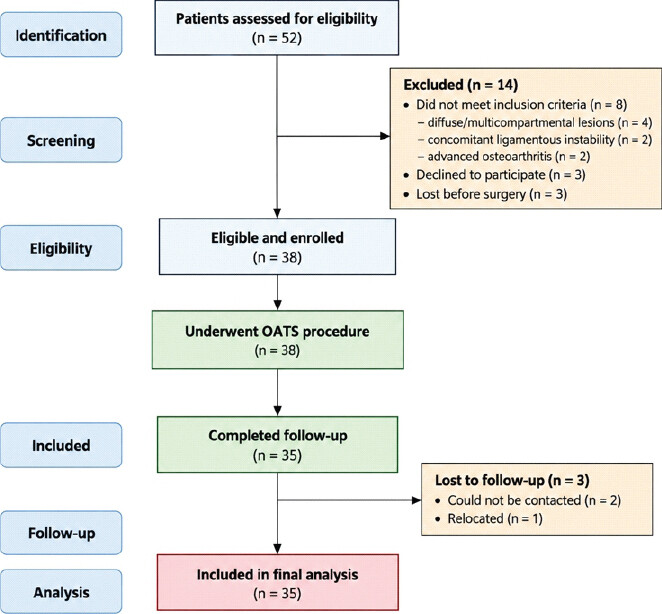
This prospective cohort study was conducted in the Department of Orthopedics at a tertiary-care postgraduate teaching institute. Ethical approval was obtained, and all participants provided written consent. This study expands on a previous 20-patient OATS series from the same institution and supervisor (Agrawal et al., Int J Acad Med Pharm. 2026;8(2):952-958; doi: 10.47009/jamp.2026.8.2.175). Fifty-two patients were screened for eligibility. Of these, 14 were excluded: Eight because they did not fulfill the eligibility criteria (diffuse or multicompartmental lesions, n = 4; concomitant ligamentous instability, n = 2; advanced osteoarthritis, n = 2); three declined to participate; and three were lost before surgery. The remaining 38 patients underwent OATS and formed the enrolled cohort. Of these, 35 completed follow-up and provided complete paired clinical outcome data for the final functional analysis (Fig. 1).
Eligibility criteria included age 15–40, an isolated ICRS Grade 4 osteochondral knee defect measuring 2.0–4.0 cm2, a clinically normal contralateral knee, and at least 18 months of post-operative follow-up. Patients were excluded if they had prior surgery involving the index knee, concurrent osteoarthritis, injuries to the anterior cruciate ligament, posterior cruciate ligament, lateral collateral ligament, or posterolateral complex, Grade III or IV medial collateral ligament injury, or associated meniscal injury.
Patients were assessed at regular follow-up intervals at 6, 12, and 18 months postoperatively. We recorded the Lysholm, IKDC, and Tegner scores at these visits.[9] Patients also underwent MRI scans at these intervals to assess osteochondral graft structural integrity and healing. The radiological assessment covered graft incorporation and healing, articular surface congruity, cystic degeneration or delamination, graft survival or failure, donor-site abnormalities when reported, and signs of degenerative osteoarthritis.[10] Follow-up MRI data were available for 35 patients with complete clinical follow-up.
All procedures were performed under spinal anesthesia with the patient supine on a standard operating table. A lateral post and foot support maintained the knee at approximately 90° of flexion, with the option to increase flexion for better access to posterior lesions.[11] A pneumatic tourniquet was applied to the proximal thigh. The procedure began with diagnostic arthroscopy to confirm the size, location, and shape of the osteochondral defect and to identify any additional intra-articular issues that could complicate cartilage restoration. The lesion was then accessed through a small medial or lateral parapatellar approach, depending on its location. This involved a short longitudinal incision alongside the patella, followed by a limited parapatellar arthrotomy, preserving a cuff of capsular tissue adjacent to the patella for future repair. If needed, we performed a slight resection of the infrapatellar fat pad to improve visualization [12–14].
Defect size and depth measured on pre-operative MRI were verified intraoperatively with a calibrated probe, and the stability of the surrounding cartilage was assessed. We placed a cannulated OATS guide perpendicular to the joint surface, adjusting knee flexion to ensure a perpendicular approach. We inserted a guide pin to a depth of 10–12 mm, then used a matched-size cannulated reamer to reach the required depth, typically 8–10 mm or matching the depth of cystic changes seen on MRI. We then used a cannulated dilator to confirm the size and depth of the recipient socket. We prepared osteochondral plugs of 6, 8, or 10 mm, using multiple plugs for larger defects. Harvesting of osteochondral autografts was performed from the lateral edge of the femoral trochlea, with the harvester positioned perpendicular to the donor surface and advanced to approximately 10 mm. The osteochondral cylinder was released from its bony attachment by controlled rotation of the harvester and carefully withdrawn to preserve the cartilage surface.
Each harvested plug was examined and its circumference measured. The osseous end was trimmed as needed, and the bone edges were gently chamfered to ease insertion. The graft was prepared about 1 mm shorter than the recipient socket to ensure a flush articular reconstruction and prevent proud graft placement. If necessary, the graft orientation was adjusted to match the angulation of the recipient surface. The graft was inserted perpendicular to the socket and carefully impacted with a bone tamp until the cartilage surface was level with the surrounding native cartilage. When multiple plugs were needed, they were placed sequentially to maximise defect coverage and maintain articular congruity. The donor site was either left empty or backfilled with a suitably sized plug. The tourniquet was then released, hemostasis was achieved, the arthrotomy and incision were closed, and a sterile soft dressing was applied.
Post-operative rehabilitation was initiated to protect graft incorporation while progressively restoring knee mobility, strength, and function. Full active range of motion was encouraged immediately after surgery, with no routine requirement for a knee brace. Closed-chain exercises and straight-leg raises were commenced in the immediate post-operative period. Partial weight bearing was permitted from 2 to 4 weeks postoperatively, progressing to full weight bearing after 4 weeks, depending on pain, clinical findings, and knee range of motion. Patients were permitted to resume light recreational sporting activities at 4–6 months, while return to high-impact sports was deferred until approximately 6 months.[15] Recovery milestones recorded for analysis included the time to pain-free full weight-bearing and the time to return to routine physical or sporting activity [16].
Continuous variables were summarized using mean ± standard deviation for approximately normally distributed variables and median with interquartile range (IQR) for non-normally distributed variables. Categorical variables were presented as frequencies and percentages. We evaluated longitudinal changes in Lysholm knee score, IKDC subjective knee evaluation score, and Tegner activity scale across the four assessment points (pre-operative, 6 months, 12 months, and 18 months) using the Friedman test for repeated measures. For post hoc pairwise comparisons, we applied a Bonferroni adjustment. Statistical analyses were performed using Microsoft Excel and IBM Statistical Package for the Social Sciences Statistics version 26.0. A two-sided P < 0.05 was considered statistically significant.
Results
Fifty-two patients were assessed for eligibility. Fourteen were excluded: Eight did not meet eligibility criteria (diffuse or multicompartmental lesions, n = 4; concomitant ligamentous instability, n = 2; advanced osteoarthritis, n = 2); three declined participation; and three were lost before surgery. Thirty-eight patients underwent OATS, forming the operative cohort. Three additional patients were lost during post-operative follow-up; therefore, 35 patients had complete paired clinical outcome data and follow-up MRI available for the final outcome analysis.
The operative cohort comprised 38 patients with a mean age of 25.8 ± 5.6 years; 29 (76.3%) were male. Their mean body weight was 71.6 ± 12.0 kg, mean height was 169.7 ± 7.7 cm, and median body mass index (BMI) was 24.2 kg/m2 (IQR, 22.6–27.2). Before symptom onset, 27 patients (71.1%) participated in recreational or competitive sports. Fourteen patients (36.8%) reported a prior injury managed non-surgically. The right knee was affected in 23 patients (60.5%), and the dominant limb was involved in 24 patients (63.2%). The median symptom duration was 5 weeks (IQR, 3–8 weeks) (Table 1).
Baseline demographic and clinical characteristics (n=38)
| Variable | Value (%) |
|---|---|
| Age, years | 25.8±5.6 |
| Male sex | 29 (76.3) |
| Female sex | 9 (23.7) |
| Weight, kg | 71.6±12.0 |
| Height, cm | 169.7±7.7 |
| BMI, kg/m2 | 24.2 (22.6–27.2) |
| Pre-onset activity: Sedentary | 11 (28.9) |
| Pre-onset activity: Recreational sports | 13 (34.2) |
| Pre-onset activity: Competitive sports | 14 (36.8) |
| Preceding injury | 14 (36.8) |
| Right knee affected | 23 (60.5) |
| Dominant limb affected | 24 (63.2) |
| Duration of symptoms, weeks | 5 (3–8) |
| History of massage/home remedies | 15 (39.5) |
Values are mean±SD, median (IQR), or n (%), as appropriate. IQR: Interquartile range, BMI: Body mass index
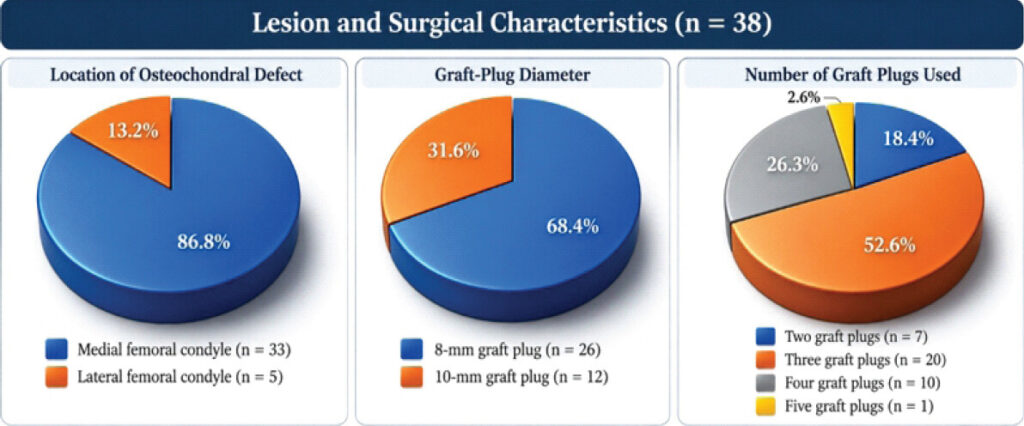
The medial femoral condyle was the most common lesion site, accounting for 33 of 38 defects (86.8%), with five lesions (13.2%) involving the lateral femoral condyle. The median defect size was 2.43 cm2 (IQR, 2.24–2.56). The median graft-plug diameter was 8 mm (IQR, 8–10), with an 8-mm plug used in 26 patients (68.4%) and a 10-mm plug in 12 (31.6%). The most common graft configuration was three plugs (20 patients, 52.6%). All donor sites were located at the lateral edge of the femoral trochlea (Fig. 2).
Longitudinal analysis showed substantial, sustained improvement in knee function after OATS. The mean pre-operative Lysholm knee score was 50.29 ± 10.90, indicating considerable functional impairment at baseline. At 6 months postoperatively, the mean score rose markedly to 93.31 ± 4.77, an absolute improvement of approximately 43 points. This improvement was maintained at subsequent assessments, with mean scores of 93.34 ± 3.90 at 12 months and 93.03 ± 4.11 at 18 months [17].
The median score also increased from 54.0 (IQR 38.0–59.0) preoperatively to 93.0 (IQR 90.5–97.5) at 6 months. At 12 months, the median score remained stable at 93.0 (IQR 92.0–96.5), and at 18 months it was 92.0 (IQR 90.0–96.0). The relatively narrow IQRs during follow-up suggest consistent functional recovery among the study participants. The most significant improvement took place in the first 6 months post-surgery, after which scores stabilized with little change through 18 months. This indicates that OATS offers quick functional recovery, with knee function remaining stable afterward (Fig. 3).
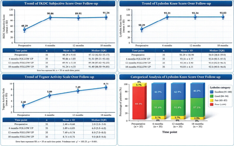
Categorical analysis showed a marked shift toward favorable functional outcomes after OATS. Preoperatively, 33 patients (94.3%) had poor Lysholm scores (≤64), while only 2 patients (5.7%) had fair scores; no patients demonstrated good or excellent function. At 6 months, there was a substantial improvement, with 18 patients (51.4%) achieving good scores (84–94) and 15 (42.9%) achieving excellent scores (95–100), while only 2 patients (5.7%) remained in the fair category. This distribution was unchanged at 12 months, indicating early attainment and maintenance of functional recovery. At 18 months, 40.0% (14 patients) had excellent scores, 57.1% (20 patients) had good scores, and only one patient (2.9%) remained in the fair category. No patient had a poor score at any post-operative assessment. Overall, the categorical distribution shows a pronounced shift from predominantly poor pre-operative function to good or excellent functional status after OATS, with these favorable outcomes maintained throughout the 18-month follow-up period (Fig. 3).
Longitudinal analysis showed marked improvement in patient-reported knee function after OATS. The mean IKDC subjective score rose substantially from 48.29 ± 9.45 preoperatively to 90.86 ± 3.85 at 6 months, an improvement of 42.57 points. This early functional gain was largely maintained throughout follow-up, with mean scores of 89.91 ± 5.18 at 12 months and 91.24 ± 4.55 at 18 months. The median IKDC score likewise increased from 47.12 (IQR 42.52–55.17) preoperatively to 91.51 (IQR 89.21–93.10) at 6 months, with favorable values sustained thereafter. The small variation in scores between 6 and 18 months suggests maintenance of functional improvement after the initial post-operative recovery. The Friedman test showed a statistically significant difference across the four assessment points (P < 0.001) (Fig. 3) [18].
The Tegner activity scale showed progressive improvement in activity levels after OATS. Among 35 patients with complete longitudinal data, the mean score rose from 2.40 ± 0.60 preoperatively to 5.89 ± 0.83 at 6 months, 7.49 ± 0.74 at 12 months, and 8.71 ± 0.75 at 18 months. The corresponding median scores rose from 2.0 (IQR 2.0–3.0) to 6.0 (5.0–6.0), 8.0 (7.0–8.0), and 9.0 (8.0–9.0), respectively. The overall longitudinal difference was statistically significant (Friedman χ2 = 103.25, P < 0.001). The greatest improvement occurred in the first 6 months, with continued gains at 12 and 18 months. Compared with baseline, the mean increase was 3.49, 5.09, and 6.31 points at 6, 12, and 18 months, respectively. All pairwise comparisons were significant (Bonferroni-adjusted P < 0.001) (Fig. 3) [19,20].
At 18 months, all three functional outcome measures improved substantially after OATS. The mean Lysholm knee score increased from 50.29 ± 10.90 preoperatively to 93.03 ± 4.11 at 18 months, with a median improvement of 39.0 points (IQR, 38.5–50.5). Similarly, the mean IKDC subjective score improved from 48.29 ± 9.45 to 91.24 ± 4.55 at 18 months, with a median increase of 42.54 points (IQR, 36.78–48.38). The Tegner activity scale increased from 2.40 ± 0.60 to 8.71 ± 0.75 at 18 months, with a median improvement of 6.0 points (IQR, 6.0–7.0) (Table 2).
Pre-operative and 18-month functional outcomes (n=35)
| Outcome measure | Pre-operative mean±SD | 18-month mean±SD | Median change (IQR) |
|---|---|---|---|
| Lysholm knee score | 50.29±10.90 | 93.03±4.11 | 39.0 (38.5–50.5) |
| IKDC subjective score | 48.29±9.45 | 91.24±4.55 | 42.54 (36.78–48.38) |
| Tegner activity scale | 2.40±0.60 | 8.71±0.75 | 6.0 (6.0–7.0) |
IKDC: International knee documentation committee, IQR: Interquartile range, SD: Standard deviation
Follow-up MRI scans were performed at 6, 12, and 18 months after surgery for all 35 patients in the final analysis. A smooth, congruent articular surface was observed in 32 patients (91.4%), whereas three patients (8.6%) showed cystic degeneration and/or delamination. All patients showed radiological signs of graft healing, with no cases of non-union or graft failure. Mild degenerative osteoarthritic changes were detected in three patients (8.6%), consistent with those with structural abnormalities. No follow-up patients reported donor-site complications.
At final radiological follow-up,[Table 3] 32 patients had a congruent articular surface and three showed cystic degeneration and/or delamination. Because only three patients showed structural abnormalities, functional comparisons between MRI subgroups were descriptive, and no inferential subgroup testing was performed [21,22].
Radiological findings at final follow-up (n=35)
| Radiological finding | n (%) |
|---|---|
| Congruent articular surface | 32 (91.4) |
| Cystic degeneration/delamination | 3 (8.6) |
| Graft healed | 35 (100) |
| Graft non-union/failure | 0 (0) |
| Degenerative osteoarthritic changes | 3 (8.6) |
| No degenerative osteoarthritic changes | 32 (91.4) |
| Donor-site complication | 0 (0) |
Discussion
This study revealed that OATS notably enhanced knee function in young patients with isolated ICRS grade IV osteochondral defects. Our cohort had a mean age of 25.8 ± 5.6 years, with 76.3% male. The median BMI was 24.2 kg/m2, and the median defect size was 2.43 cm2. Most lesions affected the medial femoral condyle (86.8%).[23] These demographic features align with those reported by Solheim et al., in which the patient group was predominantly young adults. Richter et al.’s series of 152 patients reported a mean age of 24.8 ± 4.6 years, 82.9% male, and an average defect size of 2.7 ± 0.7 cm2, which is similar to our cohort in age, sex, and lesion size.[24,25] Fodor et al.’s study of 63 primary OAT patients reported a mean age of 27.4 ± 12.4 years, 63.5% male, a BMI of 25.5 ± 5.0 kg/m2, and a lesion size of 2.3 ± 1.6 cm2. Rowland et al. reported a mean age of 29.7 years, highlighting that young adults received OATS. Overall, our cohort’s age and lesion size are comparable, and excluding ligamentous, meniscal, and degenerative issues resulted in a more uniform group [26].
The lesion characteristics and operative parameters in this study were similar to those in previous OATS series, though our cohort was limited to moderate-sized, isolated ICRS grade IV lesions. The median defect size was 2.43 cm2 (IQR 2.24–2.56), with the medial femoral condyle involved in 86.8% of cases and the lateral in 13.2%. The median plug diameter was 8 mm, with three plugs most common (52.6%). These findings align with current OAT literature. Kelms et al.’s series of 63 patients reported a mean treated area of 2.3 ± 1.6 cm2, with the medial femoral condyle involved in 47.6% of cases, and 6-, 8-, and 10-mm plugs most commonly used. Treme et al.’s series of Grade IV femoral condylar lesions found an average defect diameter of 15 mm, with 79% on the medial femoral condyle and an average of 2.3 grafts per defect .
Our study’s approach – perpendicular site preparation, use of 6-, 8-, or 10-mm plugs, and press-fit implantation flush with the cartilage – aligns with established OATS principles. Current literature also recommends 6–10 mm plugs and perpendicular preparation for surface congruity. The small defect size and limited number of plugs in our cohort indicate appropriate patient selection for autologous osteochondral transplantation.
The study showed significant improvements across all three outcome measures after OATS. At 18 months, the mean Lysholm score was 93.03 ± 4.11 compared with 50.29 ± 10.90 preoperatively, the IKDC subjective score was 91.24 ± 4.55 compared with 48.29 ± 9.45 preoperatively, and the Tegner activity scale was 8.71 ± 0.75 compared with 2.40 ± 0.60 preoperatively; longitudinal analyses showed statistically significant improvement (P < 0.001). These gains align with existing research. Shekhar et al., reviewing 610 patients over 10.2 years, reported mean improvements of 42.4 points in IKDC and 21.1 in Lysholm, with a non-significant Tegner improvement of 0.76. Our mean IKDC improvement at 18 months was 42.95 points, which closely matches Pareek’s et al. pooled result, while Lysholm and Tegner scores also showed substantial improvement. Keszeg et al. reported similar gains after OATS, supporting functional improvement. A recent meta-analysis by Robinson et al. confirmed significant gains in Lysholm, IKDC, and Tegner scores, with pooled improvements of about 35.2 points (IKDC) and 2.7 (Tegner). The greater improvements observed in our cohort may reflect its younger age, moderate lesion size, and exclusion of advanced pathology. These findings support OATS as effective for focal, full-thickness osteochondral defects .
Post-operative MRI showed successful OATS results, with 100% graft healing at 18 months and no failures. A smooth, congruent articular surface was present in 91.4% of patients, while 8.6% showed cystic degeneration and/or delamination; mild degenerative osteoarthritic changes were also reported in 8.6%. No donor-site complications were observed. MRI studies indicate bone integration and surface restoration, although appearances may vary over time despite good clinical outcomes. Because the structural-abnormality subgroup included only three patients, we did not interpret functional subgroup comparisons inferentially. Overall, the high rate of graft incorporation supports effective structural restoration after OATS, although longer-term MRI follow-up is needed to monitor cystic or degenerative changes .
This study has limitations, including a small sample size, a single-center non-randomised design, and no comparator group. Strict eligibility improved cohort homogeneity but limited applicability to other lesion types, locations, and patients with knee issues. The 18-month follow-up was enough for short-term results but not long-term graft durability or osteoarthritis progression. While rehabilitation was mostly standardised with minor patient modifications, the study did not fully assess outcomes such as MCID, satisfaction, return-to-sport, and reoperation rates. Future studies should include larger, multicenter cohorts with longer follow-up to assess long-term graft survival and osteoarthritis, using standardised MRI scoring and broader patient-centered outcomes.
Conclusion
In young patients with isolated, moderate-sized ICRS grade IV osteochondral knee defects, OATS significantly improved knee function and activity over 18 months, with high rates of graft incorporation and restored joint surface congruity. The improvements in Lysholm, IKDC, and Tegner scores support OATS as a promising joint-preserving option in selected patients. However, because of the small sample size, single-center design, lack of a comparator, and limited long-term data, these results should be seen as evidence of favorable outcomes rather than proof of superiority over other cartilage-restoration methods.
Clinical Message
OATS is a reliable, single-stage, joint-preserving procedure for young patients with isolated, moderate-sized ICRS Grade IV osteochondral knee lesions. It provides significant functional improvement and good graft integration when patients are appropriately selected and graft placement is precise.
Conflict of Interest:
Nil
Source of Support:
Nil
Consent:
The authors confirm that informed consent was obtained from the patient for publication of this article
How to Cite this Article
Kumar V, Dhankhar S, Devgan R, Singh A, Devgan A, Aggarwal C. A Prospective Clinical and Radiological Assessment of Osteochondral Autograft Transfer for International Cartilage Repair Society Grade IV Osteochondral Lesions of the Knee. Journal of Orthopaedic Case Reports 2026 October;16(10): 384-391.
References
- Howell M, Liao Q, Gee CW. Surgical management of osteochondral defects of the knee: An educational review. Curr Rev Musculoskelet Med 2021;14:60-6. [Google Scholar] | [PubMed]
- Erickson BJ, Chalmers PN, Yanke AB, Cole BJ. Surgical management of osteochondritis dissecans of the knee. Curr Rev Musculoskelet Med 2013;6:102-14. [Google Scholar] | [PubMed]
- Solheim E, Hegna J, Inderhaug E. Clinical outcome after mosaicplasty of knee articular cartilage defects of patellofemoral joint versus tibiofemoral joint. J Orthop 2020;18:36-40. [Google Scholar] | [PubMed]
- Richter DL, Tanksley JA, Miller MD. Osteochondral autograft transplantation: A review of the surgical technique and outcomes. Sports Med Arthrosc Rev 2016;24:74-8. [Google Scholar] | [PubMed]
- Pascual-Garrido C, McNickle AG, Cole BJ. Surgical treatment options for osteochondritis dissecans of the knee. Sports Health 2009;1:326-34. [Google Scholar] | [PubMed]
- Mithoefer K, McAdams T, Williams RJ, Kreuz PC, Mandelbaum BR. Clinical efficacy of the microfracture technique for articular cartilage repair in the knee: An evidence-based systematic analysis. Am J Sports Med 2009;37:2053-63. [Google Scholar] | [PubMed]
- Hangody L, Füles P. Autologous osteochondral mosaicplasty for the treatment of full-thickness defects of weight-bearing joints: Ten years of experimental and clinical experience. J Bone Joint Surg Am 2003;85-A Suppl 2:25-32. [Google Scholar] | [PubMed]
- Chow JC, Hantes ME, Houle JB, Zalavras CG. Arthroscopic autogenous osteochondral transplantation for treating knee cartilage defects: A 2- to 5-year follow-up study. Arthroscopy 2004;20:681-90. [Google Scholar] | [PubMed]
- Oztürk A, Ozdemir MR, Ozkan Y. Osteochondral autografting (mosaicplasty) in grade IV cartilage defects in the knee joint: 2- to 7-Year results. Int Orthop 2006;30:200-4. [Google Scholar] | [PubMed]
- Marcacci M, Kon E, Delcogliano M, Filardo G, Busacca M, Zaffagnini S. Arthroscopic autologous osteochondral grafting for cartilage defects of the knee: Prospective study results at a minimum 7-year follow-up. Am J Sports Med 2007;35:2014-21. [Google Scholar] | [PubMed]
- Reverte-Vinaixa MM, Joshi N, Diaz-Ferreiro EW, Teixidor-Serra J, Dominguez-Oronoz R. Medium-term outcome of mosaicplasty for grade III-IV cartilage defects of the knee. J Orthop Surg (Hong Kong) 2013;21:4-9. [Google Scholar] | [PubMed]
- Gudas R, Gudaitė A, Mickevičius T, Masiulis N, Simonaitytė R, Cekanauskas E. Comparison of osteochondral autologous transplantation, microfracture, or debridement techniques in articular cartilage lesions associated with anterior cruciate ligament injury: A prospective study with a 3-year follow-up. Arthroscopy 2013;29:89-97. [Google Scholar] | [PubMed]
- Pareek A, Sanders TL, Wu IT, Larson DR, Saris DBF, Krych AJ. Incidence of symptomatic osteochondritis dissecans lesions of the knee: A population-based study in Olmsted County. Osteoarthritis Cartilage 2017;25:1663-71. [Google Scholar] | [PubMed]
- Shekhar A, Reddy S, Patil S, Tapasvi S. Mid-term outcomes of arthroscopic osteochondral autograft transplantation for focal chondral defects of the knee. J Arthrosc Surg Sports Med 2021;2:41-6. [Google Scholar] | [PubMed]
- Keszég M, Hangody L, Egyed Z, Tóth G, Pánics G. Long-term (10-25 years) outcomes of knee osteochondral autologous transplantation in soccer players. J Cartil Joint Preserv 2022;2:100062. [Google Scholar] | [PubMed]
- Bengtsson J, Möllborg J, Werner S. A study for testing the sensitivity and reliability of the Lysholm knee scoring scale. Knee Surg Sports Traumatol Arthrosc 1996;4:27-31. [Google Scholar] | [PubMed]
- Collins NJ, Misra D, Felson DT, Crossley KM, Roos EM. Measures of knee function: International knee documentation committee (IKDC) Subjective knee evaluation form, knee injury and osteoarthritis outcome score (KOOS), Knee injury and osteoarthritis outcome score physical function short form (KOOS-PS), Knee outcome survey activities of daily living scale (KOS-ADL), Lysholm knee scoring scale, Oxford knee score (OKS), Western ontario and mcmaster universities osteoarthritis index (WOMAC), Activity rating scale (ARS), and tegner activity score (TAS). Arthritis Care Res (Hoboken) 2011;63 Suppl 11:S208-28. [Google Scholar] | [PubMed]
- Hambly K. The use of the Tegner activity scale for articular cartilage repair of the knee: A systematic review. Knee Surg Sports Traumatol Arthrosc 2011;19:604-14. [Google Scholar] | [PubMed]
- Robinson A, Lindsay A, Vidal A, Frank RM. Osteochondral autograft transfer (OATS). Oper Tech Sports Med 2020;28:150781. [Google Scholar] | [PubMed]
- Sherman SL, Thyssen E, Nuelle CW. Osteochondral autologous transplantation. Clin Sports Med 2017;36:489-500. [Google Scholar] | [PubMed]
- Fodor P, Sólyom Á, Ivănescu A, Fodor R, Băţagă T. Prevalence of chondral lesions in knee arthroscopy. J Interdiscip Med 2018;3:21-4. [Google Scholar] | [PubMed]
- Rowland R, Colello M, Wyland DJ. Osteochondral autograft transfer procedure: Arthroscopic technique and technical pearls. Arthrosc Tech 2019;8:e713-9. [Google Scholar] | [PubMed]
- Treme G, Milller MD. Autograft osteochondral transfer. Oper Tech Sports Med 2008;16:81-8. [Google Scholar] | [PubMed]
- Dahmen J, Hollander JJ, Butler JJ, Emanuel KS, Rikken QG, Stufkens SA. What are these cysts doing in my graft? A meta-analysis on cystic occurrence after autografting and allografting for osteochondral lesions of the talus. Cartilage 2026;17:408-19. [Google Scholar] | [PubMed]
- Vellios EE, Jones KJ, Williams RJ. Osteochondral autograft transfer for focal cartilage lesions of the knee with donor-site back-fill using precut osteochondral allograft plugs and micronized extracellular cartilage augmentation. Arthrosc Tech 2021;10:e181-92. [Google Scholar] | [PubMed]
- Amoo-Achampong K, Kunze KN, Ramkumar PN, Khorana A, Williams RJ 3rd. Midterm outcomes of primary osteochondral autograft transfer for symptomatic chondral defects of the knee. Orthop J Sports Med 2026;14:23259671251399796. [Google Scholar] | [PubMed]
© 2026 Journal of Orthopaedic Case Reports - Published by Indian Orthopaedic Research Group
About the Authors
-
Virender Kumar
-

Sarita Dhankhar
-
Radhika Devgan
-
Amitoj Singh
-
Ashish Devgan
-

Chetan Aggarwal
Related Articles in Journal of Orthopaedic Case Reports
 September 1, 2026 Fixed Flexion Deformity due to Displaced Hoffa Fracture: A Case Report
 August 1, 2026 The Boba Tea Sign: A Novel Magnetic Resonance Imaging Finding in Synovial Chondromatosis of the Knee – A Case Report
 August 1, 2026 Intra-articular Ganglionic Cysts of Knee: A Cause for Anterior Knee Pain
 July 1, 2026 A Case of In Situ Fixation of Large Lateral Condyle Femur Osteochondral Defect Secondary to Trauma in a Young Male – A Case Report