


Introduction
Patellar fractures account for approximately 1% of all skeletal injuries and can significantly affect the extensor mechanism of the knee [1]. The primary goals of surgical treatment are restoration of the extensor mechanism, anatomical reduction of the articular surface, and stable fixation that permits early knee mobilization while minimizing complications such as joint stiffness and post-traumatic functional impairment [2].
For displaced transverse fractures, tension band wiring (TBW) is a widely used method of fixation. The principle of TBW is based on converting tensile forces generated across the anterior aspect of the patella during knee flexion into compressive forces at the fracture site. This provides stable fixation and facilitates early post-operative range of motion (ROM) and rehabilitation [3,4,5].
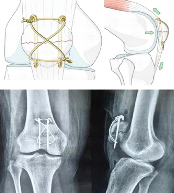
In 1979, Müller et al. and the Arbeitsgemeinschaft für Osteosynthesefragen/Association for the Study of Internal Fixation popularized the modified TBW using Kirschner wires (K-wires) and anterior figure-of-eight TBW (Fig. 1). Biomechanical studies demonstrated that appropriately positioned K-wires, combined with anterior TBW, provided effective stabilization of transverse patellar fractures [6]. The technique subsequently became widely accepted because of its relative simplicity and ability to provide fixation adequate for early mobilization. However, K-wire-based fixation has several recognized disadvantages. Prominent hardware may cause anterior knee pain and soft-tissue irritation, while K-wire migration, backing out, and loosening can result in loss of fixation or the need for subsequent implant removal [7,8,9].
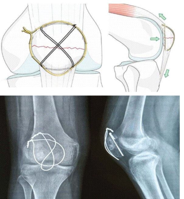
To address complications linked to K-wires, surgeons from Rowley Bristow Orthopaedic Hospital (Pyrford, United Kingdom) introduced a technique combining cerclage and longitudinal TBW in a 1990 cadaveric study. Evaluated across 10 knee specimens, this Pyrford technique demonstrated significantly greater fixation strength than the modified AO method, providing sufficient stability to support early mobilization [10]. Agarwala et al. reported good functional outcomes with the combined cerclage technique, with all fractures achieving union by 12 weeks and full ROM and unrestricted walking, squatting, and stair climbing at follow-up. The authors specifically attributed the advantages to greater fixation strength, early mobilization, applicability to comminuted fractures, and reduced reoperation rates [11]. A modification of this technique, using figure-of-eight TBW in combination with cerclage wiring and avoiding K-wires, has been adopted in the present study (Fig. 2). The absence of K-wires may potentially reduce hardware-related complications, particularly implant prominence, migration, and irritation, while maintaining adequate fracture stability.
The present study aims to compare the functional and radiological outcomes and complications of transverse patellar fractures treated with K-wire TBW versus combined cerclage and figure-of-eight TBW without K-wires.
Materials and Methods
The study was conducted at the Department of Orthopedics at Seth Gordhandas Sunderdas Medical College and King Edward Memorial Hospital, Mumbai.
Study design and population
A prospective, non-randomized, non-blinded, single-center comparative study was conducted among patients who underwent surgical fixation of patellar fractures between June 2019 and December 2024 A total of 156 patients were included and divided into two groups for analysis. Group A comprised 82 patients treated with modified TBW using K-wires, while Group B comprised 74 patients treated with combined cerclage and figure-of-eight TBW without K-wires.
Inclusion and exclusion criteria
Only two-part transverse patella fractures were included in the study. Minor comminution was permitted but the principal fracture pattern remained transverse. Patients having a post-operative follow-up of at least 18 months were included in the study. Patients with open fractures, pathological fractures, bilateral injuries, or associated neurovascular damage were excluded.
Procedure and rehabilitation protocol
Procedures were performed by multiple treating surgeons whose choice of fixation method was guided by their clinical assessment of fracture pattern, soft-tissue envelope, and intraoperative feasibility. Both groups followed a standardized post-operative rehabilitation protocol. Knee ROM exercises were initiated on post-operative day 1, restricted to 0–30° for the 1st week and 0–60° during the 2nd week, before progressing as tolerated. Patients were permitted immediate full weight-bearing mobilization on post-operative day using a long knee brace and walker. Implant removal was not performed routinely. The criteria for recommending hardware removal required radiographic evidence of complete fracture union and persistent, clinically significant hardware-related symptoms, such as localized anterior pain, skin irritation, hardware migration, or flexion restriction. For patients meeting these criteria, the final decision to proceed with surgery was based on a shared decision-making process weighing symptom severity against secondary surgical risks.
Data collection
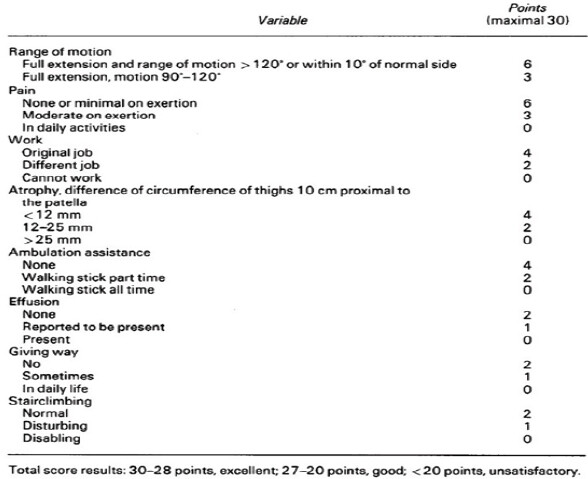
Post-operative follow-up assessments at 6 weeks, 3, 6, 12, and 18 months were taken for analysis. Clinical assessment was done by knee ROM in degrees, extension lag in degrees, and presence of local tenderness. Visual Analog Scale 0–10 was used for assessment of knee pain. Functional recovery was evaluated using the Bostman knee score, a validated, disease-specific grading scale tailored to patellar fracture outcomes (Fig. 3) [12]. This scale comprehensively assesses eight clinical and functional parameters: Knee ROM, pain, quadriceps muscle atrophy, walking ability, stair climbing, work capacity, joint effusion, and knee stability. Scores are interpreted as excellent (28–30), good (20–27), and poor (<20). Radiographs assessed parameters such as articular surface reduction, fracture site union, implant migration, and breakage. Complications such as hardware prominence and irritation, implant displacement and breakage, delayed union, infection, and implant removal were assessed in the study.
Ethical clearance
This study was approved by the Institutional Ethics Committee (Letter No. IEC 2 /OUT/450/19 Dated May 19th, 2019). This research was conducted ethically in accordance with the World Medical Association Declaration of Helsinki.
Statistics
IBM Statistical Package for the Social Sciences Statistics for Windows was used for statistical analysis. Categorical data were presented as counts and corresponding percentages, whereas continuous data were reported as mean value with standard deviation. The Chi-square test and Fisher’s exact test were used to assess association between categorical variables, and an independent-samples t-test was used to assess association between continuous variables. A two-tailed probability value of <0.05 was considered statistically significant.
Results
A total of 156 patients were included in the study, with 82 patients in the conventional K-wire TBW group (Group A) and 74 in the combined cerclage TBW group (Group B). The mean age was 44.8 ± 14.2 years in Group A and 46.0 ± 13.6 years in Group B (P = 0.32). The proportion of male patients was 60% and 66% in Groups A and B, respectively (P = 0.44). There were no significant differences between the groups in body mass index, duration of postoperative follow-up, or mechanism of injury, indicating comparable baseline characteristics (Table 1).
Baseline demographic and mechanism of injury characteristics.
| S. No. | Characteristics | K-wire TBW Group A (n=82) | Combined cerclage TBW Group B (n=74) | P-value |
|---|---|---|---|---|
| 1 | Age, Mean±SD, years | 44.80±14.2 years | 46±13.6 years | 0.32 |
| 2 | Gender, n (%) | |||
| Male | 49 (60) | 50 (66) | 0.44 | |
| Female | 33 (40) | 24 (34) | ||
| 3 | BMI | 24.66±1.92 | 23.86±1.56 | 0.12 |
| 4 | Post op follow-up (months) | 20.26±3.2 | 22.32±4.2 | 0.16 |
| 5 | Mechanism of trauma, n(%) | |||
| Direct | 36 (44) | 30 (40) | 0.26 | |
| Indirect | 46 (56) | 44 (60) | ||
SD: Standard deviation, TBW: Tension band wiring, BMI: Body mass index, K-wire: Kirschner wire
Pain scores were significantly lower in the combined cerclage group at 6 weeks (5.66 ± 1.10 vs. 6.27 ± 1.20; P = 0.046), 3 months (4.40 ± 0.90 vs. 4.66 ± 0.92; P = 0.035), and 6 months (2.20 ± 0.80 vs. 2.60 ± 0.87; P = 0.023). At 1 year, pain remained lower in the combined cerclage group, although the difference was not statistically significant (1.60 ± 0.62 vs. 2.60 ± 0.64; P = 0.055). Knee ROM and extension lag improved progressively in both groups, with numerically better outcomes in the combined cerclage group, but between-group differences were not statistically significant. Knee tenderness was significantly less frequent with combined cerclage TBW at 6 weeks (40% vs. 51%; p = 0.018) and 3 months (36% vs. 43%; P = 0.034) (Table 2).
Comparison of clinical findings between both groups
| S. No. | Characteristics | K-wire TBW Group A (n=82) | Combined Cerclage TBW Group B (n=74) | P-value |
|---|---|---|---|---|
| 1 | Pain VAS Score, mean±SD (Range 0–10) | |||
| At 6 weeks | 6.27±1.2 | 5.66±1.1 | 0.046 | |
| At 3 months | 4.66±0.92 | 4.4±0.90 | 0.035 | |
| At 6 months | 2.6±0.87 | 2.2±0.80 | 0.023 | |
| At 1 year | 2.6±0.64 | 1.6±0.62 | 0.055 | |
| 2 | ROM Knee in degrees, mean±SD | |||
| At 6 weeks | 92±3.44 | 91 ±4.48 | 0.058 | |
| At 3 months | 100±3.77 | 108±4.58 | 0.066 | |
| At 6 months | 105±4.66 | 118±5.52 | 0.062 | |
| At 1 year | 118±5.96 | 124±6.10 | 0.088 | |
| 3 | Extension Lag in degrees, mean±SD | |||
| At 6 weeks | 12.2±1.22 | 7.7±1.20 | 0.067 | |
| At 3 months | 3.05±0.96 | 2.5±0.92 | 0.078 | |
| At 6 months | 2.7±0.86 | 1.3±0.82 | 0.088 | |
| At 1 year | 1.6±0.76 | 1.3±0.74 | 0.076 | |
| 4 | Tenderness, n(%) | |||
| At 6 weeks | 42 (51) | 30 (40) | 0.018 | |
| At 3 months | 36 (43) | 27 (36) | 0.034 | |
| At 6 months | 24 (29) | 22 (29) | 0.044 | |
| At 1 year | 10 (12) | 5 (6) | 0.067 | |
SD: Standard deviation, TBW: Tension band wiring, VAS: Visual Analog Scale, ROM: Range of motion, K-wire: Kirschner wire
Functional outcomes improved progressively in both groups. The mean Bostman score was significantly higher in the combined cerclage group at 6 months (24.8 ± 1.96 vs. 23.2 ± 2.24; P = 0.046) and 1 year (26.8 ± 2.21 vs. 24.2 ± 2.25; P = 0.034). At 1 year, excellent outcomes were achieved in 20% of patients in the combined cerclage group compared with 6% in the conventional group, while unsatisfactory outcomes occurred in 2% and 7%, respectively. The pain and stair-climbing components of the Bostman score were significantly better in the combined cerclage group at selected follow-up intervals, particularly at 3 months, 6 months, and 1 year. In contrast, work modification and thigh atrophy component scores did not demonstrate statistically significant differences between the groups at any assessment (Table 3).
Details of comparison of Bostman score
| S. No. | Characteristics | K-wire TBW Group A (n=82) | Combined Cerclage TBW Group B (n=74) | P-value |
|---|---|---|---|---|
| 1 | Total Bostman Score, mean±SD (Range 0–30) | |||
| At 6 weeks | 21.4±2.5 | 21.6±2.4 | 0.065 | |
| At 3 months | 22.6±2.22 | 23.2±2.21 | 0.075 | |
| At 6 months | 23.2±2.24 | 24.8±1.96 | 0.046 | |
| At 1 year | 24.2±2.25 | 26.8±2.21 | 0.034 | |
| 2 | Pain component score, mean±SD (Range 0–6) | |||
| At 6 weeks | 2.62±0.12 | 2.65±0.15 | 0.064 | |
| At 3 months | 3.12±0.20 | 3.40±0.25 | 0.042 | |
| At 6 months | 4.62±0.22 | 4.82±0.26 | 0.048 | |
| At 1 year | 5.22±0.30 | 5.56±0.33 | 0.044 | |
| 3 | Work modification component score, mean±SD (Range 0–4) | |||
| At 6 weeks | 0.42±0.0.012 | 0.44±0.016 | 0.086 | |
| At 3 months | 1.12±0.23 | 1.33±0.26 | 0.092 | |
| At 6 months | 2.65±0.25 | 2.72±0.26 | 0.12 | |
| At 1 year | 3.42±0.25 | 3.55±0.28 | 0.1 | |
| 4 | Atrophy of thigh component score, mean±SD (Range 0–4) | |||
| At 6 weeks | 2.12±0.18 | 2.18±0.16 | 0.058 | |
| At 3 months | 2.76±0.18 | 2.88±0.18 | 0.068 | |
| At 6 months | 3.12±0.22 | 3.18±0.24 | 0.078 | |
| At 1 year | 3.62±0.22 | 3.72±0.24 | 0.11 | |
| 5 | Stair climbing component score, mean±SD (Range 0–2) | |||
| At 6 weeks | 0.22±0.03 | 0.24±0.03 | 0.052 | |
| At 3 months | 0.62±0.13 | 0.76±0.21 | 0.042 | |
| At 6 months | 1.32±0.26 | 1.52±0.23 | 0.036 | |
| At 1 year | 1.62±0.22 | 1.82±0.17 | 0.044 | |
| 6 | Bostman score at 1 year, n(%) | |||
| Excellent (28–30 points) | 5 (6) | 15 (20) | – | |
| Good (20–27 points) | 62 (86) | 57 (78) | – | |
| Unsatisfactory (<20 points) | 6 (7) | 2 (2) | – | |
SD: Standard deviation, TBW: Tension band wiring, K-wire: Kirschner wire
The incidence of articular incongruity or arthrosis at 1 year was 8% in the conventional K-wire group and 5% in the combined cerclage group (P = 0.096). Implant migration or breakage occurred significantly less frequently with combined cerclage TBW (2% vs. 17%; P = 0.038). Similarly, hardware prominence or irritation was significantly lower in the combined cerclage group (10% vs. 17%; P = 0.046). Infection occurred in 2% of patients in the conventional K-wire group and in none of the patients in the combined cerclage group (P = 0.082). Bursitis occurred in 9% and 5% of patients, respectively (P = 0.064). Revision surgery was required in 2% of patients in the conventional group and none in the combined cerclage group (P = 0.085). Patient preference for implant removal was significantly lower in the combined cerclage group, with 16% expressing willingness to undergo implant removal at 1 year compared with 43% in the conventional K-wire group (P = 0.036). By 18 months, actual implant removal surgery had been performed in 13% of patients in the combined cerclage group compared with 31% in the conventional group (P = 0.034) (Table 4).
Comparison of radiological and clinical complications between both groups
| S. No. | Characteristics | K-wire TBW Group A (n=82) | Combined Cerclage TBW Group B (n=74) | P-value |
|---|---|---|---|---|
| 1 | Articular incongruity/arthrosis at 1 year, n(%) | 7 (8) | 4 (5) | 0.096 |
| 2 | Implant migration/breakage by 1 year, n(%) | 14 (17) | 2 (2) | 0.038 |
| 3 | Hardware prominence/Irritation, n(%) | 14 (17) | 8 (10) | 0.046 |
| 4 | Infection, n(%) | 2 (2) | 0 (0) | 0.082 |
| 5 | Bursitis n (%) | 8 (9) | 4 (5) | 0.064 |
| 6 | Revision surgery | 2 (2) | 0 (0) | 0.085 |
| 7 | Willingness for implant removal surgery at 1 year, n(%) | 36 (43) | 12 (16) | 0.036 |
| 8 | Implant removal surgery done by 18 months, n(%) | 26 (31) | 10 (13) | 0.034 |
SD: Standard deviation, TBW: Tension band wiring, K-wire: Kirschner wire
Discussion
The present study demonstrates that K-wire-free cerclage TBW yields comparable functional recovery to conventional K-wire TBW, with favorable trends in early pain reduction, ROM recovery, and lower rates of implant-related complications.
These findings are consistent with those reported by Ong et al., who described four patients treated with combined cerclage and TBW without K-wires [13]. All patients achieved full knee ROM and were able to climb stairs and squat without difficulty by 3 months, with a mean Activity of Daily Living Scale score of 92%. No infection, malunion, non-union, or implant failure was reported. Similarly, Agarwala et al. reported favorable outcomes in 51 patients treated with combined cerclage and box-pattern TBW without K-wires [11]. At 12 weeks, all patients had achieved full knee ROM and were able to walk, squat, and negotiate stairs without restriction, with no cases of early loss of fixation. Sun et al. reported a mean Bostman score of 28.7 following modified cerclage wiring using anterior and posterior cerclage constructs, with 84.2% of patients achieving excellent results at a mean follow-up of 16 months [14]. Collectively, these studies support the ability of cerclage-based constructs to provide adequate fixation while facilitating early mobilization and functional recovery.
Curtis et al. demonstrated in a cadaveric biomechanical study that combined cerclage and TBW without K-wires resisted loads up to 25 kg with significantly less fracture displacement, proving superior construct strength compared to the modified AO TBW technique [10]. The proposed biomechanical mechanism of this technique relies on the synergistic effect of its dual-wiring construct. The circumferential cerclage wire maintains overall interfragmentary compression and prevents peripheral displacement of fragments, while the anterior longitudinal tension-band wire converts tensile forces generated during knee flexion into dynamic articular compression across the fracture site. Together, this construct provides stable circumferential fixation sufficient for early active mobilization without requiring K-wires.
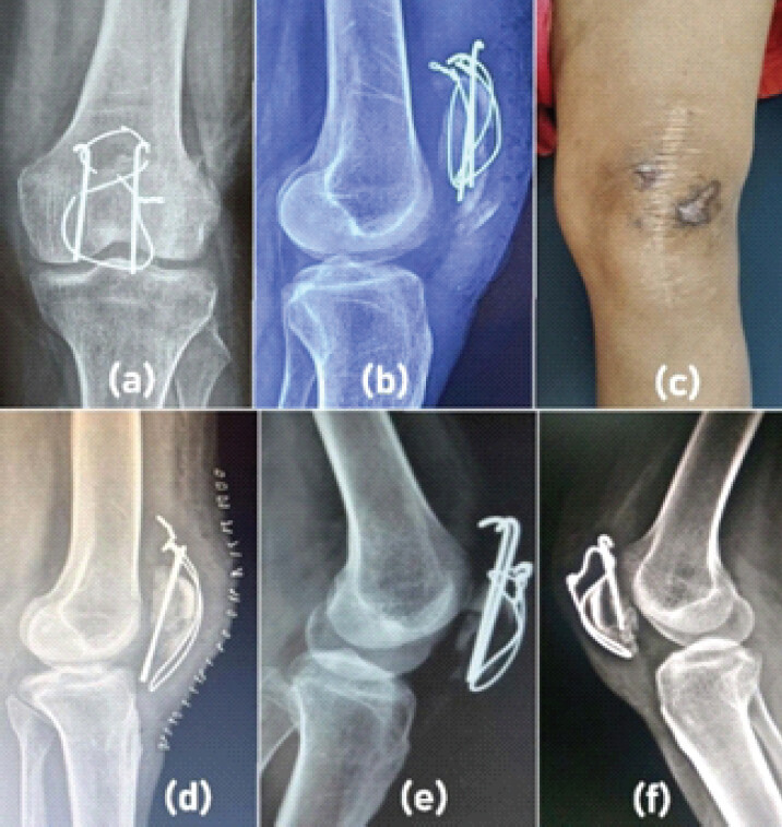
Biomechanical studies have demonstrated that modified anterior tension-band fixation using transosseous K-wires provides substantial mechanical strength compared with several alternative constructs [3]. However, this biomechanical advantage may be offset clinically by complications related to K-wire migration, slippage, prominence, and breakage. In the present study, slippage of the tension band through the K-wires occurred in 8 patients, of whom 2 required revision surgery. Thus, although K-wires may contribute to construct stability, their use may introduce implant-related complications (Fig. 4) that can compromise the clinical outcome and necessitate additional intervention.
The frequency of hardware-related complications following K-wire TBW has been documented in previous studies. Chawda et al. reported hardware impingement in 38% of patients, with wire breakage and knee effusion occurring in 10% and 12%, respectively [15]. Similarly, Hsu et al., in a series of 170 patients, identified implant irritation as the predominant complication, with nearly half of affected patients subsequently requiring implant removal [16]. K-wire migration is also a recognized complication of TBW, with implant-removal rates reported to be as high as 36.8% in some series [17].
Gupta et al. reported an overall complication rate of 15.2% following patellar fracture fixation, with the modified TBW group accounting for 71.4% of complications, predominantly due to K-wire migration and infection [18]. Although the modified TBW group demonstrated a numerically higher proportion of excellent Bostman scores, the modified cerclage wiring group had the lowest complication rate. The conventional TBW group demonstrated the greatest complication burden, particularly with respect to K-wire migration and infection.
In the present study, the lower incidence of implant-related complications in the combined cerclage group was accompanied by a substantially lower preference for implant removal and a reduced rate of actual secondary surgery. At 1 year, only 16% of patients in the combined cerclage group expressed willingness to undergo implant removal compared with 43% in the conventional K-wire TBW group. More importantly, by 18 months, actual implant removal surgery had been performed in 13% and 31% of patients, respectively, representing an absolute reduction of 18% points and a relative reduction of approximately 58% in secondary surgery. These findings suggest that reducing K-wire-related complications may translate into a clinically meaningful reduction in subsequent operative interventions. Because pain threshold, functional demands, and hardware tolerance vary significantly across individuals, the decision for hardware removal is inherently shared between the treating surgeon and the patient. While this introduces clinical variability, it reflects real-world pragmatic orthopedic practice rather than methodological oversight.
This observation is particularly relevant because symptomatic hardware is a well-recognized indication for secondary surgery following patellar fracture fixation. Lazaro et al. reported a 37% rate of hardware removal, primarily related to prominent or symptomatic implants resulting from wire breakage or persistent soft-tissue irritation [7]. Hung et al. reported hardware-removal rates of up to 10% within the first 12 months [4], while Hoshino et al. found that symptomatic implants were approximately twice as frequent with K-wires compared with cannulated screws [17]. Avoiding secondary surgery may reduce additional operative exposure, anesthetic requirements, healthcare expenditure, and patient inconvenience.
Several modifications of conventional K-wire TBW, including the use of cannulated screws, bending the K-wires at both ends, and creating loops in the K-wires, have been described to reduce implant-related complications [16,17,19,20]. However, these modifications do not completely eliminate problems such as hardware prominence, migration, soft-tissue irritation, and subsequent implant removal. In this context, the combined cerclage TBW technique provides a relatively simple alternative that eliminates the K-wire component.
We observed that avoiding passage of K-wires through the comminuted segment may reduce displacement or loss of small fracture fragments during fixation. This may therefore help preserve the articular bone stock and local vascularity of the comminuted fragments, potentially providing a more favorable biological environment for fracture healing and union. This may be one of the reasons for the favorable clinical and functional outcomes observed with combined cerclage TBW without K-wires in our study.
Overall, the findings of the present study suggest that K-wire-free combined cerclage TBW may provide a clinically effective balance between fracture stability, early functional recovery, and reduced implant-related complications. Its principal advantage appears to be the reduction in hardware-related morbidity and the consequent decrease in secondary implant-removal procedures, while maintaining functional outcomes comparable to K-wire TBW technique.
Limitations of the study
This study has several limitations. It is a single-center study and the choice of fixation method was guided by clinical assessment of multiple surgeons who performed the procedure, which might introduce selection bias, potentially limiting the generalizability of the findings. Variations in surgical experience and technical proficiency could not be completely controlled. The study included only two-part transverse patellar fractures with minimal comminution; therefore, the findings may not be directly generalizable to highly comminuted or complex fracture patterns. Blinding of patients and outcome assessors was not performed, which may have introduced observer or assessment bias.
The indications for implant removal were driven by patient-reported hardware discomfort and joint shared decision-making rather than a rigid protocol. Consequently, surgeon-dependent variations in recommending hardware removal and individual patient thresholds for undergoing secondary surgery may have introduced potential selection bias into the hardware removal outcomes.
Although follow-up extended to 18 months, late complications, including patellofemoral arthritis, delayed infection, and delayed implant-related problems, could not be assessed. While the Bostman score is specifically validated for patellar fractures, it does not encompass broader multidimensional patient-reported outcome measures (PROMs) such as the Knee Injury and Osteoarthritis Outcome Score or general health-related quality of life metrics. Future studies incorporating these comprehensive PROMs will provide deeper insights into patient-perceived recovery, long-term joint symptoms, and return to high-demand activities.
Conclusion
Both techniques were effective in achieving fracture union and restoring knee function. The study demonstrates that combined cerclage TBW without K-wires provides functional outcomes comparable to those of conventional modified TBW with K-wires at 1 year, while potentially reducing K-wire-related complications such as implant migration, prominence, irritation, and subsequent implant removal surgery. The findings support the use of combined cerclage TBW without K-wires, particularly in displaced transverse patellar fractures and fractures with comminution, as it provides stable fixation sufficient to permit early knee mobilization while avoiding the complications associated with K-wire fixation. Given these findings, cautious interpretation is warranted, and larger, multicenter randomized studies with longer follow-up, standardized surgical protocols, blinded outcome assessment, and comprehensive PROMs are needed to validate the intermediate and long-term utility of this approach.
Clinical Message
K-wire-free combined cerclage TBW is a useful alternative for appropriately selected transverse patellar fractures, offering stable fixation, early rehabilitation, and fewer symptomatic hardware-related complications.
Conflict of Interest:
Nil
Source of Support:
Nil
Consent:
The authors confirm that informed consent was obtained from the patient for publication of this article
How to Cite this Article
Pawar AV, Desai MM, Pawar KA. Combined Cerclage and Figure-of-eight Tension-band Wiring without K-wire Versus K-wire Tension Band Wiring Fixation for Transverse Patellar Fractures: A Prospective Comparative Study. Journal of Orthopaedic Case Reports 2026 October;16(10): 368-375.
References
- Boström A. Fracture of the patella. A study of 422 patellar fractures. Acta Orthop Scand Suppl 1972;143:1-80. [Google Scholar] | [PubMed]
- Levack B, Flannagan JP, Hobbs S. Results of surgical treatment of patellar fractures. J Bone Joint Surg Br 1985;67:416-9. [Google Scholar] | [PubMed]
- Bel JC, Lefèvre C. Reconstruction of patella fractures with the tension band technique: A review on clinical results and tips and tricks. Injury 2024;55 Suppl 1:111401. [Google Scholar] | [PubMed]
- Hung LK, Chan KM, Chow YN, Leung PC. Fractured patella: Operative treatment using the tension band principle. Injury 1985;16:343-7. [Google Scholar] | [PubMed]
- Dudani B, Sancheti KH. Management of fracture patella by tension band wiring. Indian J Orthop 1981;15:43-8. [Google Scholar] | [PubMed]
- Müller ME, Allgöwer M, Schneider R, Willenegger H. Manual of Internal Fixation: Techniques Recommended by the AO Group. Berlin: Springer-Verlag; 1979. 248-53. [Google Scholar] | [PubMed]
- Lazaro LE, Wellman DS, Sauro G, Pardee NC, Berkes MB, Little MT. Outcomes after operative fixation of complete articular patellar fractures: Assessment of functional impairment. J Bone Joint Surg Am 2013;95:e961-8. [Google Scholar] | [PubMed]
- Smith ST, Cramer KE, Karges DE, Watson JT, Moed BR. Early complications in the operative treatment of patella fractures. J Orthop Trauma 1997;11:183-7. [Google Scholar] | [PubMed]
- Pushpasekaran N, Karthik CA, Muthukannan HS. Migration of patella tension band metal wires to popliteal fossa: Retrieval by posterior knee arthroscopy - a case report and literature review. J Arthrosc Surg Sports Med 2023;4:53-5. [Google Scholar] | [PubMed]
- Curtis MJ. Internal fixation for fractures of the patella. A comparison of two methods. J Bone Joint Surg Br 1990;72:280-2. [Google Scholar] | [PubMed]
- Agarwala S, Agrawal P, Sobti A. A novel technique of patella fracture fixation facilitating early mobilization and reducing re-operation rates. J Clin Orthop Trauma 2015;6:207-11. [Google Scholar] | [PubMed]
- Böstman O, Kiviluoto O, Nirhamo J. Comminuted displaced fractures of the patella. Injury 1981;13:196-202. [Google Scholar] | [PubMed]
- Ong T, Chee EK, Wong CL, Thevarajan K, Khiam OT. Fixation of comminuted patellar fracture with combined cerclage and tension band wiring technique. Malays Orthop J 2008;2:40-2. [Google Scholar] | [PubMed]
- Sun Y, Sheng K, Li Q, Wang D, Zhou D. Management of comminuted patellar fracture fixation using modified cerclage wiring. J Orthop Surg Res 2019;14:324. [Google Scholar] | [PubMed]
- Chawda VR, Tank PM, Patel VJ, Shah YS. A prospective study of 50 cases of patella fractures treated with different modalities. Int J Res Orthop 2018;4:783-9. [Google Scholar] | [PubMed]
- Hsu KL, Chang WL, Yang CY, Yeh ML, Chang CW. Factors affecting the outcomes of modified tension band wiring techniques in transverse patellar fractures. Injury 2017;48:2800-6. [Google Scholar] | [PubMed]
- Hoshino CM, Tran W, Tiberi JV, Black MH, Li BH, Gold SM. Complications following tension-band fixation of patellar fractures with cannulated screws compared with Kirschner wires. J Bone Joint Surg Am 2013;95:653-9. [Google Scholar] | [PubMed]
- Gupta RK, Kalme B, Kumar A. A prospective study of comminuted patellar fractures treated with different fixation modalities: Functional outcomes and complications. J Orthop Case Rep 2026;14:174-81. [Google Scholar] | [PubMed]
- Berg EE. Open reduction internal fixation of displaced transverse patella fractures with figure-eight wiring through parallel cannulated compression screws. J Orthop Trauma 1997;11:573-6. [Google Scholar] | [PubMed]
- Lefaivre KA, O'Brien PJ, Broekhuyse HM, Guy P, Blachut PA. Modified tension band technique for patella fractures. Orthop Traumatol Surg Res 2010;96:579-82. [Google Scholar] | [PubMed]
© 2026 Journal of Orthopaedic Case Reports - Published by Indian Orthopaedic Research Group
About the Authors
-

Akshay V Pawar
-

Mohan M Desai
-

Kapil A Pawar
Related Articles in Journal of Orthopaedic Case Reports
 October 1, 2026 Association between Coronal Mechanical Axis Correction and Early Functional Outcomes after Primary Total Knee Arthroplasty: A Retrospective Observational Study
 October 1, 2026 A Study of Spinopelvic Relationship and its Impact on Functional Outcome of Total Hip Arthroplasty
 October 1, 2026 Outcomes of Primary Definitive Internal Fixation of Compound Fractures within 72 h of Injury: A Prospective Cohort Study
 October 1, 2026 Minimally Invasive J-Nail Fixation for Neer Type 2, 3, and 4 Proximal Humerus Fractures in Elderly Patients: A Prospective Clinical and Radiological Outcome Study