


Introduction
The anterior cruciate ligament (ACL) is a principal stabilizer of the knee, resisting anterior tibial translation and rotational load. ACL rupture is among the most frequently encountered sports-related knee injuries, with a reported annual incidence of 68.6/100,000 person-years [1]. Among 363 athletes presenting with sports-related knee injuries to a tertiary sports injury clinic in northern India, ACL tears were the most common injury, seen in 86.5%, followed by meniscal tears in 78.24% [2]. Arthroscopic reconstruction has become the standard of care for the symptomatically unstable knee and reliably restores mechanical stability. Functional recovery, however, does not follow automatically. Quadriceps atrophy and arthrogenic muscle inhibition begin within days of surgery and are most pronounced over the first 12 weeks [3]. Arthrogenic muscle inhibition – reflex inhibition of the quadriceps driven by effusion, pain, and altered afferent input from the injured joint – compounds simple disuse atrophy and is itself resistant to conventional strengthening [4]. Quadriceps weakness may persist for years after surgery and is associated with poorer functional performance, delayed return to sport, higher re-injury risk, and accelerated joint degeneration [5,6]. Early restoration of quadriceps bulk and strength is therefore one of the central objectives of rehabilitation after ACL reconstruction.
This objective is difficult to meet in practice. Muscle hypertrophy conventionally requires resistance training at loads of at least 65–70% of one-repetition maximum (1RM) [7]. Such loads are rarely tolerable in the first post-operative weeks, when pain, effusion, graft protection constraints, and arthrogenic inhibition all limit what a patient can perform. The result is a window in which atrophy progresses precisely when intervention would be most valuable, and within which conventional rehabilitation has limited means of intervening.
Blood flow restriction (BFR) therapy was developed to address this gap. First described in Japan in 1966 as KAATSU training – from KA, meaning “additional,” and ATSU, meaning “pressure” – the technique was initially applied in athletic conditioning and has since been adapted to clinical rehabilitation [8]. A pneumatic cuff applied to the proximal limb permits arterial inflow while restricting venous outflow, producing a hypoxic, metabolite-rich intramuscular environment during low-load exercise. The resulting accumulation of lactate and hydrogen ions, together with cell swelling, drives preferential recruitment of fast-twitch fibers and stimulates anabolic signaling and growth hormone release [9,10]. Through these mechanisms, BFR elicits hypertrophy and strength gains at external loads of only 20–30% of 1RM, comparable in magnitude to those achieved with high-load resistance training [11,12]. This makes the technique particularly suited to the early post-operative period, when high-load training is not feasible.
Evidence for BFR following ACL reconstruction has accumulated over the past few years but remains mixed. Recent meta-analyses of randomized controlled trials report improvements in pain, quadriceps strength, and functional scores relative to standard rehabilitation alone [13], and greater gains in quadriceps muscle volume and Lysholm score when BFR is applied during the early post-operative weeks [14]. A randomized trial using serial dual-energy X-ray absorptiometry demonstrated preservation of lower-limb lean mass and bone mass in the BFR group, both of which declined in controls [15]. Other systematic reviews, however, conclude that the evidence supporting benefits in muscle size and strength remains of limited and inconsistent quality [16]. Reported adverse events are uncommon, and the technique is generally well tolerated, although BFR is contraindicated in malignancy, thromboembolic disease, and active infection [17,18].
Most published work has assessed BFR using isokinetic strength testing and patient-reported outcome scores, whereas objective imaging-based quantification of muscle bulk has been reported less frequently, and data from Indian populations remain scarce. The present study was therefore undertaken to evaluate the clinical and radiological outcomes of adding BFR therapy to a standard rehabilitation protocol following arthroscopic ACL reconstruction, using quadriceps cross-sectional area (CSA) measured on magnetic resonance imaging (MRI) as the primary outcome, together with the Numeric Pain Rating Scale (NPRS), thigh girth, and Lysholm knee score.
Materials and Methods
This was a prospective observational cohort study with pre–post comparison, conducted at a tertiary care center after obtaining approval from the Institutional Ethics Committee (IEC/VMMC/SJH/Thesis/06/2022/CC-278; dated July 20, 2022). All participants provided written informed consent before enrollment. This research was conducted ethically in accordance with the World Medical Association Declaration of Helsinki. BFR therapy was part of the established post-operative rehabilitation protocol at our institution and was already used in patients undergoing ACL reconstruction independently of this study; it was not assigned to participants for research purposes. We enrolled and prospectively assessed consecutive eligible patients receiving this protocol at predefined time points, and the investigators did not influence operative or rehabilitative decision-making. The study was undertaken as a postgraduate thesis project and was approved by the IEC prior to commencement. The study was conducted at a government tertiary care institution, where imaging services are provided free of charge or at nominal subsidized cost as a matter of routine. Pre-operative MRI formed part of the standard diagnostic evaluation for suspected ACL injury. The post-operative MRI at 16 weeks, performed for the purposes of the study, was carried out in the institution’s radiology department at no cost to participants, with institutional approval. No participant incurred any additional financial expense, and the additional imaging involved no ionizing radiation or contrast administration. The study included 40 patients aged 18–45 years who underwent arthroscopic isolated ACL reconstruction. Patients with multiligamentous knee injury, meniscus injury, arthritic knee changes, chondral injury, or fractures around the knee were excluded. We also excluded patients who did not meet the inclusion criteria or in whom BFR therapy was contraindicated.
Sample size calculation
Based on the results of the study by Ohta et al. [19], at 95% confidence level and 80% power, taking the mean CSA of the knee extensor and flexor muscle before restriction of blood flow as 92 ± 11 and 99 ± 3, respectively, and the CSA of knee extensor and flexor muscle after restriction of blood flow as 92 ± 12 and 105 ± 19, respectively, the maximum sample size was calculated as 40 using the formula: n = ([Zα + Zβ] σ)2/δ2.
Pre-operative assessment
Patients presenting with symptoms of ACL injury underwent detailed clinical workup including thorough history taking and physical examination, followed by MRI for further evaluation and confirmation. Patients with ACL injury who fulfilled the inclusion criteria and were willing to participate were enrolled. Written informed consent was obtained. Axial-section plain MRI of the mid-thigh was performed to calculate CSA of the quadriceps muscle preoperatively. Pre-operative values of the Lysholm score and NPRS were assessed, and thigh girth was measured 10 cm above the superior pole of the patella preoperatively.
Limb occlusion pressure calculation
To calculate limb occlusion pressure for BFR therapy, a pneumatic tourniquet was applied to the proximal thigh postoperatively. The tourniquet was inflated, and the dorsalis pedis and tibialis posterior pulses were palpated simultaneously until they disappeared. We noted the cuff pressure at which pulses disappeared for each patient. We used 60–80% of that occlusion pressure for BFR therapy, depending on the patient’s tolerance.
Surgical procedure
All patients underwent arthroscopic single-bundle ACL reconstruction using a hamstring graft. The present study did not influence any operative aspects or decision-making in the pre-operative, intraoperative, or immediate post-operative periods, nor did it influence the rehabilitation protocol. Postoperatively, patients were monitored for vitals and to ensure painless recovery from anesthesia. Patients were advised to keep the operated limb elevated with cryotherapy in the form of ice packing. An extension brace was applied to protect the graft against involuntary movements. The rehabilitation program followed at our institute aligns with standard international guidelines.
BFR therapy protocol
On post-operative day 1, patients were assessed for NPRS and thigh girth. As per the institutional protocol, BFR therapy was initiated with the tourniquet inflated to 60% of the occlusion pressure. The tourniquet was inflated for 5 min during exercise, followed by deflation for 3 min. Patients underwent 5 cycles of inflation and deflation (40 min of therapy) twice a day, 3 times a week, for 16 weeks. Patients were followed up at 2, 6, and 16 weeks for NPRS and thigh girth. After 16 weeks, an axial MRI was obtained to calculate the quadriceps CSA and compare it with pre-operative values.
Outcome measures
Primary outcome
Quadriceps CSA. Axial T1-weighted and T2 Short Tau Inversion Recovery sequences of the thigh were acquired preoperatively and at 16 weeks postoperatively. The midpoint of the femur was localized on a coronal scout image so that the identical anatomical level was reproduced on both scans, and CSA was measured on the corresponding axial section by manually tracing the outline of the quadriceps muscle group; values are expressed in mm2. A single senior radiology faculty member performed all measurements and was blinded to the study details and clinical outcome data, although the patient’s post-operative status was necessarily apparent on the images.
Secondary outcomes
-
(I). NPRS – assessed preoperatively and on post-operative day 1 and at 2, 6, and 16 weeks. An eleven-point self-report scale in which the patient rates current pain intensity from 0 (“no pain”) to 10 (“worst pain imaginable”); a validated, widely used measure of pain intensity in musculoskeletal practice.
-
(ii). Thigh girth – assessed at the same time points. The circumference of the operated thigh, measured with a flexible non-elastic tape at a fixed point 10 cm proximal to the superior pole of the patella, with the patient supine and the knee in full extension; a simple clinical surrogate for quadriceps bulk.
-
(iii). Lysholm knee score – assessed preoperatively and at 16 weeks. A validated patient-reported instrument comprising eight domains (limp, use of support, locking, instability, pain, swelling, stair climbing, and squatting), scored from 0 to 100, with higher scores indicating better knee function.
Knee range of motion was recorded at each visit as part of routine clinical monitoring but was not analyzed as a study outcome.
Statistical analysis
Data was recorded and compiled using Microsoft Excel (version 2019) and analyzed using the Statistical Package for the Social Sciences (version 21.0; IBM, Armonk, NY, USA). Categorical data were expressed as frequencies and percentages. Quantitative data were expressed as mean and standard deviation. Paired t-tests were used for comparison with baseline values. P < 0.05 was considered statistically significant.
Results
A total of 40 patients were enrolled and followed up for 16 weeks postoperatively. The mean age was 28.25 ± 7.15 years. Most patients were in the 21–30-year age group, with 90% being male.
NPRS
As depicted in Table 1, the mean pre-operative NPRS score was 1.90 ± 1.24. A statistically significant increase in pain was observed on post-operative day 1 (4.40 ± 1.09; P < 0.0001), reflecting the expected immediate post-operative response. Thereafter, pain scores showed a significant and sustained reduction at 2 weeks (1.17 ± 0.66; P = 0.001), 6 weeks (1.05 ± 0.77; P < 0.001), and 16 weeks (1.07 ± 0.90; P = 0.001), all lower than baseline values. The effect size was large for the immediate post-operative increase and subsequent reduction (Cohen’s d > 2), whereas the long-term effect size remained small to moderate.
NPRS scores at each time point
| Time point | NPRS score (Mean±SD) | P-value (vs. pre-operative) |
|---|---|---|
| Pre-operative | 1.90±1.24 | — |
| Day 1 | 4.40±1.09 | <0.0001 |
| 2 weeks | 1.17±0.66 | 0.001 |
| 6 weeks | 1.05±0.77 | <0.001 |
| 16 weeks | 1.07±0.90 | 0.001 |
NPRS: Numeric pain rating scale, SD: Standard deviation
Lysholm score
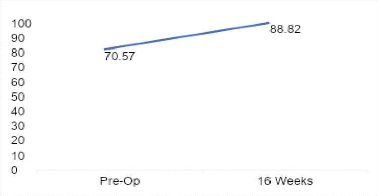
The Lysholm score shown in Table 2 improved from 70.57 ± 7.37 preoperatively to 88.82 ± 5.38 at 16 weeks, which was statistically highly significant (P < 0.0001). The mean improvement of approximately 18 points exceeds the established minimal clinically important difference (MCID) for the Lysholm score (8–10 points), indicating a clinically meaningful enhancement in knee function. The effect size was very large (Cohen’s d ≈ 2.8), suggesting a robust functional benefit.
Lysholm scores preoperatively and at 16 weeks (P<0.0001)
| Time point | Lysholm score (Mean±SD) |
|---|---|
| Pre-operative | 70.57±7.37 |
| 16 weeks | 88.82±5.38 |
SD: Standard deviation
Thigh girth
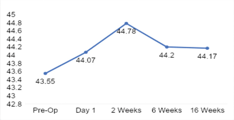
Thigh girth measurements showed no statistically significant differences at any post-operative time point compared to pre-operative values (P > 0.05 for all comparisons). The measurements remained relatively stable from baseline (43.55 ± 3.74 cm) through 16 weeks. Effect sizes were small (Cohen’s d < 0.3), indicating preservation of muscle bulk as shown in Table 3.
Thigh girth measurements at each time point
| Time point | Thigh girth in cm (Mean±SD) | P-value (vs. preoperative) |
|---|---|---|
| Pre-operative | 43.55±3.74 | — |
| Day 1 | 44.07±3.53 | 0.524 |
| 2 Weeks | 44.78±3.91 | 0.154 |
| 6 Weeks | 44.20±3.83 | 0.444 |
| 16 Weeks | 44.17±4.22 | 0.488 |
SD: Standard deviation
Quadriceps CSA on MRI
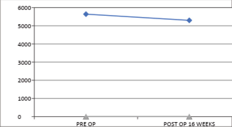
Quadriceps CSA measured on MRI decreased from 5637.40 ± 1043.88 mm2 preoperatively to 5295.25 ± 916.24 mm2 at 16 weeks, as shown in Table 4; however, this difference was not statistically significant (P = 0.123). The reduction of approximately 6% is below the commonly accepted threshold for clinical relevance (10–15%), indicating that the change was not clinically meaningful. The effect size was small (Cohen’s d ≈ 0.35).
Quadriceps CSA on MRI preoperatively and at 16 weeks
| Time point | Quadriceps CSA on MRI (mm2) (Mean±SD) | P-value |
|---|---|---|
| Pre-operative | 5637.40±1043.88 | 0.123 |
| 16 weeks | 5295.25±916.24 |
MRI: Magnetic resonance imaging, CSA: Cross-sectional area, SD: Standard deviation
Discussion
Return to sports or a pre-injury level of physical activity post-ACLR largely depends on the strength and stability of the knee joint and associated muscles, especially the quadriceps. In the present study, the addition of BFR therapy to the standard rehabilitation protocol was intended to hasten recovery and return to the pre-injury level of physical activity. Traditional resistance training for muscle hypertrophy typically involves high-load exercises (65–70% of 1RM) [7,20]. However, in the initial post-surgical period, patients are unable to tolerate such intensities due to pain, muscle inhibition, proprioceptive deficits, and psychological barriers. Hence, interest in BFR therapy as an adjunct to standard rehabilitation has increased. BFR enables muscle growth at significantly lower loads (25–50% of 1RM), which is more tolerable for post-operative patients [21]. The mean age in the current study was 28.25 ± 7.15 years, with most patients between 21 and 30 years. However, BFR therapy has also demonstrated benefits in older patients with osteoarthritis of the knee, with meta-analytic evidence of reduced pain and improved quadriceps strength [22].
The NPRS was used to assess pain. A significant increase in pain was observed on post-operative day 1, followed by a steady decline over the 16-week follow-up. Joseph et al. demonstrated that patients undergoing post-ACLR rehabilitation experienced a consistent decrease in NPRS scores as the rehabilitation progressed [23]. Although some studies have reported no significant differences in long-term pain between BFR and control groups, its potential to facilitate more intense rehabilitation with reduced discomfort remains promising [21].
In the present study, significant improvement in Lysholm scores was observed at 16 weeks postoperatively, supporting BFR’s role in enhancing functional recovery when combined with standard rehabilitation protocols. Jung et al. found significant improvements in Lysholm scores for both BFR and general rehabilitation groups, with slightly better outcomes in the BFR cohort (scores of 86.5 vs. 81 at 16 weeks) [24]. De Andrade et al. demonstrated that Lysholm scores correlated with improvements in peak torque of knee extensors in patients following ACL reconstruction, reinforcing the validity of this outcome measure in post-ACLR rehabilitation [25]. The outcome of the present study reiterates the functional benefits of incorporating BFR into standard protocols.
In the current study, no significant difference was found in thigh girth between pre-operative values and those at 16 weeks, suggesting that muscle bulk was maintained during recovery. This is important as disuse atrophy is a common and rapid consequence of post-operative immobilization. Ohta et al. concluded that BFR therapy significantly diminished post-operative atrophy of the knee extensors, a finding comparable to the current study [19]. While no increase in thigh girth was observed in our study, maintaining baseline values suggests that BFR therapy helped preserve muscle mass during a period typically associated with decline. To objectively quantify muscle preservation, the CSA of the quadriceps muscles was measured using MRI. At 16 weeks, there was no significant difference in CSA compared to baseline. The reduction of approximately 6% observed in our cohort may be placed in context against published serial imaging data following conventional rehabilitation. Garcia et al., using serial MRI, reported a 21.5% reduction in quadriceps CSA at 9 weeks after reconstruction, with a deficit of 10.1% persisting at return to activity approximately 9 months postoperatively [26]. The two studies are not directly comparable, in that Garcia et al. measured rectus femoris CSA whereas the present study measured the quadriceps group as a whole; nonetheless, the magnitude of reduction in our cohort appears smaller than that reported at comparable and later time points following standard rehabilitation alone. This observation is consistent with a muscle-preserving effect of BFR therapy, although in the absence of a control group it cannot establish one.
Limitations
The current study has several limitations. The sample size was small (n = 40). The absence of a control group and blinding limits the ability to attribute outcomes specifically to BFR therapy. The follow-up period of 16 weeks was relatively short. This duration was selected because quadriceps atrophy is most marked during the first 12 weeks after reconstruction [3], and because BFR therapy is intended to act during this early window – indeed, most published trials of BFR after ACL reconstruction have applied the intervention within the first 6 weeks [14]. Sixteen weeks therefore encompasses the period over which the intervention would be expected to exert its principal effect on muscle bulk and corresponds to the point at which rehabilitation at our institution transitions from impairment resolution and strengthening toward proprioceptive, balance, and agility training [27]. Nonetheless, quadriceps deficits after ACL reconstruction may persist well beyond this period, and functional recovery, sustained muscle preservation, and return to sport cannot be assessed within this timeframe. Longer follow-up is required to determine whether the early benefits observed here are maintained.
Conclusion
The results of BFR therapy are promising for improved functional outcomes (Lysholm score), preserved muscle mass, and reduced pain in the post-operative period. BFR therapy may therefore be used as part of the standard rehabilitation protocol following ACLR to preserve muscle bulk, improve function, prevent patellofemoral pain syndrome, and facilitate an earlier, faster return to sport in athletes.
Clinical Message
BFR therapy, when added to standard post-operative rehabilitation following ACL reconstruction, is safe, well-tolerated, and associated with preservation of muscle mass and improvement in functional outcomes. Clinicians should consider incorporating BFR therapy into rehabilitation protocols, particularly during the early post-operative phase when high-load training is not feasible.
Conflict of Interest:
Nil
Source of Support:
Nil
Consent:
The authors confirm that informed consent was obtained from the patient for publication of this article
How to Cite this Article
Gahlawat H, Kumar J, Hegde A, Sharma P, Gupta H, Joshi D. Outcomes of Blood Flow Restriction Therapy as an Adjunct to Standard Rehabilitation Following Anterior Cruciate Ligament Reconstruction: A Prospective Observational Cohort Study. Journal of Orthopaedic Case Reports 2026 October;16(10): 485-492.
References
- Sanders TL, Maradit Kremers H, Bryan AJ, Larson DR, Dahm DL, Levy BA. Incidence of anterior cruciate ligament tears and reconstruction: A 21-year population-based study. Am J Sports Med 2016;44:1502-7. [Google Scholar] | [PubMed]
- John R, Dhillon MS, Syam K, Prabhakar S, Behera P, Singh H. Epidemiological profile of sports-related knee injuries in northern India: An observational study at a tertiary care centre. J Clin Orthop Trauma 2016;7:207-11. [Google Scholar] | [PubMed]
- Grapar Žargi T, Drobnič M, Vauhnik R, Koder J, Kacin A. Factors predicting quadriceps femoris muscle atrophy during the first 12weeks following anterior cruciate ligament reconstruction. Knee 2017;24:319-28. [Google Scholar] | [PubMed]
- Sonnery-Cottet B, Saithna A, Quelard B, Daggett M, Borade A, Ouanezar H. Arthrogenic muscle inhibition after ACL reconstruction: A scoping review of the efficacy of interventions. Br J Sports Med 2019;53:289-98. [Google Scholar] | [PubMed]
- Thomas AC, Wojtys EM, Brandon C, Palmieri-Smith RM. Muscle atrophy contributes to quadriceps weakness after anterior cruciate ligament reconstruction. J Sci Med Sport 2016;19:7-11. [Google Scholar] | [PubMed]
- Schmitt LC, Paterno MV, Hewett TE. The impact of quadriceps femoris strength asymmetry on functional performance at return to sport following anterior cruciate ligament reconstruction. J Orthop Sports Phys Ther 2012;42:750-9. [Google Scholar] | [PubMed]
- Kraemer WJ, Adams K, Cafarelli E, Dudley GA, Dooly C, Feigenbaum MS. American college of sports medicine position stand. Progression models in resistance training for healthy adults. Med Sci Sports Exerc 2002;34:364-80. [Google Scholar] | [PubMed]
- Sato Y. The history and future of KAATSU training. Int J KAATSU Train Res 2005;1:1-5. [Google Scholar] | [PubMed]
- Pope ZK, Willardson JM, Schoenfeld BJ. Exercise and blood flow restriction. J Strength Cond Res 2013;27:2914-26. [Google Scholar] | [PubMed]
- Ladlow P, Coppack RJ, Dharm-Datta S, Conway D, Sellon E, Patterson SD. Low-load resistance training with blood flow restriction improves clinical outcomes in musculoskeletal rehabilitation: A single-blind randomized controlled trial. Front Physiol 2018;9:1269. [Google Scholar] | [PubMed]
- Pearson SJ, Hussain SR. A review on the mechanisms of blood-flow restriction resistance training-induced muscle hypertrophy. Sports Med 2015;45:187-200. [Google Scholar] | [PubMed]
- Lixandrão ME, Ugrinowitsch C, Berton R, Vechin FC, Conceição MS, Damas F. Magnitude of muscle strength and mass adaptations between high-load resistance training versus low-load resistance training associated with blood-flow restriction: A systematic review and meta-analysis. Sports Med 2018;48:361-78. [Google Scholar] | [PubMed]
- Gopinatth V, Garcia JR, Reid IK, Knapik DM, Verma NN, Chahla J. Blood flow restriction enhances recovery after anterior cruciate ligament reconstruction: A systematic review and meta-analysis of randomized controlled trials. Arthroscopy 2025;41:1048-60. [Google Scholar] | [PubMed]
- Lin Q, Zhang Y, Qin J, Wu F. Effects of Low-Load Blood Flow Restriction Training on Muscle Volume After Anterior Cruciate Ligament Reconstruction: A Systematic Review and Meta-analysis. Orthop J Sports Med. 2024 Dec 13;12(12):23259671241301731. doi: 10.1177/23259671241301731. PMID: 39678440; PMCID: PMC11645764. [Google Scholar] | [PubMed] | [CrossRef]
- Jack RA 2nd, Lambert BS, Hedt CA, Delgado D, Goble H, McCulloch PC. Blood flow restriction therapy preserves lower extremity bone and muscle mass after ACL reconstruction. Sports Health 2023;15:361-71. [Google Scholar] | [PubMed]
- Colapietro M, Portnoff B, Miller SJ, Sebastianelli W, Vairo GL. Effects of blood flow restriction training on clinical outcomes for patients with ACL reconstruction: A systematic review. Sports Health 2023;15:260-73. [Google Scholar] | [PubMed]
- Koc BB, Truyens A, Heymans MJ, Jansen EJ, Schotanus MG. Effect of low-load blood flow restriction training after anterior cruciate ligament reconstruction: A systematic review. Int J Sports Phys Ther 2022;17:334-46. [Google Scholar] | [PubMed]
- Minniti MC, Statkevich AP, Kelly RL, Rigsby VP, Exline MM, Rhon DI. The safety of blood flow restriction training as a therapeutic intervention for patients with musculoskeletal disorders: A systematic review. Am J Sports Med 2020;48:1773-85. [Google Scholar] | [PubMed]
- Ohta H, Kurosawa H, Ikeda H, Iwase Y, Satou N, Nakamura S. Low-load resistance muscular training with moderate restriction of blood flow after anterior cruciate ligament reconstruction. Acta Orthop Scand 2003;74:62-8. [Google Scholar] | [PubMed]
- Schoenfeld BJ. The mechanisms of muscle hypertrophy and their application to resistance training. J Strength Cond Res 2010;24:2857-72. [Google Scholar] | [PubMed]
- Scott BR, Loenneke JP, Slattery KM, Dascombe BJ. Blood flow restricted exercise for athletes: A review of available evidence. J Sci Med Sport 2016;19:360-7. [Google Scholar] | [PubMed]
- Lin Q, Yu D, Zhang Y, Chen X, Qin J, Wu F. Impact of low-load blood flow restriction training on knee osteoarthritis pain and muscle strength: A systematic review and meta-analysis of randomized controlled trials. Front Physiol 2025;16:1524480. [Google Scholar] | [PubMed]
- Joseph M, Fulkerson J, Nissen C, Sheehan TJ. Short-term recovery after anterior cruciate ligament reconstruction: A prospective comparison of three autografts. Orthopedics 2006;29:243-8. [Google Scholar] | [PubMed]
- Jung WS, Kim SH, Nam SS, Kim JW, Moon HW. Effects of rehabilitation exercise with blood flow restriction after anterior cruciate ligament reconstruction. Appl Sci 2022;12:12058. [Google Scholar] | [PubMed]
- de Andrade ALL, Castro A, Livani B, Belangero WD. Association between Lysholm score and muscular torque deficit after anterior cruciate ligament reconstruction. J Orthop Surg (Hong Kong). 2020 Jan-Apr;28(2):2309499020933485. doi: 10.1177/2309499020933485. PMID: 32618222. [Google Scholar] | [PubMed] | [CrossRef]
- Garcia SA, Curran MT, Palmieri-Smith RM. Longitudinal assessment of quadriceps muscle morphology before and after anterior cruciate ligament reconstruction and its associations with patient-reported outcomes. Sports Health 2020;12:271-8. [Google Scholar] | [PubMed]
- Wilk KE, Macrina LC, Cain EL, Dugas JR, Andrews JR. Recent advances in the rehabilitation of anterior cruciate ligament injuries. J Orthop Sports Phys Ther 2012;42:153-71. [Google Scholar] | [PubMed]
© 2026 Journal of Orthopaedic Case Reports - Published by Indian Orthopaedic Research Group
About the Authors
-

Happy Gahlawat
-

Jaswant Kumar
-

Amit Hegde
-

Prashant Sharma
-

Himanshu Gupta
-

Deepak Joshi
Related Articles in Journal of Orthopaedic Case Reports
 September 1, 2026 Comparative Analysis of Functional Outcomes of Accelerated versus Conventional Rehabilitation after Anterior Cruciate Ligament Reconstruction Surgery
 August 1, 2026 Comparative Outcomes of Hamstring Tendon versus Peroneus Longus Tendon Autograft in Arthroscopic Single-Bundle Anterior Cruciate Ligament Reconstruction: A Prospective Comparative Study
 July 1, 2026 Clinical and Donor-site Outcomes of Full-thickness Peroneus Longus Autograft in Arthroscopic Anterior Cruciate Ligament Reconstruction: A Prospective Indian Cohort Study
 February 1, 2026 Knee Joint Dislocation in a Difficult, Low-Resource Himalayan Setting: A Longitudinal Functional Outcome Study