


Introduction
Knee osteoarthritis (OA) is a major cause of pain, disability, and reduced mobility worldwide, with its burden increasing with population aging and rising obesity rates. A global population-based meta-analysis estimated the prevalence of knee OA at 22.9% among adults aged ≥40 years and the incidence at 203 cases per 10,000 person-years among adults aged ≥20 years [1]. Global Burden of Disease data have also demonstrated substantial increases in the prevalence and incidence of knee OA over recent decades [2]. In India, estimates vary according to population characteristics and diagnostic criteria; recent systematic reviews have reported substantial variation in the prevalence of knee OA among Indian adults and older populations [3, 4].
For patients with advanced symptomatic OA who continue to experience pain and functional limitation despite appropriate conservative treatment, total knee arthroplasty (TKA) is an established treatment that provides substantial improvements in pain, physical function, and quality of life [5]. Nevertheless, some patients experience residual symptoms or dissatisfaction after TKA, suggesting that successful arthroplasty depends not only on implant survivorship but also on appropriate limb alignment, restoration of knee biomechanics, and patient-reported functional outcomes [5, 6].
Coronal alignment has traditionally been regarded as an important determinant of TKA outcomes. Mechanical alignment aims to restore the mechanical axis of the lower limb toward a predefined post-operative target, with the objective of achieving balanced load transmission and reproducible femoral and tibial component positioning. Mechanical axis deviation (MAD), assessed using standing weight-bearing long-leg radiographs, provides an objective measure of coronal limb alignment and allows quantification of the change in alignment between pre-operative and post-operative assessments.
These findings suggest that the magnitude and direction of post-operative correction may be more clinically relevant than achieving neutral alignment alone. A prospective Indian study evaluating 60 knees found significant correction of pre-operative mechanical-axis deviation but no significant difference in range of motion or Knee Society Scores between patients with mechanical-axis alignment within conventional limits and those with mild residual malalignment at 6 months [6]. Conversely, a 2024 study of 409 primary TKAs demonstrated that patients with pre-operative varus alignment who were overcorrected into valgus had inferior Knee Injury and Osteoarthritis Outcome Scores and reduced early range of motion compared with patients corrected to neutral or who remained mildly varus [7].
Recent evidence has further challenged the concept of a single universal alignment target. Analysis using the coronal plane alignment of the knee (CPAK) classification has demonstrated that mechanically aligned TKA may not produce uniform clinical outcomes across different pre-operative coronal phenotypes. Patients with varus arithmetic hip-knee-ankle alignment have been reported to have inferior functional scores at 1 year [8]. Similarly, contemporary systematic reviews and meta-analyses comparing mechanical and kinematic alignment have generally reported comparable patient-reported outcomes, although selected studies have suggested advantages in specific functional parameters with individualized alignment strategies.
These findings highlight an important clinical gap. Although post-operative alignment is routinely assessed after TKA, the clinical relevance of the magnitude of change from pre-operative deformity to post-operative alignment remains incompletely defined, particularly in Indian clinical cohorts. Rather than classifying post-operative alignment simply as neutral or non-neutral, assessing both the final mechanical axis and the magnitude of correction may provide a more clinically meaningful understanding of their relationship with early functional recovery.
Therefore, this study aimed to evaluate pre-operative and post-operative coronal mechanical alignment in patients undergoing primary TKA and to examine its association with early functional outcomes. Specifically, we assessed the magnitude of mechanical-axis correction, compared functional outcomes according to post-operative alignment, and evaluated whether the magnitude of coronal correction was associated with WOMAC and Oxford Knee Scores (OKSs) during the first 6 months after surgery.
Materials and Methods
Study design and ethical considerations
A single-center retrospective observational study was conducted at Shri Balaji Institute of Medical Sciences, Raipur, India. Consecutive patients who underwent primary TKA between January 2024 and December 2025 were retrospectively identified from institutional medical records. Clinical, radiological, and functional data were retrieved from the electronic medical record system and picture archiving and communication system (PACS).
The study was approved by the Institutional Ethics Committee of Shri Balaji Institute of Medical Sciences, Raipur (Approval No. SBIMS/IEC/Certi./172/2025, dated September 16, 2025). The study was conducted in accordance with the ethical principles of the Declaration of Helsinki.
Study population
Medical records of patients aged ≥55 years with symptomatic OA who underwent primary TKA during the study period were reviewed. Patients meeting the predefined eligibility criteria were included. A total of 60 patients involving 85 operated knees were included; 25 patients underwent bilateral TKA and 35 underwent unilateral TKA.
Baseline demographic and clinical characteristics, including age, sex, height, weight, body mass index (BMI), and relevant comorbidities, were retrieved from the medical records. For patients who underwent bilateral TKA, the timing and sequence of the two procedures were recorded.
Inclusion and exclusion criteria
Patients were included if they were aged ≥55 years and had symptomatic bicompartmental or tricompartmental OA for which primary TKA had been performed.
Patients were excluded if they had a previous intra-articular or extra-articular fracture or ligamentous injury around the knee; previous septic arthritis, tuberculosis, or tumor involving the knee; deformity around the knee secondary to previous trauma, infection, congenital, metabolic, or neoplastic causes; neuromuscular disorders affecting lower-limb function; or previous surgery involving the affected limb that could influence lower-limb alignment or post-operative functional assessment.
Pre-operative assessment
Pre-operative clinical and radiographic data obtained approximately 1 week before surgery were retrospectively retrieved. Functional status was assessed using the Western Ontario and McMaster Universities Osteoarthritis Index (WOMAC) [9] and OKS [10]. Demographic characteristics, relevant comorbidities, and pre-operative clinical findings were obtained from institutional medical records.
Radiographic assessment
Full-length standing anteroposterior radiographs of both lower limbs were obtained preoperatively with the patient in a weight-bearing position and both lower limbs positioned symmetrically. The patellae were oriented anteriorly to minimize rotational malposition. Radiographs were retrospectively reviewed and digitally analyzed using the institutional PACS measurement software.
The lower-limb mechanical axis (Maquet line) was defined as the line connecting the center of the femoral head to the center of the talar dome. MAD was measured as the perpendicular distance between the mechanical axis and the center of the knee joint and was recorded in centimeters. The direction of deviation was recorded according to the predefined varus/valgus convention.
The femoral mechanical axis was defined as the line connecting the center of the femoral head to the appropriate distal femoral reference point, whereas the tibial mechanical axis was defined as the line connecting the center of the tibial intercondylar region to the center of the talar dome.
Pre-operative radiographic parameters included MAD, mechanical lateral distal femoral angle (mLDFA), medial proximal tibial angle (MPTA), joint-line convergence angle (JLCA), and femorotibial mechanical angle (FTMA). Post-operative radiographic parameters included MAD, femoral mechanical angle (FMA), tibial mechanical angle (TMA), and FTMA.
Varus alignment was recorded as negative and valgus alignment as positive. The exact radiographic definitions and angular conventions were applied consistently to all measurements.
All radiographic measurements were performed independently by two observers who were blinded to the post-operative functional outcomes. For assessment of measurement reliability, measurements were repeated in a randomly selected subset of radiographs after an interval of at least 2 weeks. Interobserver and intraobserver reliability were assessed using intraclass correlation coefficients.
Surgical technique
All patients underwent primary TKA using a standardized institutional surgical protocol under spinal anesthesia. A standard midline skin incision and medial parapatellar/subvastus approach were used according to the operative protocol. The anterior cruciate ligament and menisci were excised, and marginal osteophytes were removed.
Femoral preparation was performed using intramedullary alignment with a target distal femoral valgus angle of approximately 5°. Tibial preparation was performed using extramedullary alignment with the intended tibial resection and posterior slope according to the institutional surgical protocol. Femoral sizing was performed using anterior referencing, with approximately 3° of external rotation relative to the predefined femoral reference axis.
Flexion and extension gaps were assessed intraoperatively, and limited medial soft-tissue release was performed when required to achieve satisfactory balance. Trial components were used to assess range of motion, stability, and patellar tracking. Definitive components were implanted after satisfactory balancing. Cement fixation was performed using polymethylmethacrylate bone cement following pulsatile lavage and drying of the prepared bone surfaces. Excess cement was removed, and the knee was maintained in extension during cement polymerization.
Post-operative management
Post-operative analgesia and thromboprophylaxis were administered according to institutional protocols. Rehabilitation was initiated on the 1st post-operative day and included quadriceps and ankle-pump exercises, progressive knee flexion exercises, and weight-bearing ambulation with walker support as tolerated.
Follow-up and outcome assessment
Post-operative clinical data were retrieved at 2 weeks, 6 weeks, 3 months, and 6 months after surgery. Standing full-length anteroposterior radiographs obtained at 6 weeks using the same standardized technique as the pre-operative examination were retrospectively reviewed for post-operative alignment.
Post-operative radiographic parameters included MAD, FMA, TMA, and FTMA.
Functional outcomes were assessed using WOMAC and OKS at 6 weeks, 3 months, and 6 months. The primary radiological exposure was the change in mechanical-axis alignment from pre-operative assessment to 6 weeks postoperatively. The primary clinical outcomes were WOMAC and OKS scores at 6 months, with earlier post-operative scores considered secondary longitudinal outcomes.
The magnitude of coronal correction was calculated as the absolute difference between the pre-operative and post-operative FTMA values and categorized into three groups: <5°, 5°–10°, and >10°.
Statistical analysis
Statistical analyses were performed using IBM Statistical Package for the Social Sciences Statistics version 20. Continuous variables were summarized as mean ± standard deviation (SD), while categorical variables were summarized as frequencies and percentages. The distribution of continuous variables was assessed before inferential analysis.
Pre-operative and post-operative radiographic measurements were compared using paired statistical tests appropriate to the distribution of the data. Changes in WOMAC and OKS scores over time were assessed using a repeated-measures approach.
Associations between post-operative FTMA alignment groups and functional outcomes were assessed using statistical models appropriate for clustered observations, with patient identification included as a clustering variable to account for the correlation between knees from the same patient. Similarly, associations between the magnitude of FTMA correction and functional outcomes were analyzed using models accounting for within-patient clustering.
Where multiple-group comparisons were performed, an overall omnibus test was followed by appropriate multiplicity-adjusted pairwise comparisons. Effect estimates with 95% confidence intervals were reported where applicable. A two-sided P < 0.05 was considered statistically significant.
Results
Patient characteristics and post-operative mechanical alignment
A total of 60 patients undergoing primary TKA contributed 85 knees to the study. Twenty-five patients underwent bilateral knee arthroplasty, while 35 underwent unilateral arthroplasty. The mean age was 66.42 ± 7.06 years, and 44 patients (73.3%) were female. The mean BMI was 28.61 ± 3.38 kg/m². All knees received cruciate-retaining implants.
According to post-operative FTMA, 18 knees (21.2%) demonstrated valgus alignment (>+3°), 36 knees (42.4%) were within the neutral range (−3° to +3°), 20 knees (23.5%) demonstrated mild residual varus alignment (<−3° to >−6°), and 11 knees (12.9%) demonstrated severe residual varus alignment (≤−6°).
There were no significant differences among the four post-operative FTMA groups in age (P = 0.669), sex (P = 0.711), BMI (P = 0.743), pre-operative WOMAC score (P = 0.858), or pre-operative OKS (P = 0.148) after accounting for clustering of bilateral knees within patients. Baseline characteristics according to post-operative FTMA category are presented in Table 1.
Baseline demographic and clinical characteristics according to post-operative FTMA
| Variable | Valgus >+3° (n=18) | Neutral –3° to +3° (n=36) | Mild varus <−3° to >−6° (n=20) | Severe varus <−6° (n=11) | Overall P-value |
|---|---|---|---|---|---|
| Age, years | 66.00±8.45 | 66.92±7.12 | 68.05±6.04 | 63.91±6.16 | 0.669 |
| Female, n (%) | 16 (88.9) | 22 (61.1) | 15 (75.0) | 10 (90.9) | 0.711 |
| Male, n (%) | 2 (11.1) | 14 (38.9) | 5 (25.0) | 1 (9.1) | |
| BMI, kg/m2 | 29.19±3.44 | 28.03±2.93 | 28.90±3.72 | 28.76±3.37 | 0.743 |
| Pre-operative WOMAC | 77.06±5.09 | 76.28±5.68 | 75.10±5.93 | 76.64±7.10 | 0.858 |
| Pre-operative OKS | 11.78±2.53 | 10.50±2.30 | 11.35±2.81 | 10.55±2.77 | 0.148 |
WOMAC: Western Ontario and McMaster Universities Osteoarthritis Index, OKS: Oxford Knee Score
Pre-operative radiographic characteristics
The mean pre-operative FTMA was −10.13 ± 4.82°. The mean pre-operative MAD was 4.85 ± 2.17 cm, medial distal femoral angle (mLDFA) was 93.15 ± 2.81°, medial proximal tibial angle (MPTA) was 83.13 ± 3.04°, and joint-line convergence angle (JLCA) was 3.90 ± 1.68°.
Pre-operative FTMA differed significantly according to the post-operative alignment category (P < 0.001). Similarly, significant differences were observed in pre-operative MAD (P < 0.001), mLDFA (P < 0.001), and MPTA (P = 0.019). JLCA did not differ significantly among the post-operative FTMA groups (P = 0.943). Greater pre-operative deformity was observed among knees that subsequently demonstrated residual varus alignment (Table 2).
Pre-operative radiographic parameters according to post-operative FTMA
| Radiographic parameter | Valgus >+3° | Neutral −3° to +3° | Mild varus <−3° to >−6° | Severe varus <−6° | Overall P-value |
|---|---|---|---|---|---|
| Pre-operative FTMA, ° | −9.44±2.51 | −8.58±3.31 | −10.76±4.63 | −15.18±8.17 | <0.001 |
| Pre-operative MAD, cm | 4.25±1.73 | 3.91±1.73 | 5.61±1.89 | 7.55±2.02 | <0.001 |
| Pre-operative mLDFA, ° | 92.52±2.63 | 92.18±2.61 | 93.96±2.57 | 95.87±2.16 | <0.001 |
| Pre-operative MPTA, ° | 83.61±2.52 | 84.11±2.24 | 82.11±3.81 | 80.95±3.24 | 0.019 |
| Pre-operative JLCA, ° | 3.46±1.39 | 3.75±1.49 | 3.68±1.04 | 5.48±2.70 | 0.943 |
FTMA: Femorotibial mechanical axis, MAD: Mechanical axis deviation, mLDFA: Mechanical lateral distal femoral angle, JLCA: Joint-line convergence angle, MPTA: Medial proximal tibial angle
Correction of mechanical axis and component alignment
Substantial correction of the mechanical axis was observed following surgery. Mean MAD decreased from 4.85 ± 2.17 cm preoperatively to 1.61 ± 1.24 cm postoperatively, representing an absolute mean reduction of 3.24 cm (66.8%). In a patient-clustered mixed-effects analysis, the estimated mean change was −3.31 cm (95% CI, −3.71 to −2.90; P < 0.001).
Mean FTMA improved from −10.13 ± 4.82° preoperatively to −1.49 ± 3.95° postoperatively, corresponding to a mean correction of 8.64°. The patient-clustered estimated mean correction was 8.47° (95% CI, 7.31–9.63; P < 0.001).
The mean pre-operative mLDFA was 93.15 ± 2.81°, compared with 90.02 ± 2.08° postoperatively. The mean change was −3.12°, with a patient-clustered estimated change of −3.11° (95% CI, −3.79 to −2.44; P < 0.001). The mean post-operative FMA was 89.98 ± 2.08°, while the mean post-operative TMA was 87.67 ± 2.55° (Fig. 1 and Table 3).
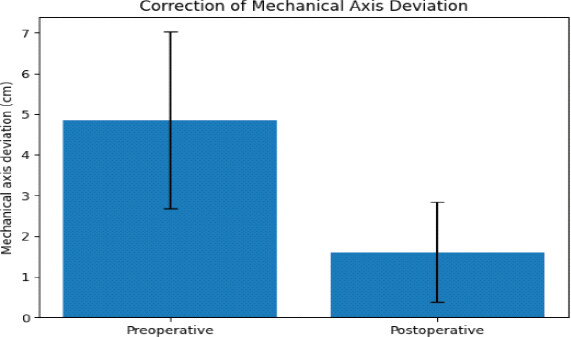
Pre-operative and post-operative radiographic parameters
| Parameter | Preoperative | Postoperative | Mean change | 95% CI for change | P-value |
|---|---|---|---|---|---|
| FTMA, ° | −10.13±4.82 | −1.49±3.95 | +8.47°* | 7.31 to 9.63 | <0.001 |
| MAD, cm | 4.85±2.17 | 1.61±1.24 | −3.31 cm* | −3.71 to −2.90 | <0.001 |
| mLDFA, ° | 93.15±2.81 | 90.02±2.08 | −3.11°* | −3.79 to −2.44 | <0.001 |
| MPTA, ° | 83.13±3.04 | — | — | — | — |
| JLCA, ° | 3.90±1.68 | — | — | — | — |
| FMA, ° | — | 89.98±2.08 | — | — | — |
| TMA, ° | — | 87.67±2.55 | — | — | — |
FTMA: Femorotibial mechanical axis, MAD: Mechanical axis deviation, mLDFA: Mechanical lateral distal femoral angle, JLCA: Joint-line convergence angle, MPTA: Medial proximal tibial angle, FMA: Femoral mechanical angle, TMA: Tibial mechanical angle
Functional outcomes over time
The mean WOMAC score decreased from 76.21 ± 5.75 preoperatively to 42.25 ± 6.44 at 6 weeks, 24.73 ± 5.55 at 3 months, and 11.58 ± 4.35 at 6 months. The corresponding mean reductions from baseline were 33.96, 51.48, and 64.64 points, respectively. Thus, the 6-month WOMAC score represented an 84.8% reduction from baseline. The overall effect of time was statistically significant (P < 0.001).
The mean OKS increased from 10.98 ± 2.55 preoperatively to 27.21 ± 3.64 at 6 weeks, 38.39 ± 3.10 at 3 months, and 41.95 ± 3.35 at 6 months. The corresponding mean improvements from baseline were 16.24, 27.41, and 30.98 points, respectively. The overall effect of time was statistically significant (P < 0.001) (Fig. 2 and Table 4).
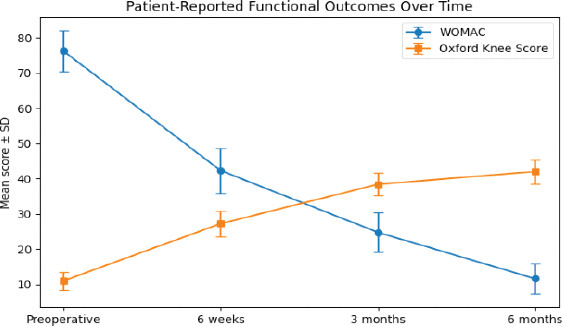
Functional outcomes at different post-operative time points
| Outcome | Preoperative | 6 weeks | 3 months | 6 months | Overall P-value |
|---|---|---|---|---|---|
| WOMAC | 76.21±5.75 | 42.25±6.44 | 24.73±5.55 | 11.58±4.35 | <0.001 |
| Change from baseline | — | −33.96 | −51.48 | −64.64 | |
| Oxford knee score | 10.98±2.55 | 27.21±3.64 | 38.39±3.10 | 41.95±3.35 | <0.001 |
| Change from baseline | — | 16.24 | 27.41 | 30.98 |
WOMAC: Western Ontario and McMaster Universities Osteoarthritis Index
Functional outcomes according to post-operative mechanical alignment
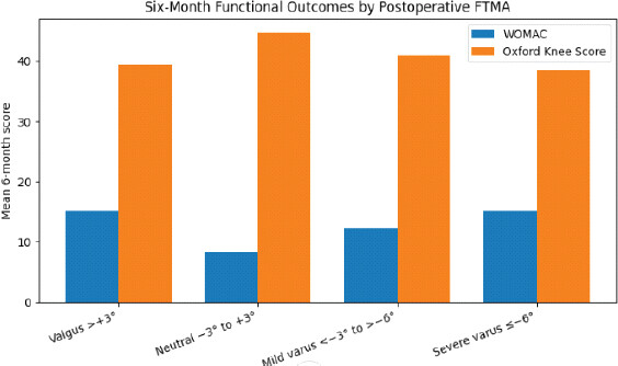
At 6 months, the mean WOMAC score was 14.60 ± 5.56 in the valgus group, 8.33 ± 2.13 in the neutral group, 12.50 ± 2.22 in the mild-varus group, and 15.18 ± 1.88 in the severe-varus group. Corresponding mean OKS values were 39.90 ± 3.05, 44.80 ± 1.65, 40.61 ± 2.45, and 38.54 ± 1.75, respectively.
After adjustment for age, sex, BMI, baseline outcome score, pre-operative FTMA, and pre-operative MAD, post-operative FTMA category was significantly associated with both 6-month WOMAC and OKS scores (overall P < 0.001 for both outcomes).
Compared with the neutral group, the adjusted mean WOMAC score was 5.31 points higher in the valgus group (95% CI, 2.85–7.77; Holm-adjusted P < 0.001), 2.21 points higher in the mild-varus group (95% CI, 1.14–3.27; Holm-adjusted P < 0.001), and 5.00 points higher in the severe-varus group (95% CI, 3.49–6.51; Holm-adjusted P < 0.001).
Similarly, compared with the neutral group, the adjusted mean OKS was 5.55 points lower in the valgus group (95% CI, −6.90 to −4.20; Holm-adjusted P < 0.001), 3.89 points lower in the mild-varus group (95% CI, −5.25 to −2.53; Holm-adjusted P < 0.001), and 6.16 points lower in the severe-varus group (95% CI, −7.58 to −4.74; Holm-adjusted P < 0.001). These findings indicate that knees with neutral post-operative mechanical alignment had the most favorable 6-month functional outcomes (Fig. 3).
Functional outcomes according to femoral and tibial component alignment
Post-operative FMA was classified as varus (<87°), neutral (87–93°), or valgus (>93°). Five knees (5.9%) were classified as varus, 71 knees (83.5%) as neutral, and nine knees (10.6%) as valgus.
At 6 months, mean WOMAC scores were 12.40 ± 2.70, 11.01 ± 3.92, and 15.56 ± 6.31 in the varus, neutral, and valgus FMA groups, respectively. Corresponding OKS values were 38.60 ± 2.61, 42.46 ± 3.18, and 39.78 ± 3.35.
The overall difference in 6-month WOMAC scores according to FMA category was not statistically significant (P = 0.238). In contrast, 6-month OKS differed significantly among FMA categories (P < 0.001), with the neutral FMA group demonstrating higher scores than both the varus and valgus groups.
Post-operative TMA was classified as varus (<87°), neutral (87–93°), or valgus (>93°). Thirty-eight knees (44.7%) were classified as varus, 45 knees (52.9%) as neutral, and two knees (2.4%) as valgus.
At 6 months, mean WOMAC scores were 12.39 ± 2.86, 10.73 ± 5.16, and 15.00 ± 5.66 in the varus, neutral, and valgus TMA groups, respectively. Corresponding OKS values were 40.32 ± 2.84, 43.38 ± 3.17, and 41.00 ± 2.83.
TMA category was significantly associated with both 6-month WOMAC (p<0.001) and OKS (p<0.001). The neutral TMA group demonstrated more favorable functional scores than the varus group. However, interpretation of comparisons involving the valgus TMA subgroup is limited because only two knees were classified as valgus (Table 5).
Six-month functional outcomes according to component alignment
| Component alignment | n | WOMAC, mean±SD | OKS, meantSB |
|---|---|---|---|
| FMA | |||
| Varus <87° | 5 | 12.40±2.70 | 38.60±2.61 |
| Neutral 87−93° | 71 | 11.01±3.92 | 42.46±3.18 |
| Valgus >93° | 9 | 15.56±6.31 | 39.78±3.35 |
| Overall P value | 0.238 | <0.001 | |
| TMA | |||
| Varus <87° | 38 | 12.39±2.86 | 40.32±2.84 |
| Neutral 87-93° | 45 | 10.73±5.16 | 43.38±3.17 |
| Valgus >93° | 2 | 15.00±5.66 | 41.00±2.83 |
| Overall P value | <0.001 | <0.001 |
FMA: Femoral mechanical angle, TMA: Tibial mechanical angle, OKS: Oxford knee score
Functional outcomes according to magnitude of mechanical-axis correction
The absolute magnitude of mechanical-axis correction was calculated as the absolute difference between pre-operative and post-operative FTMA. Twenty-four knees (28.2%) demonstrated correction of <5°, 32 knees (37.6%) demonstrated correction of 5–10°, and 29 knees (34.1%) demonstrated correction of >10°. The mean absolute correction was 8.64 ± 5.01°.
At 6 months, mean WOMAC scores were 11.46 ± 3.12, 10.25 ± 4.06, and 13.14 ± 5.09 in the <5°, 5–10°, and >10° correction groups, respectively. Corresponding OKS values were 41.54 ± 3.39, 43.31 ± 2.91, and 40.79 ± 3.35.
After adjustment for age, sex, BMI, baseline outcome score, pre-operative FTMA, and pre-operative MAD, correction magnitude was not significantly associated with 6-month WOMAC scores overall (P = 0.168). However, correction magnitude was significantly associated with 6-month OKS scores overall (P = 0.009).
For OKS, the 5–10° correction group demonstrated a higher score than the <5° group by 1.69 points (95% CI, 0.05–3.33; unadjusted P = 0.043) and a higher score than the >10° group by 2.20 points (95% CI, 0.43–3.97; unadjusted P = 0.015). After Holm correction for multiple pairwise comparisons, the corresponding adjusted p-values were 0.086 and 0.044, respectively. Thus, only the comparison between the 5–10° and >10° correction groups remained statistically significant after multiplicity adjustment.
For WOMAC, none of the pairwise comparisons remained statistically significant after multiplicity adjustment (Table 6).
Six-month functional outcomes according to magnitude of mechanical-axis correction
| Correction magnitude | n | WOMAC, meantSD | OKS, mean±SD |
|---|---|---|---|
| <5° | 24 | 11.46±3.12 | 41.54±3.39 |
| 5-10° | 32 | 10.25±4.06 | 43.31±2.91 |
| >10° | 29 | 13.14±5.09 | 40.79±3.35 |
| Overall P value | 0.168 | 0.009 | |
| Pairwise comparisons | |||
| Outcome | Comparison | Mean difference (95% CI) | Holm-adjusted P value |
| WOMAC | <5° versus 5−10° | −1.38 (−2.87 to 0.10) | 0.203 |
| WOMAC | <5° versus >10° | −0.25 (−2.03 to 1.54) | 0.785 |
| WOMAC | 5-10° versus >10° | +1.13 (−0.69 to 2.96) | 0.448 |
| OKS | <5° versus 5-10° | +1.69 (0.05 to 3.33) | 0.086 |
| OKS | <5° versus >10° | −0.51 (−2.84 to 1.81) | 0.666 |
| OKS | 5–10° versus >10° | −2.20 (−3.97 to −0.43) | 0.044 |
FMA: Femoral mechanical angle, TMA: Tibial mechanical angle, OKS: Oxford knee score
Discussion
Coronal alignment after total knee arthroplasty (TKA) has traditionally been regarded as an important determinant of component positioning, load distribution, and postoperative function. Mechanical alignment, with restoration of the lower-limb mechanical axis toward a predefined target, has therefore remained a widely used surgical strategy. However, the optimal degree of coronal correction remains debated, particularly in patients presenting with preoperative varus deformity. Contemporary evidence increasingly supports consideration of individual preoperative alignment characteristics rather than reliance on a single universal postoperative target [1–4].
In the present cohort, primary TKA was associated with substantial correction of preoperative coronal mechanical-axis deviation, with mean MAD decreasing from 4.79 ± 2.20 cm preoperatively to 1.62 ± 1.62 cm postoperatively. This finding demonstrates substantial radiographic correction of the preoperative deformity, although the magnitude of change should be interpreted alongside the statistical comparison of paired preoperative and postoperative measurements. Previous studies have similarly emphasized the importance of accurate coronal alignment for rehabilitation and functional recovery [5,6]. Longstaff et al., for example, reported that better component alignment was associated with improved function and faster rehabilitation, while Choong et al. demonstrated that more accurate alignment was associated with improved clinical outcomes in selected patients [5,6].
The postoperative mechanical alignment pattern in our cohort was heterogeneous. Neutral alignment (−3° to +3°) was achieved in 36 of 85 knees (42.4%), while 20 knees (23.5%) demonstrated valgus alignment, 18 (21.2%) mild varus alignment, and 11 (12.9%) severe varus alignment. Interestingly, patients with neutral postoperative FTMA demonstrated the most favorable six-month functional outcomes. The mean six-month WOMAC score was 8.33 ± 2.13 in the neutral group compared with 14.60 ± 5.56 in the valgus group, 12.50 ± 2.22 in the mild varus group, and 15.18 ± 1.88 in the severe varus group (p=0.049). Similarly, the neutral group demonstrated the highest mean six-month Oxford Knee Score (44.80 ± 1.65) compared with the valgus, mild varus, and severe varus groups (p=0.030).
The concept that neutral alignment is not necessarily the ideal target for every patient has subsequently gained increasing attention. Bellemans et al. demonstrated substantial constitutional varus in the normal population, challenging the assumption that neutral mechanical alignment represents the native state for all individuals [9]. More recent research using the Coronal Plane Alignment of the Knee (CPAK) classification has further demonstrated that outcomes after mechanically aligned TKA may vary according to preoperative alignment phenotype [10]. In a South Indian cohort, CPAK phenotypes were similarly characterized and evaluated following mechanical-alignment TKA, emphasizing the relevance of population-specific coronal alignment patterns [11]. Thus, postoperative alignment should be interpreted in the context of the patient’s preoperative phenotype rather than solely against a universal neutral target.
An important finding of the present study was the association between the magnitude of coronal correction and six-month functional outcomes. The change in FTMA from preoperative to postoperative assessment was categorized as <5°, 5°–10°, or >10°. At six months, significant differences were observed between the 5°–10° and >10° correction groups for both WOMAC and Oxford Knee Scores, whereas no significant differences were observed at 6 weeks or 3 months. The >10° correction group demonstrated less favourable six-month functional scores than the 5°–10° group. This finding suggests that larger changes in coronal alignment may be associated with less favourable medium-term early functional recovery.
The relevance of correction magnitude is supported by recent evidence. Gurusamy et al. reported that crossing from preoperative varus into postoperative valgus may be associated with worse early patient-reported outcomes and range of motion, emphasizing the potential importance of avoiding excessive coronal correction [12]. More importantly, Kawaguchi et al. recently investigated whether the magnitude of change in coronal plane alignment affects clinical outcomes after functional and mechanical alignment TKA, directly highlighting the concept that the change from the native alignment may be clinically relevant [13]. Although the design and population of that study differ from ours, the underlying question is closely aligned with our findings.
Component-specific findings in our study provide additional insight. Neutral femoral component alignment was associated with better selected six-month functional outcomes than valgus or varus positioning. WOMAC scores differed significantly between the neutral and valgus femoral groups (p=0.015), whereas Oxford Knee Scores differed between neutral and varus (p=0.027) and neutral and valgus (p=0.049) groups. For tibial component alignment, WOMAC did not demonstrate significant group differences, but the neutral group had significantly better Oxford Knee Scores than the varus group at three months (p=0.014) and six months (p=0.001). The component-specific findings should nevertheless be interpreted cautiously. Several component-alignment categories contained relatively few knees, particularly the valgus tibial group, reducing statistical power and increasing the uncertainty of subgroup estimates. These findings are therefore better considered exploratory rather than definitive evidence of a specific component-alignment threshold for improved function.
These observations are broadly consistent with previous literature demonstrating that component-level malalignment may influence both function and long-term survivorship. Magnussen et al. found poorer functional scores with varus tibial and valgus femoral positioning [7]. Berend et al. reported that tibial component varus beyond approximately 3° was associated with medial bone collapse and component failure, particularly in the presence of increased BMI [14]. In a large cohort of 6070 knees, Ritter et al. found the lowest failure rate when both femoral and tibial components were maintained within neutral coronal orientation, while compensatory malalignment of one component to neutralize the other increased failure risk [15]. These studies reinforce the importance of assessing component positioning independently rather than relying exclusively on the final limb mechanical axis.
The functional improvement observed across the cohort was substantial. Mean WOMAC decreased from 76.21 preoperatively to 42.25 at six weeks, 24.73 at three months, and 11.58 at six months, while mean Oxford Knee Score increased from 10.98 to 27.21, 38.39, and 41.95 at the corresponding time points. The consistent improvement in two different patient-reported outcome measures confirms the effectiveness of primary TKA in reducing symptoms and restoring knee function. The use of both WOMAC and Oxford Knee Score also strengthens the assessment by providing complementary measures of osteoarthritis-related disability and knee-specific function.
This study has several limitations. First, its retrospective single-center design and relatively small sample size limit generalizability and increase the risk of type II error in subgroup analyses. Second, 85 knees were analyzed from 60 patients, including 25 patients who underwent bilateral TKA; therefore, observations from the same patient were not statistically independent and require appropriate statistical adjustment for within-patient clustering. Third, follow-up was limited to 6 months, precluding conclusions regarding long-term implant survivorship, revision, or durability of alignment. Fourth, radiographic measurements obtained from standing full-length radiographs may be influenced by patient positioning and rotational variability. Fifth, some component-alignment subgroups contained very few knees, particularly the valgus tibial group, limiting the precision of subgroup comparisons. Sixth, potential confounding factors such as age, BMI, sex, preoperative deformity, and comorbidity burden were not comprehensively evaluated using multivariable modelling. Finally, the observed associations between alignment parameters and functional outcomes should not be interpreted as causal.
Conclusion
Primary TKA was associated with substantial correction of pre-operative coronal mechanical-axis deviation and significant improvement in WOMAC and OKSs over 6 months. Neutral post-operative FTMA was associated with more favorable 6-month functional outcomes than valgus or residual varus alignment in this cohort. In addition, correction exceeding 10° was associated with less favorable 6-month functional outcomes compared with correction of 5°–10°. These findings suggest that both final coronal alignment and the magnitude of correction may be relevant to early functional recovery. Given the retrospective design, small subgroup sizes, and clustering of bilateral knees, these findings should be considered hypothesis-generating and require confirmation in larger prospective studies using patient-specific alignment assessment and appropriate statistical methods.
Clinical Message
Neutral mechanical alignment (−3° to +3°) after primary TKA was associated with better 6-month functional outcomes. Correction >10° showed no added benefit, supporting precise alignment while avoiding excessive mechanical-axis correction.
Conflict of Interest:
Nil
Source of Support:
Nil
Consent:
The authors confirm that informed consent was obtained from the patient for publication of this article
How to Cite this Article
Qureshi A, Faatima N, Agrawal SS. Association between Coronal Mechanical Axis Correction and Early Functional Outcomes after Primary Total Knee Arthroplasty: A Retrospective Observational Study. Journal of Orthopaedic Case Reports 2026 October;16(10): 524-534.
References
- Cui A, Li H, Wang D, Zhong J, Chen Y, Lu H. Global, regional prevalence, incidence and risk factors of knee osteoarthritis in population-based studies. EClinicalMedicine 2020. 29-30:100587.. [Google Scholar] | [PubMed]
- Long H, Liu Q, Yin H, Wang K, Diao N, Zhang Y. Prevalence trends of site-specific osteoarthritis from 1990 to 2019: Findings from the global burden of disease study 2019. Arthritis Rheumatol 2022;74:1172-83. [Google Scholar] | [PubMed]
- Hazra S, Mondal K, Bhattacharya D, Handa G. Prevalence of knee osteoarthritis in India: A systematic review and meta-analysis of population-based studies. Indian J Orthop 2025;59:1785-96. [Google Scholar] | [PubMed]
- Daniel RA, Kalaivani M, Aggarwal P, Gupta SK. Prevalence of knee osteoarthritis among elderly persons in India: A systematic review and meta-analysis. J Family Med Prim Care 2025;14:1675-84. [Google Scholar] | [PubMed]
- Baker PN, Van Der Meulen JH, Lewsey J, Gregg PJ. National Joint Registry for England and Wales. The role of pain and function in determining patient satisfaction after total knee replacement. Data from the National Joint Registry for England and Wales. J Bone Joint Surg Br 2007;89:893-900. [Google Scholar] | [PubMed]
- Charaya H, Gill HS, Bhan R. Functional outcome based on mechanical axis alignment following total knee arthroplasty. Cureus 2022;14:e22553. [Google Scholar] | [PubMed]
- Gurusamy P, Liu JW, Sullivan TC, Clyburn TA, Lambert BS, Incavo SJ. Alignment in total knee arthroplasty: Avoid crossing over from varus to valgus. J Arthroplasty 2024;39:S206-11. [Google Scholar] | [PubMed]
- Franceschetti E, Campi S, Giurazza G, Tanzilli A, Gregori P, Laudisio A. Mechanically aligned total knee arthroplasty does not yield uniform outcomes across all coronal plane alignment of the knee (CPAK) phenotypes. Knee Surg Sports Traumatol Arthrosc 2024;32:3261-71. [Google Scholar] | [PubMed]
- Bellamy N, Buchanan WW, Goldsmith CH, Campbell J, Stitt LW. Validation study of WOMAC: A health status instrument for measuring clinically important patient relevant outcomes to antirheumatic drug therapy in patients with osteoarthritis of the hip or knee. J Rheumatol 1988;15:1833-40. [Google Scholar] | [PubMed]
- Murray DW, Fitzpatrick R, Rogers K, Pandit H, Beard DJ, Carr AJ. The use of the Oxford hip and knee scores. J Bone Joint Surg Br 2007;89:1010-4. [Google Scholar] | [PubMed]
- Bellemans J, Colyn W, Vandenneucker H, Victor J. The Chitranjan Ranawat award: Is neutral mechanical alignment normal for all patients? The concept of constitutional varus. Clin Orthop Relat Res 2012;470:45-53. [Google Scholar] | [PubMed]
- Shekhar A, Dungy D, Stewart SL, Jamali AA. Patient-reported outcomes of kinematic vs mechanical alignment in total knee arthroplasty: A systematic review and meta-analysis of randomized controlled trials. Arthroplast Today 2023;21:101127. [Google Scholar] | [PubMed]
- Rahman WA, Muqri KY, Suhail HM, Shajri MM, Omar YZ, Alahmari MA. Functional outcomes and patient satisfaction in kinematic vs mechanical alignment total knee arthroplasty: A systematic review. Cureus 2025;17:e93939. [Google Scholar] | [PubMed]
- Stöve J, Schrednitzki D, ReSurg, Ortwig K, Hirschmann MT, Halder AM. No difference in PROMs between kinematic and mechanical alignment in TKA: An umbrella review with secondary meta-analysis and GRADE assessment. Knee Surg Sports Traumatol Arthrosc 2026;34:988-1001. [Google Scholar] | [PubMed]
- Longstaff LM, Sloan K, Stamp N, Scaddan M, Beaver R. Good alignment after total knee arthroplasty leads to faster rehabilitation and better function. J Arthroplasty 2009;24:570-8. [Google Scholar] | [PubMed]
- Choong PF, Dowsey MM, Stoney JD. Does accurate anatomical alignment result in better function and quality of life? Comparing conventional and computer-assisted total knee arthroplasty. J Arthroplasty 2009;24:560-9. [Google Scholar] | [PubMed]
- Magnussen RA, Weppe F, Demey G, Servien E, Lustig S. Residual varus alignment does not compromise results of TKAs in patients with preoperative varus. Clin Orthop Relat Res 2011;469:3443-50. [Google Scholar] | [PubMed]
- Vanlommel L, Vanlommel J, Claes S, Bellemans J. Slight undercorrection following total knee arthroplasty results in superior clinical outcomes in varus knees. Knee Surg Sports Traumatol Arthrosc 2013;21:2325-30. [Google Scholar] | [PubMed]
- Writing Committee, Pangaud C, Siboni R, Gonzalez JF, Argenson JN, Seil R. Restoring the preoperative phenotype according to the coronal plane alignment of the knee classification after total knee arthroplasty leads to better functional results. J Arthroplasty 2024;39:2970-6. [Google Scholar] | [PubMed]
- Konishi T, Hamai S, Tsushima H, Kawahara S, Akasaki Y, Yamate S. Pre- and postoperative coronal plane alignment of the knee classification and its impact on clinical outcomes in total knee arthroplasty. Bone Joint J 2024;106-B:1059-66. [Google Scholar] | [PubMed]
- Oon IH, Lynch JT, Lindsay C, Spratford W, Coltman CE, Scarvell JM. Intraoperative coronal alignment safe zones influence patient outcomes following total knee arthroplasty: A systematic review and network meta-analysis. J Arthroplasty 2026;41:963-71. [Google Scholar] | [PubMed]
- Remer HB, Brockman BS, Osondu CU, Mosher H, Hernandez Y, Paraliticci G. Does restoration of constitutional alignment improve outcomes in total knee arthroplasty?. Arthroplast Today 2025;34:101776. [Google Scholar] | [PubMed]
- Kawaguchi K, Tay ML, Van Rooyen R, Walker ML, Farrington WJ, Bayan A. Does the magnitude of change in coronal plane alignment of the knee affect clinical outcomes in functional and mechanical alignment total knee arthroplasty?. J Arthroplasty 2026. S0883-5403(26)00477-8. doi: 10.1016/j.arth.2026.04.112. Online ahead of print.. [Google Scholar] | [PubMed] | [CrossRef]
- Berend ME, Ritter MA, Meding JB, Faris PM, Keating EM, Redelman R. Tibial component failure mechanisms in total knee arthroplasty. Clin Orthop Relat Res 2004;428:26-34. [Google Scholar] | [PubMed]
- Ritter MA, Davis KE, Meding JB, Pierson JL, Berend ME, Malinzak RA. The effect of alignment and BMI on failure of total knee replacement. J Bone Joint Surg Am 2011;93:1588-96. [Google Scholar] | [PubMed]
© 2026 Journal of Orthopaedic Case Reports - Published by Indian Orthopaedic Research Group
About the Authors
-

Aminuddin Qureshi
-

Nishat Faatima
-

Sandesh Subhash Agrawal
Related Articles in Journal of Orthopaedic Case Reports
 April 1, 2025 Beyond Hemostasis: Exploring Intra-articular Tranexamic Acid’s Analgesic Effect in Post-operative Care in Total Knee Arthroplasty – Original Article
 September 1, 2024 Functional and Radiological Outcome of Simultaneous Bilateral Total Knee Replacement in a Tertiary Care Center: A Quasi-prospective Study
 October 1, 2026 Intraoperative Discovery of Incorrect Femoral Component Packaging during Bilateral Total Knee Arthroplasty: A Near-Miss Case Report
 October 1, 2026 Comparative Study of Effect of Periarticular Fentanyl and Dexmedetomidine Injection for Pain Management in Total Knee Arthroplasty a Randomized Control Trial