Kirschner wire, cerclage wire and Olecranon hook plate surgical method for union of Olecranon Fracture.
Dr. T N Santhosh Kumar, Department of Orthopaedics, Employee’s State Insurance Corporation MC, PGIMSR and MH, Rajajinagar, Bengaluru - 560 010, Karnataka, India. E-mail: tumakuru572102@gmail.com
Introduction: Road Traffic Accidents, falls while walking and running, and Sports are the major causes of the Olecranon fracture. Early intervention is of utmost importance for the mobility of the elbow joint for early recovery of the patients so they can report to their job as soon as possible. The present study aimed to compare the clinical application of cast and surgical intervention.
Case Study: It is a prospective study carried out in Bapuji Hospital and Chigateri General Hospital attached to J. J. M Medical College, Davangere, with the technical assistance of ESIC hospital.
Results: Ten cases of fractures of the olecranon treated by Kirschner wire with tension band wiring technique for Transverse and Oblique fractures and Olecranon hook plate for Comminuted fractures. The early elbow mobility is observed in surgical intervention group when compared to cast application as yielded better results.
Conclusion: Ten cases of fractures of the olecranon treated by Kirschner wire with tension band wiring technique for Transverse and Oblique fractures and Olecranon hook plate for Comminuted fractures at the Chigateri General Hospital and Bapuji Hospital, attached to J. J. M. Medical College, Davangere, have been presented. Special attention was made to mobilize the affected elbow early. Surgical fixation for Olecranon fractures will helps in early mobility of joints and anatomical fixation of the fractures.
Keywords: Olecranon fractures, tension band wiring, olecranon hook plate, open reduction and internal fixation, road traffic accident.
Non displaced fractures can be treated with a short period of immobilization followed by gradually increasing range of motion. So keeping this in consideration, it has become important to intervene surgically. For comminuted fractures, distal fractures involving coronoid process, oblique fractures, and plate fixation are most appropriate mode of treatment. This study is directed towards the clinical evaluation of Surgical management of Olecranon fractures by tension band wiring for simple transverse fractures and plate fixation for communited fractures (Table 1).
Classification and Mechanism of Injury
Classification of olecranon fractures
No generally accepted classification of olecranon fractures has been presented in the orthopaedic literature.
A simple classification of fractures of adult olecranon is proposed by C. L. Colton and used as a basis for making recommendations about treatment
- Non-displaced or displacement less than 2mm.
- Displaced fractures
a. Avulsion
b. Oblique
c. Comminuted Fractures
d. Fracture –Discolations.
3. Major classification systems, [1, 2, 3, 4]
- The AO classification system
- The Mayo Classification System, and
- The Schatzker-Schmeling Classification System, have dominated the published data, with each system having both advantages and disadvantages.
The Schatzker-Schmeling classification system for olecranon fractures focuses specifically on fracture morphology and the biomechanical concerns related to each type of internal fixation (Table 2).
Mechanism of injury
Fractures of the olecranon are usually caused by three main types of injuries
- Direct violence, such as falling on the tip of the
- Indirect violence, such as falling on a partially flexed elbow with indirect forces generated by the strong contraction of the triceps
- Combination of direct and indirect
- Classification of olecranon fractures taken for study,
I. Undisplaced and stable fractures
To be considered un displaced and stable, the fractures must be
displaced <2 mm, exhibit no change in position with gentle
flexion to 900 with extension against gravity.
II. Displaced fractures
- Avulsion fractures
A transverse fracture line separates a small proximal fragment of the olecranon process from the rest of the ulna.
B. Oblique and transverse fractures
The fracture line runs obliquely, starting near the deepest part of the semilunar notch and running dorsally and distally to emerge on the subcutaneous crest of the proximal part of the ulna. This fracture may be a single oblique line or it may have an element of comminution caused by a fracture in the sagittal plane or a central area of depression in the articular surface.
C. Comminuted fractures
This group includes all the severely comminuted fractures of the olecranon, which usually result from direct trauma to the posterior aspect of the elbow. There are multiple fracture planes, often with severe crushing of many fragments. There may be associated fractures of the distal end of the humerus, the shafts of the forearm bones, or the head of the radius.
D. Fracture-dislocations
The olecranon fracture is at or near the level of the tip of the coronoid process, so that a plane of instability is located through the fracture site and the radiob humeral joint as well, resulting in an anterior dislocation of the ulna and radius (Fig. 1).
Treatment
Later, the practitioners slowly began to use the position of mid-flexion but, this frequently led to non-union because of wide separation of fracture fragments, resulting in decreased power of the triceps mechanism [3]. The dilemma for nonunion and stiffness led Lister to choose the fracture of olecranon to be the first fracture treated by open reduction and internal fixation using his method of asepsis with a wire loop 2. Modifications of this technique, which was the forerunner of the tension band technique advocated by the AO group are now in use. Multiple methods of internal fixation have been proposed for olecranon fractures and the commonly used are
- Open reduction and fixation with a figure- of-eight wire
- Intramedullary
- A combination of medullary pin or screw and tension
- AO plate
The choice of the method of internal fixation depends on the nature and location of the fracture, the amount of comminution and the age of the patient.
The advantages of open reduction and internal fixation include –
- This method provides an anatomical reduction of the fracture and a congruous articular
- Rigid fixation allows for an early range of
- Elbow stability is
- The extensor power of the triceps muscle is maintained.
The present study consists of 10 cases of fracture olecranon treated by Tension band wiring with Kirshner wire for Simple transverse fractures and Olecranon hook plate for Communited fractures at the Chigateri General Hospital and Bapuji Hospital, Davangere. The study was conducted with due emphasis for clinical observation and analysis of results after surgical management of fractures of olecranon by Krishner wires with Tension band wiring and Olecranon hook plate.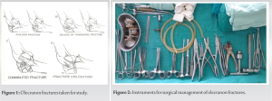
Immediate management
Immediately on arrival of the patient, if he/she was in shock, the level of shock was noted and managed accordingly. X-ray of the part was taken and the elbow was immobilized in whatever the position patients presence in a A/E POP posterior slab. The affected limb was kept elevated. Analgesics and antibiotics were given if necessary. Patient was then prepared for surgery and anesthesia after the pre-anesthetic checkup.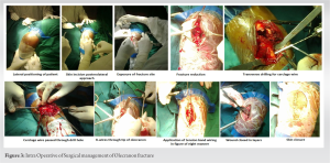
Selection of cases for Krishner wires with tension band wiring and Oleranon hook plate
Following points were considered – (a) Age of the patient, (b) extent of damage to the articular surface, and (c) degree of comminution. The patients of extremes of age and the patients in whom operative risk was great were not taken up for surgery.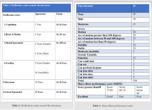
Methods
Surgical procedure
(a) Anesthesia – The operation was performed under general anesthesia or brachial block. (b) Position and Tourniquet – Mid arm tourniquet was applied with patient in supine or lateral position. Site of the surgery was thoroughly painted with iodine and spirit and draped. (c) Exposure – Exposure of the olecranon was done by Campbell’s posterolateral approach. A vertical incision was taken over the posterior aspect of the elbow about 2.5 cms proximal to olecranon, curving distally along the lateral aspect of olecranon reaching the subcutaneous border of the ulna and extending distally for about 7.5 cm distal to olecranon. Fascia was incised along the line of skin incision and fracture site was exposed. Fracture hematoma was cleared off and the fracture site was gently curettage. Accurate anatomical hairline reduction was achieved and held with either reduction clamp or long towel clip. Two K-wires are introduced parallel from the tip of the olecranon, that is, the proximal fragment across the fracture site to the distal fragment. Periosteum was stripped from the shaft of ulna distal to fracture site and a transverse hole was drilled approximately 3–5 cm distal to fracture site. A No.18 stainless steel malleable wire was passed through this transverse hole and crossed over the posterior surface of olecranon in a figure-of-eight manner and then passed around the protruding Kirschner wires and tightened using AO tensioner and then secured with a twist. Bend the proximal ends of the Krishner wires 1800 and tap the cut ends back into the proximal fragment. Accuracy of reduction was that stability was tested by moving the joint. Wound closed in layers and sterile dressing and compression bandage given. For communited olecranon fracture, exposure of the olecranon was done by Campbell’s posterolateral approach. A vertical incision was taken over the posterior aspect of the elbow about 2.5 cm proximal to olecranon, curving distally along the lateral aspect of olecranon reaching the subcutaneous border of the ulna and extending distally for about 7.5 cm distal to olecranon. Fascia was incised along the line of skin incision and fracture site was exposed. Fracture hematoma was cleared off and the fracture site was gently curettage. Accurate anatomical hairline reduction was achieved and held with either reduction clamp olecranon hook plate was applied on the posterior surface with cortical screws after drilling and tapping, through wash was given, wound closed in layers and sterile dressing was applied [5, 6].
Post-operative Management
- All the patients were treated with Inj. Cefotaxime 1gm twice daily for 5 days followed by Tab Cefixime 200 mg daily for 5 days. Some cases were treated with Amikacin 500 mg daily for 3 days.
- Anti-inflammatory analgesics, Inj. Diclofenac for 3 days followed by Tab Diclofenac 50 mg twice
- Affected limb was elevated and patient was asked to perform finger movements on day Elbow movements was advised from 3rd post-operative day.
- For comminuted fractures and unstable fixations, the limb was immobilized in A/E POP posterior slab with elbow in 900flexion for 2 For other fractures, the limb was mobilized by about 3rd post-operative day.
Follow-up
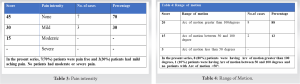
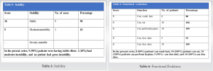
This part of the study should be done very carefully and meticulously. In our study, the patients on discharge were advised to report for follow-up after 6 weeks and 12 weeks and thereafter every 3 months. The result is assessed 3 months after the procedure. At follow-up, a detailed clinical examination was done and patient was assessed subjectively for the symptoms such as pain, swelling, and restriction of joint motion. On clinical examination, swelling of the joint, tenderness, movements of the elbow joint, prominence of head of cancellous screw, nutrition, and power of the muscles acting on the joint were noted (Table 3).
Patients were instructed to carry out physiotherapy in the form of, active flexion-extension, and pronation-supination without loading (Table 4).
Patients were instructed to carry out physiotherapy in the form of active flexion extension and pronation supination without loading (Table 5).
Check X-ray was taken and when final X-ray showed union, implant was removed. In all patients duration after which they returned to job was noted (Table 2).
Evaluation of results
Although there are many methods of evaluation of results given by many authors, the treated olecranon fractures by Tension band wiring and olecranon hook plate were evaluated in our study with Mayo Elbow Performance score (MEPS) (According to Morrey BF, An KN. Functional evaluation of the elbow) [7, 8]. for functional outcome and standard radiographs for radiological outcome (Figs. 2 and 3).
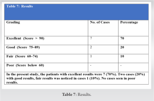
Study consists of 10 cases of fractures of the olecranon treated by Tension band wiring with Kirshner wire for simple transverse fractures and Olecranon hook plate for communited fractures in Chigateri General Hospital and Bapuji Hospital. All cases were followed up periodically during the period. The following are the observations made and the available data are analyzed as follows (Tables 6 and 7). Excellent results were achieved in 72%, good results in 16%, and fair results in 12%.
There were no poor results.
The main aim of the treatment of fracture is not only achieving union but to preserve the optimum function of the adjacent soft tissues and joints. In the management of intra articular fractures like fractures of the olecranon, a perfect anatomical reduction [9] of the fragments to obtain articular congruity and rigid fixation of the fragments is of utmost importance, if early movements are to be instituted to prevent complications like traumatic arthritis and joint stiffness. Tension band wiring with two intramedullary Kirschner wires provides the strength of fixation, that is, by converting tensile force to compressive force at the fracture site and for comminuted fractures Olecranon hook plate is used. In our study, 10 cases of fractures of the olecranon [10] were treated with Tension band wiring and Kirschner wires for simple transverse and oblique fractures and Olecranon hook plate for comminuted fractures. Our experience with this method of fixation has given favorable results. The findings, the end results and various other data is analyzed and compared in the following discussion. Ten cases of fractures of the olecranon treated by Kirschner wire with tension band wiring technique for transverse and oblique fractures and Olecranon hook plate for comminuted fractures at the Chigateri General Hospital and Bapuji Hospital, attached to J.J.M. Medical College, Davangere have been presented. Special attention was made to mobilize the affected elbow early. A review of literature on fractures of the olecranon has been presented. The anatomy of the elbow joint with particular reference to olecranon has been discussed in detail. The principle of tension band has been discussed in detail. The mechanism of injury, classification of olecranon fractures, and management have been described. Patients were diagnosed as having olecranon fractures on the basis of detailed history and thorough examination. Specific investigations such as X-ray, (anteroposterior and lateral view) of elbow in all 10 cases, which helped to confirm the diagnosis. Routine investigation such as Hb%, urine routine examination and blood urea, and serum creatinine was carried out as and when required. All patients underwent surgery, various parameters such as – age, sex, side involvement, mechanism of injury, and type of fracture were analyzed. Surgical procedure (K-wires with tension band wiring or Olecranon hook plate) was carried out in 10 patients.
All the cases were followed up and findings have been recorded regularly. Results were analyzed according to MEPS. Excellent results were achieved in 72%, good results in 16%, and fair results in 12%. There were no poor results. The complications such as superficial infection and symptomatic metal prominence were noticed in one and two cases respectively, which were treated accordingly. Surgical Fixation for Olecranon Fractures will helps in early mobilization of joint and anatomical fixation of Fractures.
In the present study, maximum number of patients were found to be in the age group between 21 and 30 years. (3 patients i.e., 30%). There was a significant male predominance in the present study (six patients 60%). Right side 6 (60%) olecranon fracture were more common than left side 4 (40%) in the present study. In the present study, 8 (80%) patients were transverse fractures and oblique fractures, 2 (20%) patients were comminuted fractures. All the patients were operated between 2 and 10 days with an average period of 3.48 days after the injury.
References
- 1.Horner SR, Sadasivan KK, Lipka JM, Saha S. Analysis of mechanical factors affecting fixation of olecranon fractures. Orthopedics 1989;12:1469-72. [Google Scholar]
- 2.Rowland SA, Burkhart SS. Tension band wiring of olecranon fractures. A modification of the AO technique. Clin Orthop Relat Res 1992;277:238-42. [Google Scholar]
- 3.Berger P. The treatment of fractures of the olecranon and particularly the Sutur of the olecranon by a process (Cedarg de L'Olecranon). GaHebd Med 1902;2:193-9. [Google Scholar]
- 4.Chaldis BE, Sachinis NC, Samoladas EP, Dimitriou CG, Pournaras JD. Is tension band wiring technique the “gold standard” for the treatment of olecranon fractures? A long term functional outcome study. J Orthop Surg Res 2008;3:9. [Google Scholar]
- 5.Cooper JL, D’Ambrosia RD. Fracture and fracture dislocation about the elbow. In: Chapman MW. Operative Orthopaedics. 2nd ed., Vol. 1., Ch. 33. Philadelphia: J.B. Lippincott Company; 1993. [Google Scholar]
- 6.Matar HE, Ali AA, Buckley S, Garlick NI, Atkinson HD. Surgical interventions for treating fractures of the olecranon in adults. Cochrane Database Syst Rev 2014;2014:CD010144. [Google Scholar]
- 7.Cabanela ME. Fractures of the proximal ulna and olecranon. In: Morrey BF, editor. The Elbow and its Disorders. Philadelphia: W.B. Saunders; 1985. [Google Scholar]
- 8.Chapman MW, Mahoney M. The role of early internal fixation in the management of open fractures. Clin Orthop Relat Res 1979;138:120-31. [Google Scholar]
- 9.Arthrology. In: Gray’s Anatomy. 36th ed., Ch. 4. London: Churchill Livingstone; 1980. p. 460-4. [Google Scholar]
- 10.Colton CL. Fractures of the olecranon in adults: Classification and management. Injury 1973;5:121-9. [Google Scholar]