Severe damage to the physis of the weight-bearing bones can be adequately managed with the aid of regular follow-ups and spontaneous healing.
Dr. Shivank Khurana, Department of Orthopaedic Surgery, Jawahar Lal Nehru Medical College, Aligarh Muslim University, Aligarh, Uttar Pradesh, India. E-mail: sshivankhurana2@gmail.com
Introduction: Gunshot injuries, while relatively uncommon in pediatric patients, can have lasting consequences, both physically and psychologically. Physeal injuries to the distal tibia are very common just second distal radius physis. Disruption of physis often leads to growth disturbances and deformities if not managed appropriately. This case report discusses the experience of a 9-year-old girl who sustained a gunshot injury to her left ankle injuring her distal tibial epiphysis. The report highlights the importance of long-term follow-up and rehabilitation in pediatric gunshot injury cases.
Case Report: The patient is a 9-year-old girl who suffered a gunshot wound to her left ankle. The bullet’s trajectory traversed her tibia and talus, miraculously avoiding any significant neurovascular injury. Immediate medical attention was sought, and she underwent surgical intervention to address the damage caused by the gunshot wound. The surgical procedure aimed to stabilize the fractured bone, remove any foreign bodies, and repair soft tissue damage. Patient has been followed up for 2 years, with remarkable recovery considering the severity of her injury. The patient has returned to her daily routine activities with slight chronic pain and some degree of the limitation of movement owing to injury and subsequent surgery. This case underscores the importance of long-term rehabilitation and follow-up care in pediatric gunshot injuries, as the effects can be far-reaching and persistent.
Conclusion: Injury to physis of weight bearing bones can be challenging to the patient as well as the surgeon. Long-term follow-up with continued medical and psychological support for the patients is necessary to ensure the better prognosis and quality of life after such traumatic events.
Keywords: Distal tibia, gunshot, pediatric, physis.
Injuries to the distal tibial epiphysis represent the second most common type of physeal injuries, with the most common ones occurring in the distal radius [1]. These injuries are of significant concern because any damage to the physis, or growth plate, can result in growth arrest, potentially leading to limb length discrepancies and deformities. Consequently, the appropriate treatment of these injuries is of paramount importance, as inadequate management can result in permanent damage to the growth plate. The significance of the distal tibial physis in the context of growth cannot be overstated. Studies, as described by Rathjen and Birch have indicated that the distal tibial physis contributes to approximately 45% of the longitudinal growth of the tibia, with the remaining 55% being attributed to the proximal physis [2]. During the most rapid growth years in childhood, the distal physis contributes to an average of about 5 mm of growth per year. Any disruption or damage to this critical area can have profound consequences for the proper development of the lower limb. One of the most severe complications following a physeal injury is growth plate arrest, which can manifest as either partial or complete cessation of growth. The management of these injuries can be approached either conservatively or through surgical means. Treatment options include methods such as the use of a shoe raise to compensate for limb length differences, excision of bone bridges that may form at the site of injury, leg lengthening procedures, epiphysiodesis (both contralateral and ipsilateral), and corrective osteotomies [3, 4]. The choice of treatment depends on various factors, including the nature and extent of the injury, the patient’s age, and their skeletal maturity. Interestingly, it is noteworthy that, based on extensive research, there appears to be a scarcity of documented cases involving gunshot injuries as a mechanism of physeal trauma specifically affecting the distal tibial epiphysis [5-10]. This underscores the rarity and uniqueness of such cases, which may require specialized assessment and management strategies to address the impact of gunshot injuries on the growth plate in the distal tibial region. Further research and documentation in this area would contribute to a deeper understanding of the potential consequences and treatment options for such uncommon but significant injuries.
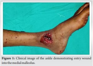
A 9-year-old female was inadvertently shot in her left ankle during an altercation between two unidentified individuals. The gunshot projectile traversed through the medial malleolus, causing a non-exiting wound (Fig. 1).
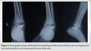
Initial assessment, including plain X-ray imaging (Fig. 2), revealed a bullet trajectory passing through the distal tibial physis and a comminuted fracture of the left talus, with the bullet remaining lodged within the anatomical structures. Fortunately, there were no concurrent vascular or neural injuries observed.
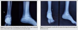
To address the injury, the patient underwent surgical intervention, which encompassed wound debridement and removal of the embedded bullet (Fig. 3) followed by slab application and immobilization for 3 weeks. Subsequently, she was prescribed a regimen of bed rest and administered antibiotics to mitigate the risk of infection. Follow ups was done first at 2 weeks and then monthly for 3 months followed by every 3 months for next 2 years. The soft tissue wounds healed uneventfully without any lasting complications. Follow-up radiographs demonstrated sclerosis of the fracture margins (Fig. 4 and 5).
However, a protracted 9-month post-operative period unveiled progressive avascular necrosis (AVN) of the talus (Fig. 6), concomitant with the chronic non-union of the medial aspect of the distal tibia.

These findings were conclusively established through magnetic resonance imaging (Fig. 7).

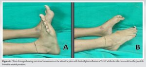
The patient has been regularly followed up for 2 years. At present, the patient experiences significant pain during weight-bearing activities and a noticeable limitation in the range of motion within the left ankle joint. Specifically, her ankle exhibits restricted plantar flexion, spanning from 0° to a mere 20°, while dorsiflexion from a neutral position remains unattainable (Fig. 8). Given the complex nature of her condition and the involvement of the growth plate, the current management strategy leans toward a conservative approach. The patient will be closely monitored until the growth plate fusion occurs, thereby stabilizing the distal tibia. This approach aims to address the aforementioned complications and optimize the long-term functionality of the affected ankle joint.
Physeal injuries resulting from gunshot wounds have been documented in medical literature, yet a notable gap exists in the existing body of knowledge regarding gunshot-induced injuries to the distal tibial growth plate. These fractures may lead to growth arrest with resultant angular deformity or leg length discrepancy [3]. Kling found that younger patients had the greatest chance of developing a growth disturbance. Younger patients were found to have a higher rate of partial physeal arrest after these fractures [11]. We present this case to shed light on a rather unique and infrequently reported injury, emphasizing the dearth of prior literature concerning gunshot injuries affecting the distal tibial physis. The necessity and timing of primary fixation to stabilize gunshot fractures are determined by a number of factors. These include the patient’s overall health, the characteristics of the fracture, the wound, any accompanying neurovascular injuries, and the type of any additional associated injuries (other than orthopedic) [12]. In the specific case at hand, the chosen course of primary treatment was surgical intervention, comprising wound debridement and removal of the embedded bullet. Following the procedure, the patient was immobilized using a below-knee plaster slab and was advised to observe bed rest. Subsequently, after a period of 6 weeks, she commenced weight-bearing activities and initiated ankle physiotherapy in an attempt to rehabilitate the affected limb. However, over time, a concerning development emerged. The patient exhibited signs of AVN within the talus and a chronic non-union of the medial aspect of the distal tibia. These findings were confirmed through clinical assessment and radiological investigations. Adults with mildly displaced or undisplaced fractures have been documented to have AVN whereas pediatric AVN of the talus body has been documented, but only in connection with talus fracture-dislocations [13, 14], hence, making the AVN of the talus in this patient another rare finding. There is still no clear consensus on the best course of action for treating talar fractures in children or the frequency of AVN that results from them. In response to this complex clinical scenario, a conservative management approach was adopted. The medical team opted to allow the body’s natural healing mechanisms to take over, avoiding any further invasive procedures. The patient is currently being monitored with the anticipation that a physeal bar will eventually form. This developmental process will be a pivotal factor in guiding the course of future treatment. The length of time spent treating a growth disturbance may have the biggest impact on the result. If children were presented later, they were more likely to have a linear or angular malformation. When compared to individuals who presented more than 2 years later, those who presented within 2 years exhibited noticeably less linear or angular abnormalities [3]. As of the present time, there is no discernible limb length discrepancy, indicating the effectiveness of the initial intervention and the patient’s continued recovery. While the patient does experience mild pain during walking, there is no evidence of significant functional limitations. This outcome, although on a conservative management strategy, reflects a positive aspect of the case, affirming that even in the absence of surgical interventions, the body has the remarkable capacity to heal and adapt. The rarity of this case serves as a reminder of the complexities and unique challenges that can arise from gunshot-induced physeal injuries, underlining the need for careful monitoring, individualized management, and the potential for favorable outcomes even in the absence of surgical correction. Future research and documentation in this area would certainly contribute to a better understanding of such exceptional cases and their optimal management.
Physeal injuries due to gunshots have been reported before, but no reference to an injury to the distal tibia was found in the literature. The primary wound management and bullet dislodgement can prove to be a good approach to manage physeal gunshots injuries with stringent clinic-radiological follow up till the physeal bar redevelops. A good follow-up and functional rehabilitation are important to steer clear of the development of deformities and limb length discrepancy. Subsequent investigations and record-keeping in this domain will undoubtedly enhance comprehension of these unique instances and their ideal handling and establishing a standard protocol.
This article summarizes the management of Salter Harris type 4 physeal injury of distal tibia with comminuted fracture talus left ankle due to a gunshot injury, their identification, diagnosis, and management. The propensity of nature to heal the injury by itself coupled with close monitoring can avoid development of deformities and inadvertent surgeries.
References
- 1.Peterson CA, Peterson HA. Analysis of the incidence of injuries to the epiphyseal growth plate. J Trauma 1972;12:275-81. [Google Scholar]
- 2.Rathjen K, Birch J. Physeal injuries and growth disturbances. In: Rockwood and Wilkins Fractures in Children. 6th ed. Philadelphia, PA: Lippincott Williams & Wilkins; 2006. p. 102-4. [Google Scholar]
- 3.Berson L, Davidson RS, Dormans JP, Drummond DS, Gregg JR. Growth disturbance after distal physeal fractures. Foot Ankle Int 2000;21:54-8. [Google Scholar]
- 4.Williamson RV, Staehli LT. Partial physeal growth arrest: Treatment by bridge resection and fat interposition. J Pediatr Orthop 1990;10:769-76. [Google Scholar]
- 5.Bartlett CS, Helfet DL, Hausman MR, Straus E. Ballistic and gunshot wounds: Effects on musculoskeletal tissues. J Am Acad Orthop Surg 2000;8:21-36. [Google Scholar]
- 6.Letts RM, Miller D. Gunshot wounds of the extremities in children. J Trauma 1976;16:807-11. [Google Scholar]
- 7.Miclau T, Gerich T, Foglar C, Lindsey RW, Krettek C. Behandlung von schussverletzungen des bewegungsapparates (gunshot wounds of the musculoskeletal system). Unfallchirurg 2000;105:188-98. [Google Scholar]
- 8.Ogden JA. Skeletal Injury in the Child. Berlin, Heidelberg, New York: Springer Verlag; 2000. p. 188-90. [Google Scholar]
- 9.Washington ER, Wayne AL, Ross WA. Gunshot wounds to the extremities in children and adolescents. Orthop Clin North Am 1995;26:19-28. [Google Scholar]
- 10.Wright DG, Levin JS, Esterhai JL, Heppenstall RB. Immediate internal fixation of low-velocity gunshot-related femoral fractures. J Trauma 1993;35:678-82. [Google Scholar]
- 11.Kling TF, Bright RW, Hensinger RN. Distal tibial physeal fractures in children that may require open reduction. J Bone Joint Surg 1984;66A:647-57. [Google Scholar]
- 12.Burg A, Nachum G, Salai M, Haviv B, Heller S, Velkes S, et al. Treating civilian gunshot wounds to the extremities in a level 1 trauma center: Our experience and recommendations. Isr Med Assoc J 2009;11:546-51. [Google Scholar]
- 13.Dimentberg R, Rosman M. Peritalar dislocations in children. J Pediatr Orthop 1993;13:89-93. [Google Scholar]
- 14.Pereles TR, Koval KJ, Feldman DS. Fracture-dislocation of the neck of the talus in a ten-year-old child: A case report and review of the literature. Bull Hosp Joint Dis 1996;55:88-91. [Google Scholar]