
Humeroradioulnar synostosis is an exceedingly rare orthopedic anomaly that is often seen alongside other orthopedic disorders, with this report describing the first case of a humeroradioulnar synostosis in a patient with congenital scoliosis and deformities of the ipsilateral shoulder, wrist and hand.
Dr. Tyler Small, Department of Orthopedics, Nemours Children’s Health, 6535 Nemours Pkwy, Orlando, Florida. E-mail: tyler.small@nemours.org
Abstract
Introduction: Synostoses, or fusions, of the bones of the elbow are rare orthopedic occurrences that result from abnormal segmentation of the humerus, radius, and/or ulna during fetal development. The upper extremity begins as a single cartilage anlage in utero that segments into a distinct humerus, radius, and ulna and when disrupted, synostoses of the elbow occur. The most uncommon synostosis reported is the humeroradioulnar synostosis, in which all three bones of the elbow are fused. These synostoses often occur in conjunction with other orthopedic anomalies, as well as defects of other body systems due to their simultaneous development in utero. There are only six previous reports of humeroradioulnar synostoses. These reports describe additional concurrent orthopedic anomalies; however, anomalies of the ipsilateral shoulder or wrist, or scoliosis of any kind have not been reported. This report adds to the limited body of literature surrounding this rare orthopedic anomaly and presents additional skeletal manifestations not previously reported.
Case Report: This patient is a 17-year-old male with a history of congenital scoliosis previously treated with Mehta casting, and no other known medical diagnoses. He presented to our orthopedic clinic with concerns about his worsening spinal deformity and moderate back pain. On imaging and clinical exam, he was found to severe thoracic and thoracolumbar curves, as well as a fixed deformity of the right elbow, due to a humeroradioulnar synostosis. He was also found to have deformities and limited motion of the ipsilateral shoulder, wrist and hand. The patient’s scoliosis was treated with posterior spinal fusion after a course of halo traction, and he has decided to forgo any further evaluation or treatment for his right humeroradioulnar synostosis. The patient has been able to maintain a high level of function, participate in daily activities and even cook as a hobby.
Conclusion: Humeroradioulnar synostosis is an exceedingly rare orthopedic condition that results from abnormal segmentation of the bones of the elbow during foetal development. Only six reports of this anomaly were found in our literature review. This report is of a 17-year-old male with a humeroradioulnar synostosis and is the first to describe this anomaly in a patient with defects of the ipsilateral shoulder and wrist with underlying congenital scoliosis. He has been able to maintain a high level of function, enjoy hobbies, such as cooking, and go to school, and is thus being treated conservatively with observation for this issue.
Keywords: Humeroradioulnar synostosis, elbow synostosis, congenital elbow deformity, congenital scoliosis.
Synostoses, or fusions, of the bones of the elbow are a rare orthopedic occurrence and can occur in congenital or posttraumatic setting; this report focuses on the congenital form. Congenital anomalies of the elbow are a result of abnormal segmentation of the humerus, radius, and/or ulna during fetal development. The upper extremity begins as a single cartilage anlage in utero that segments into a distinct humerus, radius, and ulna as development progresses in and is typically able to be identified via ultrasound around 35 days after conception [1]. This segmentation can fail to occur, or occur abnormally, for a variety of reasons, that is, genetic mutations, intrauterine exposure to teratogens and idiopathic causes. When segmentation is disrupted, abnormalities of the elbow can develop, as well as other orthopedic anomalies, such as arthrogryposis, syndactyly, symbrachydactyly, radial head dislocations and radial club hand [1]. Other body systems are commonly affected due to their simultaneous development around 8–10 weeks of gestation; this can include the neurological, cardiovascular, and renal systems as well as the gastrointestinal and respiratory tracts [2]. The most well-documented elbow synostosis is the proximal radioulnar synostosis [1,2,3,4,5], followed by the radiohumeral synostosis [6,7,8,9]. The most uncommon form of these synostoses is the humeroradioulnar synostosis, in which all three bones of the elbow are congenitally fused. Our literature review produced six previous reports of a humeroradioulnar synostosis [10,11,12,13,14,15], making this an exceedingly rare orthopedic condition. Patients presenting with a humeroradioulnar synostosis typically have limited motion of the elbow, if any; however, most patients are able to compensate well for this anomaly with motion at the shoulder, wrist and fingers, assuming no additional defects of the ipsilateral upper extremity are present. The reports produced during our literature review describe humeroradioulnar synostosis as both an isolated anomaly and an anomaly occurring in conjunction with other skeletal manifestations and defects of other body systems. Kakarala et al. describe the treatment of a 13-year-old male with bilateral humeroradioulnar synostoses and only minor hypoplasia of the lesser digits of the bilateral hands [10]. Other reports describe additional skeletal anomalies such as lambdoid stenosis and craniosynostosis [11,14], oligodactyly and syndactyly [12], distal humerus bifurcation [13,14], upper extremity phocomelia and ectrodactyly [13] and radial and ulnar hypoplasia or deficiency [11,14,15]. None of the cases currently published describes associated anomalies of the ipsilateral shoulder or wrist, or scoliosis of any kind, in patients with humeroradioulnar synostosis. Our report describes a 17-year-old male with a history of congenital scoliosis found to have a congenital humeroradioulnar synostosis and concomitant deformity of the ipsilateral shoulder, wrist, and hand. This report adds to the limited body of literature surrounding this rare orthopedic anomaly and presents additional skeletal manifestations observed in a patient with humeroradioulnar synostosis not yet reported in the available literature.
This patient is a 17-year-old male with a history of congenital scoliosis treated with Mehta casting at the age of three at an outside institution. The patient’s mother reports that the patient underwent thorough genetic testing at an early age after his IIS diagnosis, but this workup was negative, and no further investigation took place. The patient was born full term without complications of pregnancy, birth or the post-natal period, and he has no additional medical history. He met all development milestones during childhood and began walking at 15 months of age. This patient was lost to follow-up for 14 years before he presented to our orthopedic clinic with concerns about his worsening spinal deformity, as well as moderate back pain. He was also complaining about not having any motion of his right elbow and a limited range of motion of the right shoulder, which his mother states has been present since birth. Physical examination of the patient’s spine demonstrated significant truncal shift with uneven shoulder heights and a significant left-sided prominence of the thoracolumbar region. Neurologic examination was within normal limits. The patient’s right upper extremity is significantly atrophic from the shoulder to the wrist and notably shorter and smaller than the left. The patient has a limited range of motion of the right shoulder with approximately 15° of forward flexion and 20° of abduction. The right elbow is fixed at 100° of flexion with no active or passive flexion, extension, pronation, or supination. He has near full flexion, extension, radial deviation, and ulnar deviation of the ipsilateral wrist, but very limited pronation and supination. The right small finger is shortened and capable of motion, but weak against resistance. The right upper extremity is otherwise neurovascularly intact, and there are no obvious deformities or limitations to motion of the left upper extremity or bilateral lower extremities. Initial imaging of the patient’s spine demonstrated a 123° left-sided curve from T7-L2 with severe thoracic kyphosis. On magnetic resonance imaging, it was not that the patient has a cervical and thoracic syrinx, as well as vertebral anomalies at T10-T12, seen on 3D reconstruction of the patient’s spine. Radiographs of the right shoulder demonstrate a dysplastic glenoid and proximal humerus with lateral humeral bowing (Fig. 1).
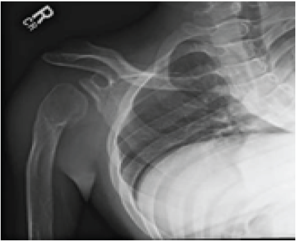
Figure 1: Anterior-posterior radiograph of the right shoulder demonstrating glenoid and proximal humerus dysplasia and lateral humeral bowing.
Anteroposterior, lateral and oblique images of the patient’s right elbow demonstrate complete fusion of the humerus, radius, and ulna with an appreciable trochlea, capitellum, radial head or trochlear notch (Fig. 2, 3, 4).
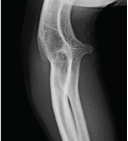
Figures 2: Anterior-posterior radiograph of the right elbow demonstrating complete bony fusion of the humerus, radius, and ulna (humeroradioulnar synostosis) without identifiable unlohumeral or radiocapitellar joints.
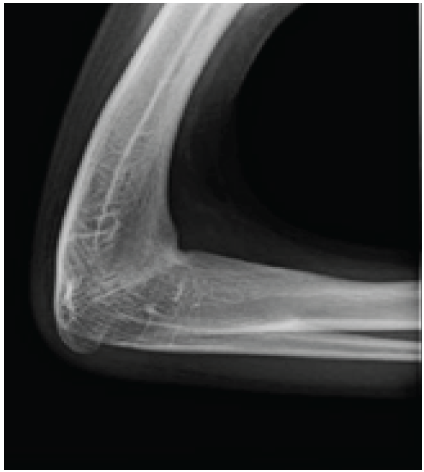
Figure 3: Lateral radiograph of the right elbow demonstrating complete bony fusion of the humerus, radius, and ulna (humeroradioulnar synostosis).
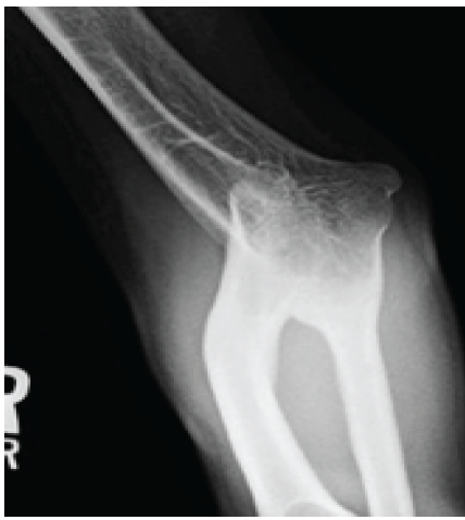
Figure 4: Oblique radiograph of the right elbow demonstrating complete bony fusion of the humerus, radius, and ulna (humeroradioulnar synostosis).
Imaging of the right forearm and hand demonstrates the patient’s humeroradioulnar synostosis of the right elbow with abnormalities of the carpal bones and an absent fifth metacarpal with a pseudo joint formed by the fifth proximal phalanx articulating with the midshaft of the fourth metacarpal (Fig. 5 and 6).
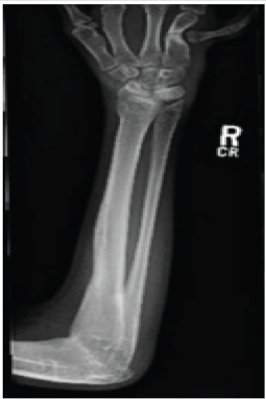
Figure 5: Anterior-posterior radiograph of the right forearm demonstrating the patient’s humeroradioulnar synostosis, as well as carpal bone and hand anomalies.
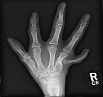
Figure 6: Posteroanterior radiograph of the right hand demonstrating the patient’s right-hand anomalies.
The patient’s scoliosis was treated in a staged fashion, with 11 weeks of halo traction followed by definitive posterior spinal fusion from T2-L4 and has recovered well from this operation. Although this patient has no range of motion of the right elbow and limited motion of the ipsilateral shoulder, he is still able to compensate well. He cooks as a hobby and has no significant limitations in extracurricular activities or activities of daily living. Due to his high level of function and lack of viable reconstruction options at this time, he and his family have chosen to forego any treatment for this issue and focused on correcting his scoliosis.
Congenital synostoses of the elbow are rare orthopedic anomalies and result from abnormal segmentation of the bones of the upper extremity during fetal development [1]. When segmentation of the cartilage anlage fails to progress normally, the bones of the elbow remain fused, resulting in congenital synostoses of the elbow. These anomalies are often associated with other orthopedic abnormalities and irregularities of other body systems due to their simultaneous development in utero [2]. The most well-documented elbow synostosis is the proximal radioulnar synostosis [1,3,4,5], followed by the radiohumeral synostosis [6,7,8,9]. The most uncommon form is the humeroradioulnar synostosis, in which all three bones of the elbow are fused. Our literature review produced only six formal reports of this anomaly [10,11,12,13,14,15]. This report describes a 17-year-old male with a history of congenital scoliosis with a congenital humeroradioulnar synostosis of the right elbow, as well as anomalies of the ipsilateral shoulder, wrist, and hand. The other reports of humeroradioulnar synostoses describe various additional skeletal manifestations, but do not describe associated deformities of the ipsilateral shoulder, wrist, or spine. Our report is the first to describe a patient with humeroradioulnar synostosis and concomitant shoulder and wrist anomalies with underlying congenital scoliosis. Although this patient’s humeroradioulnar synostosis has left him with no motion in his right elbow, he is able to compensate with his shoulder and wrist well enough to complete activities of daily living, school and even participate in cooking as a hobby. Since this patient can maintain such a high level of function, and there is a lack of viable reconstruction options at this time, his humeroradioulnar synostosis is being managed conservatively with observation and no intervention planned. This report adds to the extremely limited body of literature surrounding an exceedingly rare orthopedic anomaly and describes additional skeletal manifestations, involving the ipsilateral shoulder and wrist as well as the spine, which have not been previously presented in existing case reports.
Humeroradioulnar synostosis is an exceedingly rare orthopedic condition that results from abnormal segmentation of the bones of the elbow during fetal development. Only six reports of this anomaly were found in our literature review. This report is of a 17-year-old male with a humeroradioulnar synostosis and is the first describe this anomaly in a patient with defects of the ipsilateral shoulder and wrist with underlying IIS. Despite having no motion in his right elbow, he can maintain a high level of function, enjoy hobbies, such as cooking, and go to school. He is currently being treated conservatively with observation for this issue.
Humeroradioulnar synostosis is an exceedingly rare orthopedic condition that is often associated with additional orthopedic anomalies, as well as defects of other body systems due to their simultaneous development in utero. This report is of a 17-year-old male with a humeroradioulnar synostosis and is the first describe this anomaly in a patient with defects of the ipsilateral shoulder and wrist and an underlying diagnosis of congenital scoliosis.
References
- 1. Elliott AM, Kibria L, Reed MH. The developmental spectrum of proximal radioulnar synostosis. Skeletal Radiol 2010;39:49-54. [Google Scholar] [PubMed]
- 2. Solomon BD. VACTERL/VATER association. Orphanet J Rare Dis 2011;6:56. [Google Scholar] [PubMed]
- 3. Cleary JE, Omer GE Jr. Congenital proximal radio-ulnar synostosis. Natural history and functional assessment. J Bone Joint Surg Am 1985;67:539-45. [Google Scholar] [PubMed]
- 4. Alabau-Rodriguez S, Garrido Ferrer JF, Bulló Mir X, Martín Dominguez LA, Pardo Pol A, Soldado Carrera F. Congenital radioulnar synostosis review: Recommendations and treatment outcomes. Children (Basel) 2024;11:1317. [Google Scholar] [PubMed]
- 5. Rutkowski PT, Samora JB. Congenital radioulnar synostosis. J Am Acad Orthop Surg 2021;29:563-570. [Google Scholar] [PubMed]
- 6. Sahdi H, Rasit AH, Khoo CS, Bojeng A, Nur-Alyana BA. Modified french osteotomy for humeroradial synostosis in a child with multiple synostoses syndrome: A case report. Malays Orthop J 2019;13:52-5. [Google Scholar] [PubMed]
- 7. Nema S, Vyas G, Sirsikar A, Bhoj PK. Congenital humeroradial synostosis: A case report. Malays Orthop J 2012;6 SupplA:41-2. [Google Scholar] [PubMed]
- 8. Mahmoud EE. Bilateral congenital humeroradial synostostis presenting with bilateral proximal radius fractures: A case report. Case Rep Orthop Res 2021;4:29-32. [Google Scholar] [PubMed]
- 9. Mnaymneh WA. Congenital radio-humeral synostosis. A case report. Clin Orthop Relat Res 1978;131:183-4. [Google Scholar] [PubMed]
- 10. Kakarala G, Kavarthapu V, Lahoti O. Distraction osteogenesis to improve limb function in congenital bilateral humeroradioulnar synostosis. Acta Orthop Belg 2006;72:765-8. [Google Scholar] [PubMed]
- 11. Edwards TJ, Haan EA, Humphrey IJ. Humeroradioulnar synostosis in a patient with lambdoid synostosis. J Med Genet 1993;30:81-2. [Google Scholar] [PubMed]
- 12. Hersh JH, Joyce MR, Profumo LE. Humero-radio-ulnar synostosis: A new case and review. Am J Med Genet 1989;33:170-1. [Google Scholar] [PubMed]
- 13. Leroy JG, Speeckaert MT. Humeroradioulnar synostosis appearing as distal humeral bifurcation in a patient with distal phocomelia of the upper limbs and radial ectrodactyly. Am J Med Genet 1984;18:365-8. [Google Scholar] [PubMed]
- 14. Douma H, Abdelkrim EH. Cas rare de fracture des deux os de l’avant-bras chez un patient présentant une bifurcation de l’humérus et une déficience ulnaire: Synostose huméro-radioulnaire [Rare case of fracture of the two bones of the forearm in a patient with humerus bifurcation and an ulnar deficiency: Humero-radioulnar synostosis]. Pan Afr Med J 2022;41:316. [Google Scholar] [PubMed]
- 15. Swenson V, Spinek A. A rare case of congenital humeroradioulnar synostosis from medieval Pawłów Trzebnicki, Poland. Int J Osteoarchaeol 2020;30:256-63. [Google Scholar] [PubMed]


Related Articles in Journal of Orthopaedic Case Reports
 April 10, 2022 A Case Report of the Complete Union of an Impending Failure of Patellar Fracture Treated with Cannulated Cancellous Screws and Tension Band Wiring for Failed Modified Tension Band Wiring Technique
 June 10, 2024 Multiple Vertebral Compression Fractures Secondary to Pregnancy-induced Osteoporosis: A Case Report
 March 1, 2025 Arthroscopic Transtibial Pullout Repair for Medial Meniscus Posterior Root Tear using HitPat’s Modified All-Suture Technique
 December 6, 2020 Reviewers Acknowledgement & Photo-gallery December 2020