

A bilobed lipoma may colonize the dead space created by radial head excision years after the index procedure and should be considered in the differential diagnosis of a late soft-tissue swelling at a previous surgical site.
Dr. V. S. Subramaniyan, Department of Orthopaedics, Fortis Hospital, Bannerghatta Road, Bengaluru - 560076, Karnataka, India. E-mail: vss5950@gmail.com
Abstract
Introduction: Lipoma is the commonest benign soft-tissue tumor, but its occurrence within a surgically created dead space is exceedingly rare. Radial head excision leaves a void in the lateral compartment of the elbow that is ordinarily obliterated by hematoma and fibrous tissue. We report what we believe to be the first case in the indexed literature of a bilobed lipoma arising within this post-excision space.
Case Report: A 58-year-old Indian man presented with an 8-month history of progressive pain and swelling over the right elbow. He had undergone right radial head excision 5 years earlier for a traumatic radial head fracture and had been largely symptom-free in the interval. Examination showed a soft, compressible, lobulated swelling over the lateral aspect of the elbow with flexion restricted to 90° and no neurological deficit. Radiographs confirmed the absence of the radial head with no other bony abnormality. Magnetic resonance imaging showed a well-defined bilobed homogeneous fatty lesion occupying the ventral and lateral aspects of the post-excision space, with signal suppression on fat-saturated sequences and no features of malignancy. At exploration through the previous lateral approach, a single bilobed, well-encapsulated lipomatous mass was found in the post-excision space, with one lobe ventral and one lateral. The posterior interosseous nerve was free of the mass. The isthmus was divided to permit delivery, and both lobes were excised intact, measuring 8 × 4 and 6.5 × 4 cm. Histopathology confirmed a benign lipoma. At 6 months, the patient was pain-free, flexion had improved to 120°, and there was no recurrence.
Conclusion: A bilobed lipoma can develop within the dead space left by radial head excision after a prolonged latent interval. The bilobed configuration appears to be molded by the architecture of the post-excision void, a novel observation regarding the pathogenesis of post-surgical lipomas. Magnetic resonance imaging is diagnostic, and excision with preservation of the posterior interosseous nerve is curative. This entity should be included in the differential diagnosis of late soft-tissue swelling at a previous surgical site so that it is not mistaken for recurrence or a malignant fatty tumor.
Keywords: Bilobed lipoma, radial head excision, post-surgical lipoma, elbow, posterior interosseous nerve, dead space.

Lipoma is the most frequently encountered benign soft-tissue tumor and usually presents as a slow-growing, painless subcutaneous mass. Although lipomas may arise almost anywhere, their development within a surgically created dead space is exceedingly rare and has not been well characterized [1,2]. Radial head excision is an established procedure for comminuted radial head fractures, particularly Mason type III and type IV injuries, and remains in use where reconstruction or arthroplasty is not feasible [3]. Removal of the radial head leaves a void in the lateral compartment of the elbow. In the normal postoperative course, this void fills with hematoma and is subsequently obliterated by fibrous tissue ingrowth. Colonization of this space by proliferating adipose tissue forming a true lipoma has not, to the best of our knowledge and on the basis of the literature search detailed below, been described previously. Post-traumatic and post-surgical lipomas are recognized but uncommon entities. Proposed mechanisms include prolapse of adipose tissue through disrupted fascial planes, producing a pseudolipoma [4], and true adipocyte proliferation driven by cytokine release following trauma and hematoma formation [1,2]. Reported latent intervals between the inciting event and presentation range from approximately 6 months to 5 years [1,4,5]. Lipomas around the elbow are of particular clinical interest because of the course of the posterior interosseous nerve through the radial tunnel, and several reports describe compressive neuropathy of this nerve caused by fatty masses in the proximal forearm [6,7]. We report a case of a bilobed lipoma arising in the post-radial head excision space to alert clinicians to this unusual diagnosis and to contribute to the understanding of adipogenesis within orthopedic dead spaces.
Clinical presentation:
A 58-year-old Indian man presented to the orthopedic outpatient department with an 8-month history of gradually progressive pain and swelling over the right elbow. There was no preceding fresh injury. His past surgical history was significant for a right radial head excision performed 5 years previously for a traumatic radial head fracture. He reported a largely symptom-free interval between the index surgery and the present presentation. On examination, there was a soft, compressible, lobulated swelling over the lateral aspect of the right elbow at the site of the previous surgical scar. Elbow flexion was restricted to 90°. There was no distal neurovascular deficit, and the overlying skin was normal with no signs of inflammation.
Investigations:
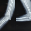
Anteroposterior and lateral radiographs of the right elbow showed the expected absence of the radial head, consistent with the previous excision. There was no heterotopic ossification, bony erosion, periosteal reaction, or soft-tissue calcification (Fig. 1).
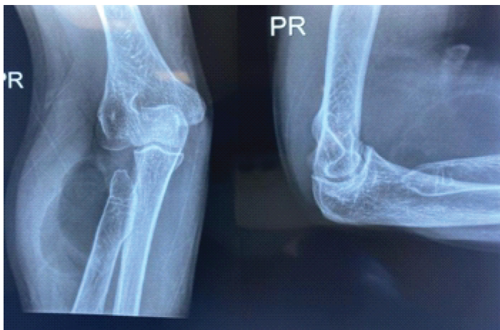
Figure 1: Anteroposterior and lateral radiographs of the right elbow demonstrating absence of the radial head consistent with prior excision. No heterotopic ossification, periosteal reaction, or bony erosion is identified. A soft tissue density is noted in the lateral compartment corresponding to the lipomatous masses.
Magnetic resonance imaging of the right elbow demonstrated a well-defined bilobed lesion occupying the ventral and lateral aspects of the radial head excision space. Both lobes were hyperintense on T1-weighted and T2-weighted sequences and showed complete signal suppression on fat-saturated sequences, appearances characteristic of mature fatty tissue. The larger lobe measured approximately 8 cm in its greatest dimension. There were no thick internal septations, nodular non-fatty components, or enhancing areas to suggest malignancy [8,9] (Figs. 2 and 3).
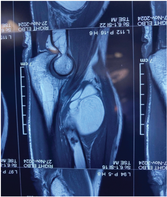
Figure 2: Magnetic resonance imaging of the right elbow (T2-weighted turbo spin echo sequence, sagittal plane) demonstrating a well-defined bilobed homogeneous hyperintense mass in the post-excision space of the radial head. The larger inferior lobe (approximately 8 cm) occupies the ventral compartment, and the smaller superior lobe is seen in the lateral recess. Both lobes demonstrate signal characteristics consistent with mature fatty tissue.
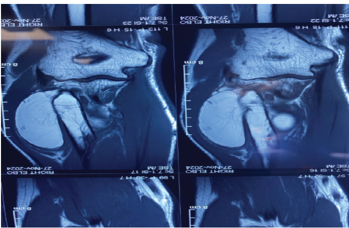
Figure 3: Magnetic resonance imaging of the right elbow (T2-weighted turbo spin echo sequence, coronal plane) showing the bilobed lipomatous mass in two sequential slices. The bilobed mass is well-circumscribed with homogeneous high signal intensity, no thick internal septations, and no involvement of adjacent neurovascular structures. The absence of enhancing nodular components excludes malignancy. Scale bars indicate lesion dimensions of up to 8 cm.
Operative findings and management:
Surgery was performed under general anesthesia with the patient supine and a pneumatic tourniquet applied to the upper arm. The previous lateral elbow scar was used. On deep dissection, a single bilobed, well-encapsulated, lobulated, yellowish-white lipomatous mass was found occupying the post-excision space of the radial head, with one lobe in the ventral compartment and the other in the lateral compartment, the two lobes joined by a narrow isthmus. The posterior interosseous nerve was identified, carefully traced along its course, and confirmed to be entirely free of the mass, with no compression or adherence. As the intact mass could not be delivered through the available exposure, the isthmus was divided and each lobe excised separately with its capsule intact. Hemostasis was secured and the wound closed in layers (Fig. 4).
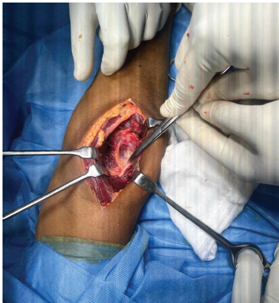
Figure 4: Intraoperative photograph through lateral elbow approach showing the surgical field following excision of both lipomatous masses. Retractors demonstrate the post-excision cavity in the lateral compartment. The posterior interosseous nerve was identified, traced, and confirmed to be entirely free of tumor involvement throughout the procedure.
Pathological findings:
Gross examination showed a bilobed, well-encapsulated, lobulated fatty specimen, received as two lobes following division of the connecting isthmus at the time of extraction (Fig. 5). The ventral lobe measured 8 × 4 cm and had a smooth, glistening, encapsulated surface with lobulated yellow adipose tissue. The lateral lobe measured 6.5 × 4 cm and had a similar appearance with visible surface vascularity. Microscopy of both lobes showed mature adipocytes arranged in lobules separated by thin fibrous septa, with no cellular atypia, lipoblasts, abnormal vascularity, or necrosis (Fig. 6). The findings confirmed a benign lipoma.
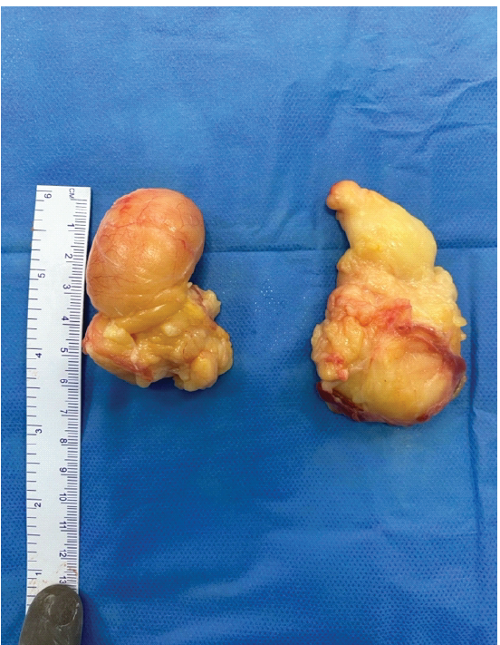
Figure 5: Gross pathological photograph of the excised bilobed lipomatous specimen laid adjacent to a ruler for scale. Left: Lobe 1 (ventral lobe, 8 × 4 cm) with a smooth, well-encapsulated, lobulated surface. Right: Lobe 2 (lateral lobe, 6.5 × 4 cm) with lobulated yellow adipose tissue and surface vascularity. Both lobes demonstrate the characteristic gross appearance of a benign bilobed lipoma.
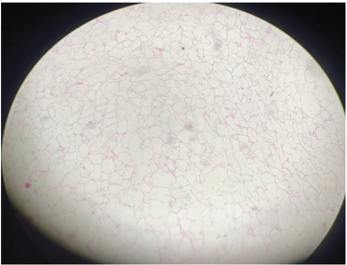
Figure 6: Photomicrograph of haematoxylin and eosin stained section of the excised lipoma (original magnification ×40). The section demonstrates sheets of mature adipocytes arranged in lobules and separated by thin fibrous septa. Individual adipocytes are large, uniform, and polygonal with abundant clear cytoplasm and small, peripherally compressed nuclei. There is no nuclear atypia, pleomorphism, lipoblast formation, or necrosis. These appearances are consistent with the histopathological diagnosis of a benign lipoma.
Outcome and follow-up:
The post-operative course was uneventful. Supervised physiotherapy was started on the 3rd post-operative day. At 6 months, the patient reported complete resolution of pain and swelling. Elbow flexion had improved from 90° pre-operatively to 120°, a gain of 30°. There was no clinical evidence of recurrence at final follow-up.
A search of Medline through PubMed using the terms “lipoma,” “radial head,” “radial head excision,” “post-traumatic lipoma,” “elbow,” and “dead space” did not identify any previous report of a lipoma arising within a post-excision space. A title search for lipoma with elbow or radial head returned 12 records, comprising lipoma arborescens of the elbow joint, intraneural and intramuscular lipomas causing nerve compression, and parosteal lipomas of the proximal radius; none arose within a surgically created space. A title search for post-traumatic lipoma returned 10 records, none involving the elbow or a previous excision site. We therefore believe this to be the first such reported case. It is notable on three counts: The bilobed morphology, the localization within a surgically created dead space, and the functional impairment produced without any neurological deficit. The pathogenesis of post-traumatic and post-surgical lipoma remains incompletely understood, and two mechanisms are described. The first proposes prolapse of adipose tissue through disrupted fascial planes, giving rise to a pseudolipoma rather than a true neoplasm, as described by Penoff [4] and reported earlier by Meggitt and Wilson as fat fractures following blunt injury [10]. The second proposes that trauma and hematoma formation release cytokines that drive differentiation of resident pre-adipocytes into mature adipocytes, forming a true lipoma. Signorini and Campiglio examined nine patients in whom a lipoma appeared within months of blunt trauma, found the prolapse mechanism incompatible with their findings, and argued for true adipose neoformation [2]. Aust et al. identified 34 post-traumatic lipomas among 170 patients, and 25 of their 31 patients reported an extensive, slowly resolving hematoma at the site of subsequent lipoma formation [1]. In the present case, the initial fracture and the subsequent excision are likely to have acted as dual inciting events, supplying both the anatomical space and the biological stimulus. Notably, the lipomas in the Aust series were all subcutaneous and superficial to the musculofascial system, whereas our lesion lay deep within the lateral compartment. The bilobed morphology is the most instructive feature of this case. The narrow isthmus joining the lobes, identified before division, establishes a single lesion rather than two independent tumors. The lobes corresponded to the ventral and lateral sub-compartments of the void left after radial head removal, with the isthmus at the point where these compartments communicate. The mass therefore appears molded by the architecture of the dead space, expanding to fill each recess and remaining narrow where the space between them is confined. This observation suggests that the shape of a postsurgical dead space may directly determine the growth pattern of any tumor that subsequently arises within it and may explain why the lesion remained clinically occult until it was large enough to restrict flexion. The 5-year latent interval in our patient lies at the upper end of the range reported for post-traumatic lipomas, in which the mean interval was 2 years with a range of 6 months to 5 years in the largest series [1]. A comparable phenomenon has been described at other previously injured sites, including a lipoma arising over a healed parietal fracture in a child [11] and a post-traumatic lipoma of the parotid gland attributed to hematoma, fat necrosis, and subsequent lipoma formation [12]. The relationship of the mass to the posterior interosseous nerve is a critical surgical consideration. The nerve traverses the radial tunnel close to the site of radial head excision, passing beneath the arcade of Frohse between the two heads of the supinator. Compressive neuropathy from fatty masses in the proximal forearm is well described. Bieber et al. reported four patients with radial nerve compression by lipomas at the elbow, all resolving completely after excision [6]. In a systematic review of 34 patients from 30 articles, Cheng et al. found age and lipoma size were not significant predictors of motor recovery, whereas symptom duration was, particularly below 18 months; 73.5% recovered complete motor function after excision alone at a mean of 9.7 months, and the mean age of 58.2 years closely matches our patient [7]. In our case, the nerve was entirely free of the mass, accounting for the absence of motor symptoms despite the size of the lesion. Scarring from the index procedure makes this dissection more demanding than in a virgin field, and we would advise identifying the nerve proximally in unscarred tissue before working distally. The principal differential diagnosis in this age group is a well-differentiated liposarcoma. Gaskin and Helms, reviewing 126 consecutive fatty masses, found magnetic resonance imaging to be 100% sensitive for well-differentiated liposarcoma and 100% specific for simple lipoma, although 63% of lesions considered suspicious for liposarcoma proved benign [8]. Kransdorf et al., in 60 histologically verified fatty tumors, identified size >10 cm, thick septa, globular or nodular non-adipose areas, and <75% fat content as features favoring malignancy, with male sex and thick septa increasing the likelihood 13-fold and nine-fold, respectively [9]. Our patient warranted care on this account, being male and 58 years old with an 8 cm lesion deep to fascia. However, the homogeneous signal, well-defined margins, and absence of thick septa or nodular components favored a benign lesion, confirmed on histopathology. Histopathological confirmation remains essential here, since imaging alone cannot wholly exclude a well-differentiated liposarcoma in an older male with a deep fatty mass [13]. Other differentials that merit consideration at a previous surgical site include a postoperative seroma, a synovial cyst, heterotopic ossification, and, rarely, an infective collection; each of these was readily excluded on imaging. Complete excision remains the definitive treatment for a symptomatic lipoma in this location. Excision with nerve preservation gave an excellent functional result in our patient, with a 30° gain in flexion and complete symptom resolution and no recurrence at 6 months, which is consistent with the uniformly good outcomes reported after complete excision of lipomas at the elbow [6,14]. The take-home message from this case is that a late, painless, slowly enlarging swelling at the site of a previous orthopedic procedure should not be assumed to be a scar, seroma, or recurrence of the original pathology; a fatty tumor arising within the surgically created dead space is a real, if rare, possibility, and magnetic resonance imaging will resolve the question before operation.
Limitations:
This is a single case with 6 months of follow-up, and the proposed mechanism of dead-space molding, while consistent with the operative and imaging findings, remains a hypothesis requiring further reports to substantiate.
A bilobed lipoma arising within the post-excision space of the radial head is an exceedingly rare and, to our knowledge, previously unreported entity. This case advances knowledge of the pathogenesis of post-surgical lipoma by demonstrating that a surgically created dead space can provide both the site and the shape for a fatty tumor arising years after the index operation. The wider principle, that a dead space left by any surgical excision may later be colonized by a benign tumor, is of relevance across surgical specialties. Magnetic resonance imaging provides near-definitive pre-operative characterization, and complete excision with preservation of the posterior interosseous nerve is curative and restores function.
A slowly enlarging, painless swelling appearing year after radial head excision may be a lipoma arising within the surgically created dead space rather than scar, seroma or recurrence. Magnetic resonance imaging is diagnostic, and complete excision with identification and preservation of the posterior interosseous nerve is curative and restores elbow flexion.
References
- 1. Aust MC, Spies M, Kall S, Gohritz A, Boorboor P, Kolokythas P, et al. Lipomas after blunt soft tissue trauma: Are they real? Analysis of 31 cases. Br J Dermatol 2007;157:92-9. [Google Scholar] [PubMed]
- 2. Signorini M, Campiglio GL. Posttraumatic lipomas: Where do they really come from? Plast Reconstr Surg 1998;101:699-705. [Google Scholar] [PubMed]
- 3. Antuna SA, Sanchez-Marquez JM, Barco R. Long-term results of radial head resection following isolated radial head fractures in patients younger than forty years old. J Bone Joint Surg Am 2010;92:558-66. [Google Scholar] [PubMed]
- 4. Penoff JH. Traumatic lipomas/pseudolipomas. J Trauma 1982;22:63-5. [Google Scholar] [PubMed]
- 5. Parinyakhup W, Boonriong T, Boonsri P, Chuaychoosakoon C. Right proximal tibia post-traumatic lipoma following a jogging fall. Oxf Med Case Reports 2024;2024:omae057. [Google Scholar] [PubMed]
- 6. Bieber EJ, Moore JR, Weiland AJ. Lipomas compressing the radial nerve at the elbow. J Hand Surg Am 1986;11:533-5. [Google Scholar] [PubMed]
- 7. Cheng C, Punjabi A, Gunther S, Chepla K. Forearm lipoma causing posterior interosseous nerve compression: Literature review and meta-analysis of predictors for motor recovery. Hand (N Y) 2024;19:149-53. [Google Scholar] [PubMed]
- 8. Gaskin CM, Helms CA. Lipomas, lipoma variants, and well-differentiated liposarcomas (atypical lipomas): Results of MRI evaluations of 126 consecutive fatty masses. AJR Am J Roentgenol 2004;182:733-9. [Google Scholar] [PubMed]
- 9. Kransdorf MJ, Bancroft LW, Peterson JJ, Murphey MD, Foster WC, Temple HT. Imaging of fatty tumors: Distinction of lipoma and well-differentiated liposarcoma. Radiology 2002;224:99-104. [Google Scholar] [PubMed]
- 10. Meggitt BF, Wilson JN. The battered buttock syndrome — fat fractures. A report on a group of traumatic lipomata. Br J Surg 1972;59:165-9. [Google Scholar] [PubMed]
- 11. Tewfik K, Covelli C, Rossini M, Burlini D. Lump on the scalp of a child arising over a previous parietal fracture: Growing skull fracture or post-traumatic lipoma? BMJ Case Rep 2022;15:e246283. [Google Scholar] [PubMed]
- 12. Rehal SS, Alibhai M, Perera E. Post-traumatic lipoma of the parotid gland. J Surg Case Rep 2016;2016:rjw041. [Google Scholar] [PubMed]
- 13. Murphey MD, Arcara LK, Fanburg-Smith J. From the archives of the AFIP: Imaging of musculoskeletal liposarcoma with radiologic-pathologic correlation. Radiographics 2005;25:1371-95. [Google Scholar] [PubMed]
- 14. Bancroft LW, Kransdorf MJ, Peterson JJ, O’Connor MI. Benign fatty tumors: Classification, clinical course, imaging appearance, and treatment. Skeletal Radiol 2006;35:719-33. [Google Scholar] [PubMed]





Related Articles in Journal of Orthopaedic Case Reports
 September 1, 2026 Distal Humerus Fracture in a 28-Year-Old Male with Achondroplasia Treated with Reconstruction Plate: Case Report and Review of Literature
 December 1, 2025 Engagement of the Radial Head in Posterior Capitellum Fracture: Defining the Critical Point
 November 1, 2025 Surgical Treatment of Neglected Elbow Dislocation with the Box-loop Technique: A Case Report
 October 1, 2025 A Rare Repair: Trochlear Fracture Fixed With Headless Screws and Medial Epicondyle Osteotomy – A Case Report