

Early computed tomography-based recognition of the terrible tetrad, followed by systematic reconstruction and supervised rehabilitation, is essential for restoring elbow stability and optimising functional outcomes.
Dr. Vinay Gangwar, Department of Orthopaedic Surgery, Dev Primus Hospital, Bareilly, Uttar Pradesh, India. E-mail: vinay.gangwar304@gmail.com
Abstract
Introduction: The “terrible tetrad” of the elbow—simultaneous posterior ulno-humeral dislocation, comminuted radial head fracture with dislocation, coronoid process fracture, and proximal ulnar fracture—is an injury pattern that simultaneously fulfils criteria for both the terrible triad and a Monteggia-type fracture-dislocation. Fewer than fifteen cases have been documented worldwide. We present two cases managed by different surgeons at different institutions and propose the formal adoption of the term “terrible tetrad".
Case Report: Case 1: A 50-year-old male painter sustained a terrible right-sided TBI following a fall from a ladder. Closed reduction failed. Single-stage reconstruction through a posterior approach comprised cementless radial head arthroplasty, ulnar shaft plating, and lateral collateral ligament (LCL) repair. At 5 months, the range of motion was 25–110° with a stable, congruent joint; limited recovery was attributed to rehabilitation non-compliance. Case 2: A 53-year-old woman sustained a left-sided terrible tetrad following a low-energy fall down stairs, with background hypertension and hepatitis C positivity. A combined posterior and lateral two-incision approach was used: Ulnar locking compression plate fixation, coronoid repair with FibreWire and Endobutton, uncemented radial head replacement, and LCL repair with suture anchors. Intraoperative stability and arc of motion were excellent; at 6 weeks, the patient was progressing well with supervised physiotherapy.
Conclusion: The terrible tetrad is extraordinarily rare and falls outside all existing classification systems. Computed tomography with three-dimensional reconstruction is essential. Systematic single-setting reconstruction—the lateral column restored first—is achievable via either a single posterior or combined two-incision approach, depending on injury configuration. Supervised physiotherapy is the principal determinant of outcome.
Keywords: Terrible tetrad, terrible triad, monteggia equivalent, complex elbow fracture- dislocation, radial head arthroplasty, locking compression plate, coronoid fracture, lateral collateral ligament repair, case series.

The “terrible triad” of the elbow, first described by Hotchkiss, refers to the combination of posterior ulno-humeral dislocation with fractures of the radial head and coronoid process [1]. It is among the most surgically demanding injuries of the upper limb, with substantial risks of persistent instability, stiffness, and post-traumatic arthritis. The Monteggia fracture-dislocation, classified by Bado into four types, is defined by an ulnar fracture with proximal radioulnar joint disruption and radial head dislocation [2]. Monteggia equivalents – in which the radial head is fractured rather than simply dislocated – have been described in small series [3]. The simultaneous fulfilment of all criteria for both injuries in a single elbow constitutes a “terrible tetrad”. This pattern has been documented in fewer than 15 cases in the entire world literature, with the term appearing in fewer than five publications [4,5,6], and no existing classification system – including the Bado and Jupiter classifications or terrible triad variant descriptions – encompasses this combination. We present two cases of the terrible tetrad managed by different surgeons at different institutions, compare their surgical approaches and outcomes, and propose the formal adoption of the term “terrible tetrad” as a new classification entity.
Case 1: Right-sided terrible tetrad – single-stage posterior approach:
A 50-year-old right-handed male painter fell from a ladder, sustaining direct impact to the right arm. He presented with severe pain, swelling, and gross deformity of the right elbow and proximal forearm. Neurovascular examination was intact throughout. Plain radiographs confirmed posterior ulno-humeral dislocation and a proximal ulnar shaft fracture (Fig. 1a and b).
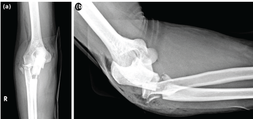
Figure 1: Pre-operative plain radiographs of the right elbow. (a) Anteroposterior view demonstrating proximal ulnar shaft fracture and disrupted elbow congruence. (b) Lateral view confirming posterior ulno-humeral dislocation.
Closed reduction under intravenous sedation was unsuccessful. Computed tomography (CT) with three-dimensional reconstruction identified four distinct injury components: (1) posterior ulno-humeral dislocation; (2) comminuted radial head fracture with dislocation; (3) coronoid process fracture; and (4) proximal one-third ulnar shaft fracture (Fig. 2a, b, c).
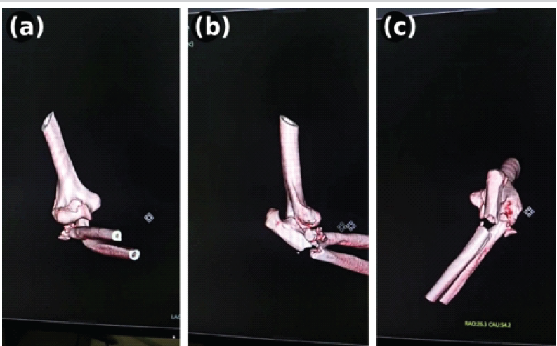
Figure 2: Three-dimensional computed tomography reconstructions confirming all four components of the “terrible tetrad.”
This pattern simultaneously fulfilled the criteria for both the terrible triad and a Monteggia-type fracture-dislocation – constituting a “terrible tetrad” of the elbow. Surgery was performed through a single posterior approach. The comminuted radial head was replaced with a cementless modular radial head prosthesis (22 mm head diameter) [7]. The proximal ulnar shaft fracture was fixed with a 3.5 mm dynamic compression plate. The lateral ulnar collateral ligament and common extensor origin were repaired to the lateral epicondyle with a suture anchor. The coronoid – a small Type I fragment – did not require separate fixation once lateral column stability was restored [8], consistent with published algorithms. Dynamic intraoperative assessment confirmed a stable, congruent elbow throughout the full arc of motion. The patient returned overseas 3 weeks postoperatively without access to formal physiotherapy. At 5 months, the range of motion was 25–110°. The elbow was stable and pain-free; post-operative radiographs confirmed concentric radial head prosthesis seating, appropriate anterior prosthetic positioning on the lateral view, and satisfactory ulnar plate alignment (Fig. 3a and b). Limited functional recovery was attributable to rehabilitation non-compliance.
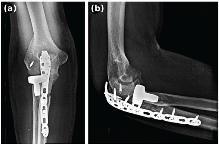
Figure 3: Post-operative plain radiographs. (a) Anteroposterior view showing concentric radial head prosthesis seating and satisfactory ulnar dynamic compression plate alignment. (b) Lateral view confirming appropriate anterior prosthetic positioning and restored ulno-humeral congruence.
Case 2: Left-sided terrible tetrad – two-incision reconstruction strategy:
A 53-year-old right-hand-dominant woman presented following a fall down a flight of stairs, sustaining a left elbow injury. Her medical history included controlled hypertension (on oral antihypertensives) and Hepatitis C virus (HCV) positivity; she was not on anticoagulation and had no prior elbow pathology. On examination, there was diffuse swelling and deformity of the left elbow with disturbance of the three-point bony relationship. Distal pulses were palpable bilaterally, and there was no neurological deficit at presentation. Pre-operative liver function tests and coagulation screen were within acceptable limits for surgery. Plain radiographs demonstrated posterior ulno-humeral dislocation, a comminuted radial head fracture, and a proximal ulnar fracture (Fig. 4a and b).
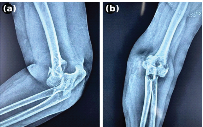
Figure 4: Pre-operative plain radiographs of the left elbow. (a) Lateral view and (b) anteroposterior view demonstrating posterior ulno-humeral dislocation, comminuted radial head fracture, and proximal ulnar fracture.
Closed reduction under anaesthesia was unsuccessful. CT with three-dimensional reconstruction confirmed: (1) posterior ulno-humeral dislocation; (2) comminuted radial head fracture; (3) coronoid process avulsion fracture; and (4) proximal ulnar fracture (Fig. 5).
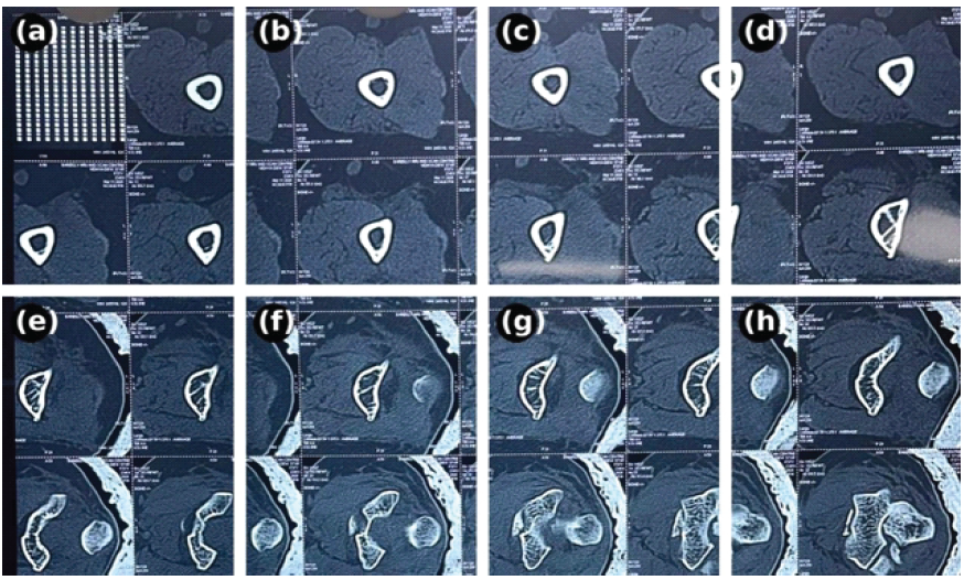
Figure 5: Computed tomography reconstructions of the left elbow confirming all four components of the “terrible tetrad” from multiple projections.
A terrible tetrad was confirmed. A combined two-incision approach was employed. Through a posterior midline incision, the proximal ulnar fracture was fixed with an anatomical locking compression plate. Via a subsequent lateral Kocher approach, the comminuted radial head was replaced with an uncemented modular radial head prosthesis. The coronoid avulsion fragment was repaired using FibreWire sutures passed through bone tunnels and secured with an Endobutton on the anterior ulnar cortex [9], providing stable anteromedial column fixation. The lateral collateral ligament was repaired to the lateral epicondyle using suture anchors from the same lateral approach. Dynamic intraoperative assessment confirmed a stable, congruent elbow with an almost full arc of motion. There were no intraoperative or post-operative complications. Intraoperative fluoroscopy confirmed satisfactory positioning of all implants with a congruent ulno-humeral joint (Fig. 6a and b).
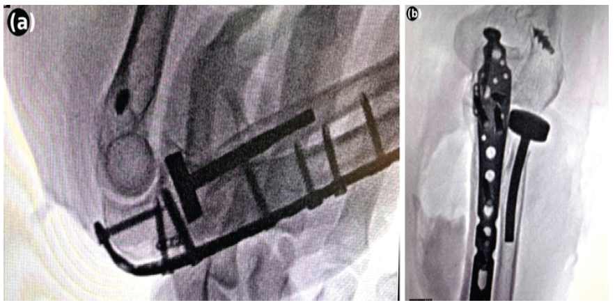
Figure 6: Intraoperative fluoroscopic images. (a) Anteroposterior view confirming satisfactory positioning of the ulnar locking compression plate and radial head prosthesis with restored ulno-humeral congruence. (b) Lateral view confirming appropriate anterior prosthetic positioning and joint congruence intraoperatively.
The arm was immobilised in an above-elbow posterior slab for 3 weeks, followed by supervised passive range-of-motion exercises. At 6-week follow-up, the patient was progressing satisfactorily, with a stable, pain-free elbow and improving range of motion (Fig. 7a, b, c, d). Post-operative radiographs at 6 weeks confirmed satisfactory implant positioning with no hardware complications (Fig. 8a and b).
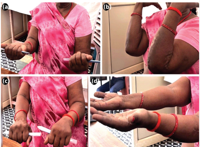
Figure 7: Clinical photographs at 6-week follow-up demonstrating range of motion of the left elbow. (a) Bilateral supination, (b) bilateral flexion, (c) pronation, and (d) extension, showing satisfactory early functional recovery with a well-healed surgical scar.
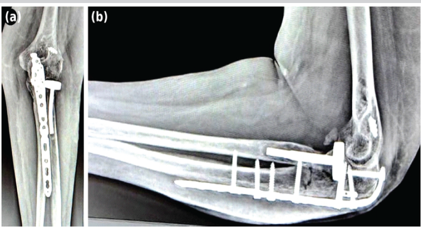
Figure 8: Post-operative plain radiographs at 6 weeks. (a) Anteroposterior view showing the ulnar locking compression plate in satisfactory position with the uncemented radial head prosthesis seated concentrically at the radiocapitellar joint. (b) Lateral view confirming maintained ulno-humeral congruence, appropriate anterior prosthetic positioning, and no hardware complications.
Both cases simultaneously fulfilled all diagnostic criteria for the terrible triad [1] and a Monteggia-type fracture-dislocation [2,3], constituting what we term the “terrible tetrad” of the elbow. The term has appeared in fewer than five publications, and total cases reported under any equivalent description do not exceed fifteen worldwide [4,5,6]. No existing classification system encompasses this pattern. To the best of our knowledge, this is the first case series to present two such cases with a comparative analysis of surgical strategy and outcome. The two cases share the quadruple-component injury pattern but differ in several clinically important respects. Case 1 involved a high-energy mechanism (fall from height) in a younger male, while Case 2 arose from a low-energy mechanism (fall down stairs) in an older female with medical comorbidities – demonstrating that the terrible tetrad can occur across a spectrum of energy levels and patient profiles. The HCV positivity and hypertension in Case 2 added perioperative complexity, requiring pre-operative optimisation, a consideration not previously highlighted in this literature. The principal surgical difference was the approach strategy. In Case 1, a single posterior incision provided access to all four injury components. The coronoid – a small Type I fragment – did not require direct fixation once lateral column stability was restored, consistent with published management algorithms [8]. In Case 2, a combined posterior and lateral two-incision approach was employed, enabling direct coronoid repair via FibreWire and Endobutton – a technique validated for avulsion fragments of sufficient size [9]. Both cases followed the established lateral-column-first principle: Radial head replacement – the pivotal step in restoring the primary valgus and axial stabiliser of the elbow – was performed before ligamentous and bony repair of the medial column [10]. Both approaches achieved intraoperative stability, validating their respective indications. The outcomes differed primarily because of rehabilitation access rather than surgical technique. Case 1 achieved a functional arc of 25–110° at 5 months despite returning overseas without physiotherapy; Case 2 was under supervised physiotherapy from 3 weeks, consistent with published protocols for terrible triad reconstruction [11,12], and was progressing well at 6 weeks. This contrast reinforces a key message in the terrible triad literature: Structured and supervised physiotherapy is the principal determinant of functional outcome after complex elbow reconstruction and must be planned and secured pre-operatively [1,6]. Surgeons encountering the terrible tetrad should follow a systematic pre-operative assessment: CT with three-dimensional reconstruction is mandatory to identify all four components; coronoid fragment size and accessibility should guide approach selection (single posterior versus combined); and physiotherapy must be arranged before the patient leaves hospital. We propose that the term “terrible tetrad” be formally adopted in the orthopaedic classification literature to facilitate recognition, case reporting, and future aggregation of outcomes in this rare injury pattern.
We present two cases of the “terrible tetrad” of the elbow – a quadruple-component injury simultaneously fulfilling criteria for both the terrible triad and a Monteggia-type fracture-dislocation – one managed through a single posterior approach and one via a combined two-incision strategy. With fewer than fifteen cases in the world literature, this is one of the rarest elbow injury configurations ever described. CT characterisation is essential; approach selection should be individualised to injury configuration; and supervised physiotherapy is the principal determinant of functional outcome. Formal adoption of the term “terrible tetrad” would facilitate recognition, reporting, and evidence-based classification of this rare but surgically demanding injury.
The “terrible tetrad” of the elbow – comprising posterior ulno-humeral dislocation, radial head fracture, coronoid fracture, and a proximal ulnar fracture – represents a distinct injury pattern that fulfils criteria for both the terrible triad and a Monteggia-type fracture-dislocation and should be recognised as such rather than classified under either entity alone. Three-dimensional computed tomography reconstruction is mandatory to delineate all four components and guide surgical planning. Reconstruction should adhere to the lateral-column-first principle, with radial head fixation or arthroplasty preceding coronoid and ligamentous repair and approach selection (single posterior versus combined two-incision) individualised according to coronoid fragment size and accessibility. Irrespective of technique, supervised physiotherapy must be secured before discharge, as post-operative rehabilitation access – rather than surgical execution alone – appears to be the principal determinant of functional recovery in this rare and technically demanding injury.
References
- 1. Hotchkiss RN. Fractures and dislocations of the elbow. In: Rockwood CA, Green DP, Bucholz RW, Heckman JD, editors. Rockwood and Green’s Fractures in Adults. 4th ed. Philadelphia, PA: Lippincott-Raven; 1996. p. 929-85. [Google Scholar] [PubMed]
- 2. Bado JL. The monteggia lesion. Clin Orthop Relat Res 1967;50:71-86. [Google Scholar] [PubMed]
- 3. Singh AP, Dhammi IK, Jain AK, Raman R, Modi P. Monteggia fracture dislocation equivalents–analysis of eighteen cases treated by open reduction and internal fixation. Chin J Traumatol 2011;14:221-6. [Google Scholar] [PubMed]
- 4. Caviglia H, Garabano G, Landro ME, Gallucci GL, Boretto JG, De Carli P. The terrible tetrad of the elbow. Rev Asoc Argent Ortop Traumatol 2010;75:281-8. [Google Scholar] [PubMed]
- 5. Ramesh S, Lim YJ. Complex elbow dislocation associated with radial and ulnar diaphyseal fractures: A rare combination. Strategies Trauma Limb Reconstr 2011;6:97-101. [Google Scholar] [PubMed]
- 6. Medina G, Keller RE, Sabbag OD, Oh LS. Terrible triad of the elbow and associated variants: A systematic review. JSES Rev Rep Tech 2022;2:205-13. [Google Scholar] [PubMed]
- 7. Preston CF, Chen AL, Wolinsky PR, Tejwani NC. Posterior dislocation of the elbow with concomitant fracture of the proximal ulnar diaphysis and radial head: A complex variant of the posterior Monteggia lesion. J Orthop Trauma 2003;17:530-3. [Google Scholar] [PubMed]
- 8. Mathew PK, Athwal GS, King GJ. Terrible triad injury of the elbow: Current concepts. J Am Acad Orthop Surg 2009;17:137-51. [Google Scholar] [PubMed]
- 9. Garrigues GE, Wray WH 3rd, Lindenhovius AL, Ring DC, Ruch DS. Fixation of the coronoid process in elbow fracture-dislocations. J Bone Joint Surg Am 2011;93:1873-81. [Google Scholar] [PubMed]
- 10. Ring D, Jupiter JB, Simpson NS. Monteggia fractures in adults. J Bone Joint Surg Am 1998;80:1733-44. [Google Scholar] [PubMed]
- 11. Pugh DM, Wild LM, Schemitsch EH, King GJ, McKee MD. Standard surgical protocol to treat elbow dislocations with radial head and coronoid fractures. J Bone Joint Surg Am 2004;86:1122-30. [Google Scholar] [PubMed]
- 12. Jupiter JB, Leibovic SJ, Ribbans W, Wilk RM. The posterior monteggia lesion. J Orthop Trauma 1991;5:395-402. [Google Scholar] [PubMed]


Related Articles in Journal of Orthopaedic Case Reports
 October 1, 2025 A Unique Case of Bilateral Elbow Terrible Triad in a Polytrauma Patient: Associated Challenges in Management
 April 1, 2025 A Case Series of Terrible Triad Elbow Injuries Treated with Lasso Suture Fixation for Coronoid, Tripod Fixation for Radial Head, and LCL Repair
 October 1, 2026 Pertrochanteric Femur Fracture in a Post-Polio Patient treated with a Pediatric Hip Plate: A Case Report
 August 1, 2026 Augmentation Plating with Intramedullary Nailing for Aseptic Long Bone Non-union: A Prospective Observational Study of Clinical, Radiological, and Functional Outcomes