

Following hemiarthroplasty, medial migration develops with a certain incidence. As delayed intervention makes treatment more challenging, regular follow-up is required.
Hiroki Kobayashi, Department of Orthopaedic Surgery, National Defence Medical College, Tokorozawa, Saitama, Japan. E-mail: supercova0205@gmail.com
Abstract
Introduction: Central migration of the prosthetic femoral head occurs in approximately 7% of cases following bipolar hemiarthroplasty (BHA) for femoral neck fractures. If treated promptly, it can be managed by cup placement; however, if left unaddressed, a large acetabular bone defect can develop. Herein, we report a case of central migration with a severe bone defect after BHA, successfully treated with conversion to total hip arthroplasty (THA) using a Burch–Schneider cage.
Case Report: An 86-year-old man underwent BHA at a local hospital for a right femoral neck fracture in Year X-8. He experienced right hip pain in Year X-4. An X-ray examination in Year X-3 revealed mild central migration of the prosthesis. However, follow-up was discontinued afterward. In Year X, he visited the hospital with complaints of worsening right hip and abdominal pain. He was subsequently referred to our hospital for further examination and treatment. Based on clinical images, he was diagnosed with central migration of the prosthetic femoral head with a Type 3B acetabular defect in the Paprosky classification. A conversion THA with a reinforcement ring was planned. The surgery was performed using a supine anterolateral approach. A Burch–Schneider cage was placed to bridge the gap, and the defect was filled with bone cement. He commenced full weight-bearing the day after surgery without experiencing any pain. He achieved independent ambulation with a cane 8 days after surgery and was discharged home. One year postoperatively, he had a favourable postoperative course without any complications.
Conclusion: Central migration of the prosthesis inevitably occurs at a certain rate after surgery. In cases involving extensive bone defects, rigid acetabular reconstruction with implants is essential. However, the procedure is technically challenging and could pose a significant surgical burden to patients. Periodic imaging evaluations and timely interventions are essential for patients with BHA to avoid this condition.
Keywords: Central migration, bipolar hemiarthroplasty, conversion total hip replacement, acetabular reinforcement ring.

Bipolar hemiarthroplasty (BHA) is a well-established surgical procedure for treating femoral neck fractures. It is minimally invasive and enables early ambulation after surgery. However, central migration of the prosthetic femoral head occurs at a certain rate due to wear of the acetabular cartilage, even in cases where there is no apparent cartilage degradation at the time of surgery. Therefore, long-term follow-up, monitoring degenerative changes in the acetabulum, is essential [1]. Central migration of the prosthesis occurs in approximately 7% of BHA cases [2]. In the early stages, the condition can be managed with standard total hip arthroplasty (THA). However, if left unaddressed, a large acetabular defect may develop. In such cases, highly invasive procedures using a reinforcement implant are often required. Therefore, early diagnosis and timely treatment are crucial to prevent additional invasive surgery for patients following BHA. We herein report a case of central migration of the prosthesis after head following BHA that was successfully treated with a conversion THA using a Burch–Schneider cage. This case underscores the importance of early detection of central migration and the undesirable consequences that can arise when treatment is delayed.
The patient was an 86-year-old man. In Year X-8, he fell and sustained a right femoral neck fracture. He underwent a BHA using a posterior approach at a local hospital (Fig. 1).
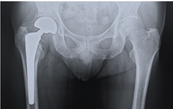
Figure 1: Radiograph in year X-8. Bipolar hemiarthroplasty has been performed.
Clinical course following surgery was uneventful, and he was able to walk independently with a cane. In Year X-4, he experienced pain in his right hip and received conservative treatment. The following year, although radiographs were taken at the same hospital due to worsening pain, no abnormalities were identified, and no treatment was provided. However, subsequent reexamination of these radiographs at our hospital revealed that an early sign of acetabular cartilage destruction by the prosthesis had already been present (Fig. 2).
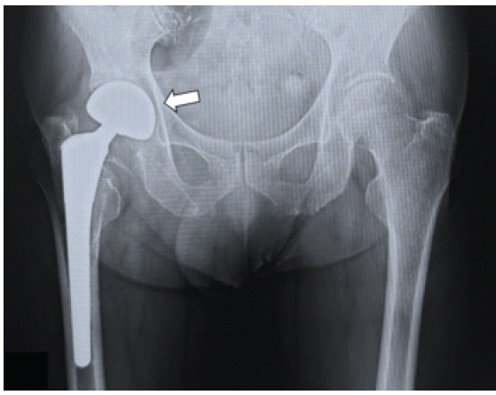
Figure 2: Radiograph in year X-3. Very mild femoral head migration is observed (white arrow).
Despite his persistent pain, his hospital visits were subsequently discontinued. The following year, his right hip pain gradually worsened, making it difficult for him to walk independently. In addition to hip pain, he developed pain in his right lower abdomen. He visited the local hospital in Year X, where he was diagnosed with central migration of the prosthesis. He was subsequently referred to our hospital for further examination and treatment. At the time of his visit, he could barely walk and required a wheelchair due to hip pain. The persistent pain was also causing sleep disturbances. Radiographs and computed tomography (CT) images showed severe bone defects in the acetabulum and intrapelvic migration of the prosthetic femoral head (Figs. 3 and 4).
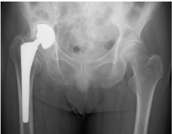
Figure 3: Pre-operative radiograph. Central migration with severe acetabular defect is observed.
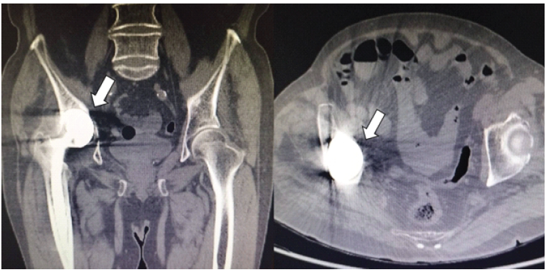
Figure 4: Plain computed tomography images. Left, coronal section; right, axial section. The prosthetic femoral head has migrated into the pelvic cavity due to an acetabular bone defect (white arrow).
The acetabular bone defect corresponded to Type 3B of the Paprosky classification. The right lower abdominal pain was assumed to be caused by direct irritation from the prosthesis. Due to the extensive acetabular bone defect, revision arthroplasty using an acetabular reinforcement ring was deemed necessary. The surgery was performed using a supine anterolateral approach. The short external rotators, including the obturator internus and obturator externus muscles, were scarred, but because they were strong factors resisting dislocation, they were preserved as much as possible during exposure. The prosthetic femoral head had migrated deeply into the acetabulum and was barely visible. The lateral acetabulum was partially removed to expose the femoral head, and it was detached from the stem to facilitate its removal (Fig. 5).
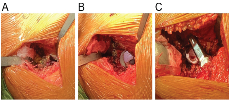
Figure 5: Intraoperative findings. (a) The prosthetic femoral head has migrated deeply into the pelvic cavity and is barely visible. (b) A part of the lateral acetabulum has been removed, exposing the underside of the femoral head. (c) The prosthetic femoral head has been detached from the stem.
The femoral stem was firmly fixed to the bone, and we decided to leave it in place. A Burch–Schneider cage was placed in the acetabular defect to bridge the gap, and the remaining defect was filled with bone cement for support. After confirming sufficient joint stability, the surgery was finished (Fig. 6).
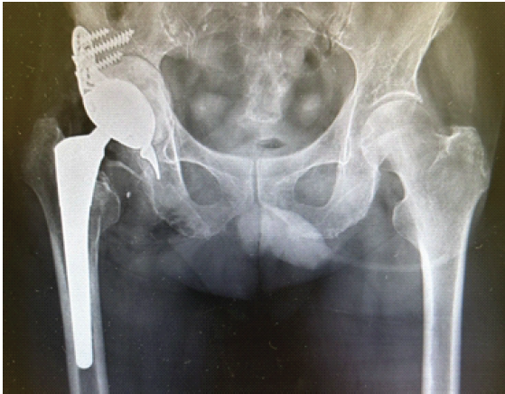
Figure 6: Post-operative radiograph. The stem was preserved, and a Burch–Schneider cage was placed for the extensive bone defect.
The patient experienced immediate relief from hip and lower abdominal pain after surgery. Gait rehabilitation with full weight-bearing was initiated on the 1st post-operative day. He was able to walk with a cane by post-operative day 8 and was discharged home. The clinical course was uneventful, and approximately a year after surgery, he is well without pain and has a stable gait.
While central migration of the prosthesis after BHA occurs at a certain rate, previous long-term observational studies suggest that it is not necessarily associated with patients’ physical activities after surgery [1,3]. Several studies have reported that the progression of central migration of the prosthesis following BHA varies greatly among cases, indicating that this condition largely depends on the patient’s physical background. In a case series study, Tsukanaka et al. reported that, although central migration of the prosthesis was not evident up to 1 year after surgery, significant wear of the acetabular cartilage was visible on subsequent radiographs in some patients. Those patients consisted mainly of elderly patients (around or over 90 years old), suggesting that bone fragility is closely associated with the development of this condition [4]. In accordance, it has also been reported that central migration of the prosthesis may occur in patients with poor bone quality, even in cases with no apparent degenerative changes in the acetabular cartilage at the time of surgery [5]. This further underscores the subchondral bone quality of the acetabulum as an important determinant of long-term clinical results. In general, the progression of central migration of the prosthesis is gradual. However, in some patients, it may present as an acute condition. Several potential causes of this rapid progression have been suggested. First, in some cases, early onset of this condition is likely associated with damage to the subchondral bone or articular cartilage caused by injury rather than long-term wear [6]. Second, the inappropriate size of the prosthetic femoral head can cause early cartilage wear and ultimately lead to central migration. If the prosthetic femoral head is too large relative to the original femoral head, it can induce wear in the acetabular rim. Conversely, if the prosthetic head is too small, it can result in uneven load distribution and accelerated cartilage wear [2]. Finally, periprosthetic joint infection has also been suggested as a potential cause for central migration, at least in some cases. Adenikinju et al. reported that cartilage wear can increase significantly due to a periprosthetic joint infection caused by low-virulence bacteria, which are difficult to detect using standard bacterial cultures. They suggest that periprosthetic joint infection should be considered in the differential diagnosis, even in cases without clear signs of infection, in patients with central migration [7]. In our patient, we found no evidence of the aforementioned conditions, indicating that cartilage wear was primarily the result of cumulative mechanical load. It is essential to carefully plan the surgery before treating the central migration of the prosthesis. In general, conversion THA is an effective treatment for this condition; however, the risk of complications is significantly higher than primary THA. According to a meta-analysis, dislocation occurs in 8.2%, and periprosthetic joint infection in 3.8% of cases after conversion to THA. Therefore, the complication ratio of conversion THA is comparable to that of revision THA [8,9]. Regarding femoral stem preservation, there is the potential advantage of reducing perioperative complications associated with stem removal. However, it has also been reported to significantly increase the risk of hip dislocation. Therefore, the decision must be made by weighing these pros and cons [8,10]. The timing of surgery is one of the most important factors for successful treatment. In general, conversion to THA should be considered when central migration of the prosthesis is identified. Minimal bone defects can be managed using an acetabular cup. However, if surgery is delayed, severe defects can develop, necessitating acetabular reconstruction with implants. These surgeries are more technically challenging than standard THA and impose a greater surgical burden on patients. Our patient experienced right hip pain in Year X-4, and the early signs of central migration of the prosthesis were evident on radiographs from Year X-3. Unfortunately, these signs were overlooked, and he was left untreated. Since there was no apparent bone defect in Year X-3, the patient could have been treated with a simple conversion THA without a reinforcement ring. However, by the time he was referred to our hospital, there was a marked bone defect that required implants to reconstruct the acetabulum. Given that the 10-year re-revision rate after conversion to THA reaches 15% [11], some suggest performing THA rather than BHA for patients with femoral neck fractures whenever possible. When BHA is selected for reasons such as the absence of a hip specialist, follow-up with regular imaging evaluation should be thoroughly performed.
We reported a case of central migration of a prosthetic femoral head, accompanied by a severe bone defect that was successfully treated with a conversion THA using a Burch–Schneider cage. Periodic radiographic evaluation is mandatory for patients after BHA, and salvage surgery should be considered once central migration is confirmed.
Following hemiarthroplasty, medial migration develops with a certain incidence. As delayed intervention makes treatment more challenging, regular follow-up is required.
References
- 1. Pace TB, Prather B, Burnikel B, Shirley B, Tanner S, Snider R. Comparative outcomes assessment: Hip hemiarthroplasty as an alternative to THA in patients with surgically pristine acetabulum-is there still a role? ISRN Orthop 2013;2013:632126. [Google Scholar] [PubMed]
- 2. Schiavi P, Pogliacomi F, Colombo M, Amarossi A, Ceccarelli F, Vaienti E. Acetabular erosion following bipolar hemiarthroplasty: A role for the size of femoral head? Injury 2019;50:S21-5. [Google Scholar] [PubMed]
- 3. Lind D, Natman J, Mohaddes M, Rogmark C. Long-term risk of reoperation after modular hemiarthroplasty: Any differences between uni- or bipolar design?BMC Musculoskelet Disord 2023;24:911. [Google Scholar] [PubMed]
- 4. Tsukanaka M, Støen RØ, Figved W, Frihagen F, Nordsletten L, Röhrl SM. Steady state acetabular cartilage wear after bipolar hemiarthroplasty: A case series of 10 patients with radiostereometric analysis. Hip Int 2017;27:193-7. [Google Scholar] [PubMed]
- 5. Muraki M, Sudo A, Hasegawa M, Fukuda A, Uchida A. Long-term results of bipolar hemiarthroplasty for osteoarthritis of the hip and idiopathic osteonecrosis of the femoral head. J Orthop Sci 2008;13:313-7. [Google Scholar] [PubMed]
- 6. Mahmoud AN, Suk M, Horwitz DS. Symptomatic acetabular erosion after hip hemiarthroplasty: Is it a major concern? A retrospective analysis of 2477 hemiarthroplasty cases. J Clin Med 2024;13:6756. [Google Scholar] [PubMed]
- 7. Adenikinju A, Slover JD, Egol KA. Rapid acetabular chondrolysis following hemiarthroplasty of the hip: A poor prognostic sign. Case Rep Orthop 2019;2019:7328526. [Google Scholar] [PubMed]
- 8. Poursalehian M, Hassanzadeh A, Lotfi M, Mortazavi SM. Conversion of a failed hip hemiarthroplasty to total hip arthroplasty: A systematic review and meta-analysis. Arthroplasty Today 2024;28:101459. [Google Scholar] [PubMed]
- 9. Hernandez NM, Fruth KM, Larson DR, Kremers HM, Sierra RJ. Conversion of hemiarthroplasty to THA carries an increased risk of reoperation compared with primary and revision THA. Clin Orthop Relat Res 2019;477:1392-9. [Google Scholar] [PubMed]
- 10. Figved W, Hallan G, Engebretsen L, Furnes O, Gjertsen JE, Havelin LI. Conversion of failed hemiarthroplasty to total hip arthroplasty: A norwegian hip arthroplasty register study of 595 cases. J Bone Joint Surg Br 2007;89:1427-30. [Google Scholar] [PubMed]
- 11. Hernandez NM, Fruth KM, Larson DR, Kremers HM, Sierra RJ. Conversion of failed hemiarthroplasty to total hip arthroplasty remains high risk for subsequent complications. J Arthroplasty 2019;34:2030-6. [Google Scholar] [PubMed]





Related Articles in Journal of Orthopaedic Case Reports
 October 1, 2026 Dual Mobility Total Hip Arthroplasty for Recurrent Dislocation After Femoral Neck Fracture in an Elderly Patient: A Case Report
 May 1, 2026 Neglected Bipolar Hemiarthroplasty Dislocation in a Young Adult with Superior Acetabular Wall Defect – A Case Report
 April 1, 2026 Dislocation and Iatrogenic Dissociation of Bipolar Hip Hemiarthroplasty: A Case Series and Literature Review
 April 1, 2025 From Trivial Trauma to Major Surgery: Navigating Skeletal Metastases with Sequential Hip Arthroplasty for Pathological Femoral Neck Fractures in Triple-Negative Breast Cancer