

Complex femoral head–neck fracture-dislocations beyond the Pipkin classification require early individualized surgical management, prioritizing anatomical reconstruction and hip preservation in young patients, while reserving arthroplasty for selected elderly patients with poor reconstructive potential.
Dr. Niraj Narain Singh, Department of Orthopaedics, ESIC Medical College and Hospital, Patna, Bihar, India. E-mail: drnirajnrsingh@gmail.com
Abstract
Introduction: Femoral head–neck fracture-dislocation with associated posterior acetabular wall and pertrochanteric fractures represents an extremely rare and severe “beyond Pipkin IV” injury pattern.
Case Report: A 14-year-old boy sustained posterior hip dislocation with femoral head, neck, acetabular, and trochanteric involvement following a road traffic accident. Emergency open reduction and internal fixation were performed using a modified Kocher–Langenbeck posterior approach with a trochanteric flip.
Results: Radiological union was achieved by 3 months. At 1-year follow-up, the patient had good hip motion without avascular necrosis or implant failure.
Conclusion: Early anatomical reconstruction using a posterior approach can produce excellent outcomes in young patients with this rare injury.
Keywords: Femoral head fracture, Pipkin IV, hip dislocation, Kocher–Langenbeck approach, adolescent, acetabular fracture, trochanteric fracture.

The prevalence of femoral head fracture (FHF) dislocation ranges from 8% to 26%. FHFs are uncommon fractures that typically result from high-energy trauma. They generally arise following posterior dislocation, with around 5–15% of posterior hip dislocations linked to FHF [1,2]. FHF may simultaneously occur with acetabular fractures, with an anticipated occurrence of 29.2% [1,2]. The incidence of ipsilateral femoral head and neck fractures (iFHNF) was negligible. Nonetheless, iFHNFs, a unique classification of fracture, demonstrate the least favorable prognostic for every FHF. Alignment of the head as well as the hip during axial stress typically dictates the nature of damage [3]. Timely diagnosis and immediate intervention are essential to avert future problems encompassing avascular necrosis (AVN) of the femoral head as well as arthritis [4]. Essential imaging modalities, such as X-ray as well as computed tomography (CT) scan, should be conducted before a reduction attempt due to the increased danger of an iatrogenic FHF after closed reduction. The appropriate treatment technique in such circumstances remains contentious. The irreducible FHF-dislocation was treated employing a flip trochanteric at fracture as well as the extended Kocher-Langenbeck method. Femoral head lesions are classified according to Pipkin (four classes) [5] and Chiron (five grades, further categorized as A if isolated, B with acetabular fracture, and C in the event of FHF) [6].
A 14-year-old male presented following a road traffic accident with pain, swelling, deformity, and inability to bear weight on the left lower limb. After initial trauma stabilization, clinical examination revealed shortening, external rotation, and flexion of the left hip, with a palpable femoral head in the gluteal region, suggesting posterior hip dislocation. All hip movements were painful and restricted. Radiological evaluation included anteroposterior (AP) radiographs of both hips and CT with three-dimensional reconstruction, which confirmed a femoral head and neck fracture with associated posterior acetabular wall involvement. The pre-operative radiographic and CT findings are shown in Fig. 1.
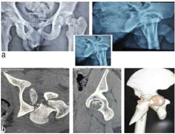
Figure 1: (a) Pre-operative X-ray – Pre-operative anteroposterior radiograph of the pelvis showing posterior dislocation of the left hip with associated femoral head and neck fracture. (b) Pre-operative computed tomography scan – Pre-operative computed tomography with three-dimensional reconstruction demonstrating femoral head and neck fracture with posterior acetabular wall involvement and per-trochanteric extension.
AP pelvic radiographs were obtained preoperatively and postoperatively and at regular follow-up intervals. Surgical intervention was performed in the lateral decubitus position using a modified Kocher–Langenbeck approach with a trochanteric flip at the fracture site. Intraoperatively, the femoral head was fractured in the anteroinferior region and displaced posteriorly against the ilium, with an associated posterosuperior acetabular rim fracture and labral avulsion. The femoral head fragments were fixed using headless compression screws. After removal of incarcerated intra-articular fragments, the hip was reduced, and the femoral neck and peritrochanteric fracture were stabilized using a dynamic hip screw with an additional anti-rotation screw. The posterior acetabular wall was fixed with cortical screws. Intraoperative assessment confirmed a stable and concentric reduction. The intraoperative findings and fixation construct are shown in Fig. 2.
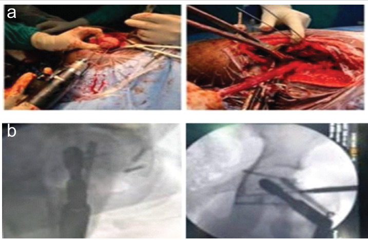
Figure 2: (a) Intra-operative. (b) Intraoperative c-arm image-Intra operative pictures and anteroposterior C-arm image showing anatomical reduction of the hip with fixation of the femoral head using headless screws, femoral neck and trochanteric region with dynamic hip screw and anti-rotation screw, and posterior acetabular wall fixation.
Postoperatively, range of motion (ROM) exercises was initiated on the 3rd day. The patient was kept non-weight-bearing for 6 weeks, followed by partial weight bearing, and progressed to full weight bearing at 12 weeks after radiographic evidence of fracture union. The patient was followed up at 6 weeks, 3 months, 6 months, and 2 years with clinical and radiological evaluation. At final follow-up, there was no implant failure, femoral head collapse, or radiological evidence of AVN or post-traumatic arthritis. The patient had a painless ROM from 0° to 110°, with only mild activity-related discomfort.
Patient operated within 24 h of injury and was admitted for 3 weeks in hospital. The 1st postoperative week started with ROM of hip exercise, and static quadriceps exercise started after 2 weeks. Follow-up after 6 weeks shows signs of union. The patient was non-weight-bearing for a period of 6 weeks. The fracture got united after 3 months. Partial weight-bearing is allowed after 3 months. At the 2-year follow-up, the patient demonstrated a good functional outcome with a satisfactory range of motion (ROM). There is no evidence of deep vein thrombosis/AVN till the 1-year follow-up. Serial follow-up radiographs at 6 weeks and 2 years, along with the final functional outcome, are shown in Fig. 3.
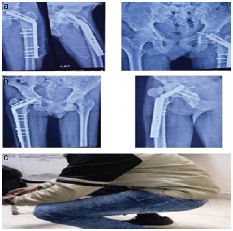
Figure 3: (a) 6-week follow-up – Radiograph at 6-week follow-up showing evidence of fracture uniting and maintenance of joint congruity. (b) 2-year follow-up – Radiograph at 2-year follow-up showing evidence of fracture union and maintenance of joint congruity. (c) 2-year follow-up: Follow-up at 2-year follow-up demonstrating healed fractures, maintained hip alignment, and absence of avascular necrosis or implant failure with functional hip.
FHFs associated with posterior hip dislocation are rare, high-energy injuries and represent true orthopedic emergencies. Although femoral neck fractures and posterior dislocations result from different biomechanical forces, they often coexist due to the magnitude and direction of trauma. Femoral neck fractures are typically produced by direct impact over the greater trochanter or rotational forces, whereas posterior dislocation occurs from an axial load applied to a flexed, abducted, and internally rotated hip [1]. Prompt reduction is critical, as delayed management significantly increases the risk of femoral head AVN. Previous studies have demonstrated that the incidence of AVN rises to approximately 14–15% when reduction is delayed beyond 12 h, reflecting disruption of the extra-osseous blood supply following traumatic dislocation. Therefore, urgent reduction followed by stable fixation remains the cornerstone of management [7,8]. Selection of the optimal surgical approach in such complex injuries is challenging. Although the Ganz surgical dislocation with trochanteric flip osteotomy provides excellent intra-articular exposure and preserves femoral head vascularity in isolated FHFs, it is not suitable in the presence of associated femoral neck or trochanteric extension, as the integrity of the medial femoral circumflex artery and trochanteric fragment cannot be reliably preserved [9,10]. In the present case, an extended posterior (modified Kocher–Langenbeck) approach was preferred, as it allows simultaneous access to the femoral head, neck, posterior acetabulum, and trochanteric–subtrochanteric region, enabling comprehensive fracture fixation or arthroplasty when required. Anterior approaches were avoided due to their limited access to posterior and trochanteric components and their higher risk of further compromising femoral head vascularity. This approach provided stable fixation, adequate visualization, and a biologically favorable environment for healing in this complex fracture-dislocation pattern [11,12,13,14].
Femoral head–neck fracture–dislocations with associated acetabular and trochanteric fractures with subtrochanteric extension involvement represent a rare and complex injury pattern that lies beyond the conventional Pipkin classification. Early reduction and an appropriately selected surgical approach are critical to preserving femoral head vascularity and optimizing outcomes. The modified Kocher–Langenbeck approach with trochanteric flip osteotomy provides comprehensive exposure while minimizing additional soft tissue and vascular compromise, making it a valuable option in managing such challenging fracture-dislocations.
This case illustrates that achieving early anatomical reduction and ensuring stable internal fixation of a highly uncommon femoral head–neck fracture-dislocation, which is linked with posterior acetabular wall and peritrochanteric fractures, can lead to outstanding functional and radiological results in adolescents. The use of a modified Kocher–Langenbeck approach allowed for sufficient exposure to reconstruct all fracture components simultaneously while maintaining the viability of the femoral head.
References
- 1. Dunseath OA, Al-Obaidi I, Ignatius L, Rudran B, Jordan C. Femoral head fractures: Anatomy, diagnosis and management. EFORT Open Rev 2026;11:175-82. [Google Scholar] [PubMed]
- 2. Ompusunggu IH, Putra YY, Auliya A. Articular femoral head fracture management: A comprehensive systematic review. J Adv Res Med Health Sci 2024;10:57-63. [Google Scholar] [PubMed]
- 3. McKee MD, Garay ME, Schemitsch EH, Kreder HJ, Stephen DJ. Irreducible fracture-dislocation of the hip: A severe injury with a poor prognosis. J Orthop Trauma 1998;12:223-9. [Google Scholar] [PubMed]
- 4. Hougaard K, Thomsen PB. Traumatic posterior fracture-dislocation of the hip with fracture of the femoral head or neck, or both. J Bone Joint Surg 1988;70:233-9. [Google Scholar] [PubMed]
- 5. Pipkin G. Treatment of grade IV fracture-dislocation of the hip. J Bone Joint Surg 1957;39-A:1027-42. [Google Scholar] [PubMed]
- 6. Golovakha M, Lisunov M, Braunsteiner T, Orljanski W. Fractures of the femoral head (clinical lecture). Orthop Traumatol Prosthet 2024;4:86-94. [Google Scholar] [PubMed]
- 7. Giannoudis PV, Gonzalez Edery E, Nemade P, Bagaria V, Femoral head impaction fracture: A new technique for closed reduction and biological stabilisation. BMJ Case Rep 2025;18:e264269. [Google Scholar] [PubMed]
- 8. Milenkovic S, Mitkovic M, Mitkovic M. Avascular necrosis of the femoral head after traumatic posterior hip dislocation with and without acetabular fracture. Eur J Trauma Emerg Surg 2022;48:613-9. [Google Scholar] [PubMed]
- 9. Hassan M, Elnosh HA, Khidir Mohamed EN, Adam YA, Ahmed AE, Mohamedalhassan Abbas DM, et al. Evaluating the outcomes of surgical hip dislocation for femoral head trauma: A systematic review. Cureus 2025;17:e97152. [Google Scholar] [PubMed]
- 10. Iglesias CJ, García BE, Valarezo JP. Controlled ganz dislocation. EPRA Int J Multidiscip Res 2025;11:1410-3. [Google Scholar] [PubMed]
- 11. Mukhopadhaya J, Bhadani JS, Shyam A. Functional outcome of Pipkin type III fracture managed by osteosynthesis through trochanteric flip osteotomy in a young patient after 5 years follow-up – a case report and literature review. J Orthop Case Rep 2021;11:101-6. [Google Scholar] [PubMed]
- 12. Konda SR, Solasz SJ, Pean CA, Lowe DT, Ganta A. Repair of a femoral head fracture dislocation with an associated posterior wall acetabular fracture (Pipkin 4) via a Kocher-Langenbeck approach. J Orthop Trauma 2022;36:S19-20. [Google Scholar] [PubMed]
- 13. Staresinic M, Lindtner RA, Krappinger D, Gänsslen A. Posterior approaches to the acetabulum. Arch Orthop Trauma Surg 2024;144:4633-40. [Google Scholar] [PubMed]
- 14. Liu D, Yu X, Chen L, Wang Z. Ipsilateral femoral neck, intertrochanteric and acetabular fractures with posterior dislocation of the hip: A case report and literature review. Medicine (Baltimore) 2023;102:e36275. [Google Scholar] [PubMed]

Related Articles in Journal of Orthopaedic Case Reports
 May 1, 2026 Atypical Hip Pain due to Intra-articular Osteoid Osteoma Treated by Surgical Hip Dislocation in an Adolescent: Case Report
 October 1, 2025 Salvaging Femur Head in a Pipkin Type III Femoral Head Fracture with Open Reduction and Internal Fixation Using Herbert Screw Fixation: A Rare Case Report
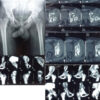 October 1, 2026 Posterior Acetabular Reconstruction Can Sometimes Lead To Retroverted Cup Placement In Complex Total Hip Arthroplasty
 October 1, 2026 Recurrent Ventral Scapular Osteochondroma Presenting with Pseudo-winging in an Adolescent: A Case Report and Follow-up of a Previously Published Case