

This study demonstrates that intra-articular growth factor concentrate is more effective than platelet-rich plasma in reducing pain, improving knee function, and preserving cartilage, making it a promising biological treatment option for knee osteoarthritis.
Dr. S Krishna Kumar, Assistant Professor, Department of Orthopedics, Teerthanker Mahaveer Medical College and Research Centre, Moradabad, Uttar Pradesh, India. E-mail: drkkasik08091995@gmail.com
Abstract
Introduction: Knee osteoarthritis (OA) is a common chronic joint disorder that affects millions of individuals worldwide, with approximately 16–25% of the population aged 50 years and above, with higher prevalence in women. Traditional treatments, including non-steroidal anti-inflammatory drugs and physical therapy, have limitations, while surgical options are invasive. Intra-articular therapies such as hyaluronic acid, platelet-rich plasma (PRP), and concentrated growth factors (CGF) have emerged as potential therapeutic options for OA of the knee (KOA). Furthermore, there is limited data on the comparative efficacy of PRP and CGF, generating a crucial gap in understanding their relative benefits in managing KOA. This study aimed to address this gap by evaluating and comparing the effects of intra-articular CGF and PRP in patients with KOA.
Materials and Methods: In the present triple-blind randomized study, a total of 180 participants were randomly divided into three groups: Group-G (CGF), Group-P (PRP), and a control group. PRP and growth factor concentrate injection were injected into the affected knee. Demographic characteristics, comorbidities, and baseline clinical parameters such as Visual Analog Scale (VAS), Western Ontario and McMaster Universities Arthritis Index (WOMAC), serum COLL 2-1 levels, and knee whole-organ magnetic resonance imaging score were recorded before treatment and at follow-up visits.
Results: The results demonstrated significant improvements in both the VAS and WOMAC scores for both CGF and PRP groups, with the CGF group showing superior outcomes in pain reduction (VAS: 1.68 ± 0.76) and functional improvement (WOMAC: 34.11 ± 9.98). Serum Coll2-1NO2 levels significantly decreased in Group-G (P < 0.0001) and showed a non-significant reduction in Group-P (P = 0.40), whereas the control group exhibited a slight increase in levels (P = 0.32). In terms of WORM scores, Group-G showed notable improvements, especially in the medial femorotibial joint and patellofemoral joint, whereas the control group had no significant changes. The CGF group also demonstrated superior efficacy in cartilage regeneration and overall knee function when compared to PRP and the control group.
Conclusion: Intra-articular CGF injections appear to be more effective than PRP in reducing pain, improving knee function, and promoting cartilage regeneration in patients with knee OA. These findings suggest that CGF may be a promising alternative to PRP in the management of KOA.
Keywords: Intra-articular growth factor concentrate, platelet-rich plasma injections, knee osteoarthritis.

Osteoarthritis (OA) is a chronic joint disease characterized by the progressive loss of articular cartilage, inflammation, soft-tissue damage, and remodeling of subchondral bone. It is a significant cause of pain and disability, particularly affecting weight-bearing joints such as the knees and hips [1].
It is the second most common rheumatologic condition, with an estimated prevalence in India ranging from 22% to 39%, showing a higher predominance among women compared to men. OA of the knee (KOA) is a leading cause of mobility impairment, especially among women. Globally, OA has been ranked as the 10th leading cause of non-fatal health burden [2].
The management of KOA often involves conservative approaches such as exercise, physiotherapy, pharmacotherapy, and joint injections [3]. Recently, intra-articular injections of platelet-rich plasma (PRP), concentrated growth factor (CGF), and hyaluronic acid (HA) have emerged as promising treatment options for KOA [4,5]. These therapies are minimally invasive, safe, and effective in reducing pain and improving joint function [6].
Various meta-analyses have evaluated the effectiveness of PRP compared to other procedures, revealing superior pain relief and functional improvement at various time points following injection. PRP is obtained from autologous peripheral blood and is rich in platelets containing regenerative factors, including platelet-derived growth factor, transforming growth factor-beta, insulin-like growth factor-1, vascular endothelial growth factor, epidermal growth factor, and fibroblast growth factor [7].
Furthermore, CGF, a third-generation platelet concentrate, offers higher levels of growth factors than PRP and features a denser fibrin structure [8,9]. Studies suggest that CGF enhances osteogenesis and angiogenesis, which contribute to improved bone regeneration [10]. Its unique properties may offer advantages over PRP in tissue repair and regeneration.
While both PRP and CGF have shown promising results in improving outcomes for KOA patients, direct comparative studies between the two remain limited. Previous investigations have primarily focused on evaluating either PRP or CGF individually against other treatments such as HA, leaving a critical gap in understanding their relative effectiveness.
Addressing these gaps, the present study aimed to provide a comprehensive comparison of the therapeutic effects of intra-articular CGF and PRP in patients with KOA. By offering a direct comparison, this study contributes valuable insights into the efficacy of these biological treatments, facilitating the way for more informed clinical decisions.
Study design
This triple-blind randomized study was conducted in the Department of Orthopaedics at Teerthanker Mahaveer Medical College and Research Centre (TMMC &RC), Moradabad. Before the initiation of the study, informed consent was obtained from the patients. Approval from the ethical committee was also secured.
Inclusion criteria
The study included patients who met the American College of Rheumatology criteria for the diagnosis of KOA and radiological evidence of Grade II and III KOA joints, as determined by the Kellgren–Lawrence scale.
Exclusion criteria
Patients with malignancy, drug abuse, gouty arthritis, inflammatory or secondary OA, active knee infections, anemia, bleeding disorders, rheumatoid arthritis, or genu varum/valgum > 5 degrees were excluded from the study. In addition, individuals with uncontrolled diabetes mellitus, ligamentous instability of the knee (confirmed by clinical examination), OA of any other joint, and those for whom magnetic resonance imaging (MRI) was contraindicated were not included in the study.
Methodology
A total of 180 patients were randomized into three groups: Group P, Group G, and the control group. The preparation of PRP and growth factor concentrate (GFC) was carried out according to a standardized protocol (Fig. 1 and 2).
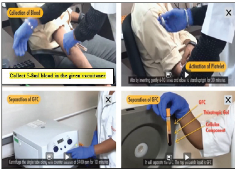
Figure 1: Preparation of growth factor concentrate.
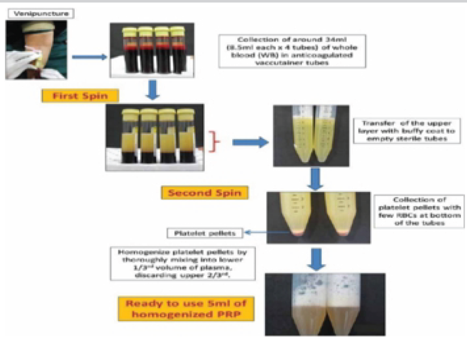
Figure 2: Preparation of platelet-rich plasma.
For the intra-articular injection, patients were positioned with their legs hanging from the table at a 90° flexion angle. No local anesthesia was administered before the procedure. PRP and GFC were injected into the affected knee joint using a 22-G needle, following standard approaches. After the injection, patients were instructed to flex and extend their knees a few times to ensure the even distribution of the injected substances within the joint. We have used commercially available GFC; “OSSISURE GFC therapy kit” {Pattern Pharmaceuticals Pvt. Ltd}.
Post-procedure, patients were observed for 15–20 min before being discharged. They were advised to apply cold packs to the treated area 3–4 times daily for 10 min each time, for up to 72 h following the injection. Quadriceps strengthening exercises were recommended as part of the recovery process, and patients were prescribed tramadol 50 mg to be taken as needed for pain relief.
Baseline assessments included recording the VAS and Western Ontario and McMaster Universities Arthritis Index (WOMAC) scores before the first injection. Serum Coll2-1NO2 levels were analyzed to establish baseline values, and an MRI of the most affected knee was performed using the whole-organ MRI score (WORMS) protocol to assess cartilage condition. These evaluations were repeated before the second injection and at the 6th and 9th month follow-ups. At the final follow-up, serum Coll2-1NO2 levels and knee MRI (WORMS protocol) were reassessed to compare the results with baseline measurements.
The study included 180 participants divided equally into three groups. The mean age and body mass index (BMI) were comparable across groups (P = 0.29 and P = 0.65, respectively). Females constituted the majority in all groups (P = 0.5). Bilateral knee involvement was most frequent in Group-P (40%) compared to Group-G (23.33%) and Group-C (33.33%), with borderline significance (P = 0.05) (Table 1).
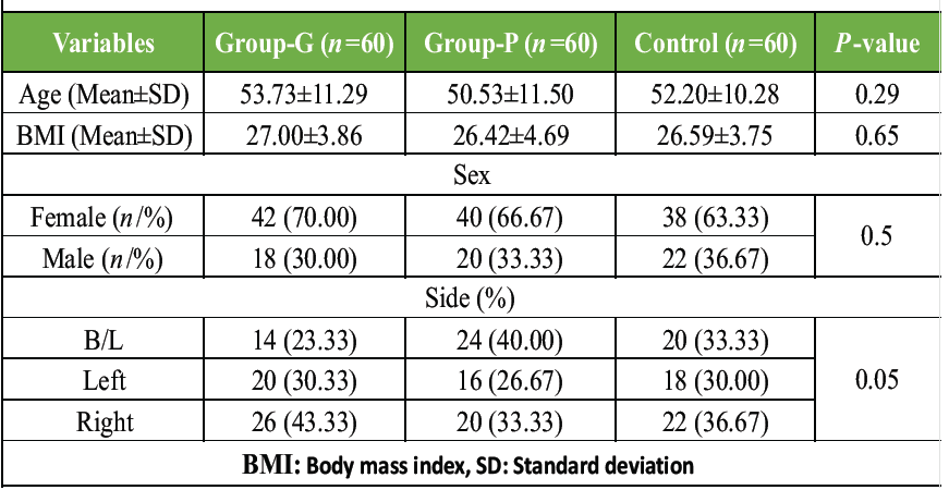
Table 1: Demographic characteristics in the study groups
Among the three groups, comorbidities varied. Group-G had the highest prevalence of hypertension (26.67%) and controlled T2DM (25.00%), whereas COPD was more common in Group-P (15.00%). Group-C had the highest proportion of patients with no comorbidities (41.67%) (Table 2).
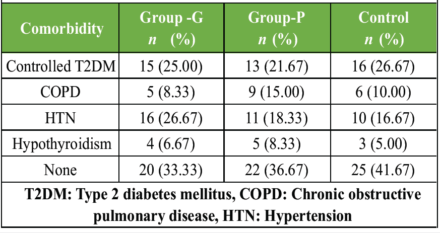
Table 2: Comorbidity among three groups of patients
Group-G (GFC) showed the most significant improvement, with a reduction in serum COLLE 2-1 levels (P < 0.0001). Group-P (PRP) showed a non-significant reduction (P = 0.40), whereas the control group had a slight, non-significant increase (P = 0.32). Thus, GFC was more effective (Table 3).
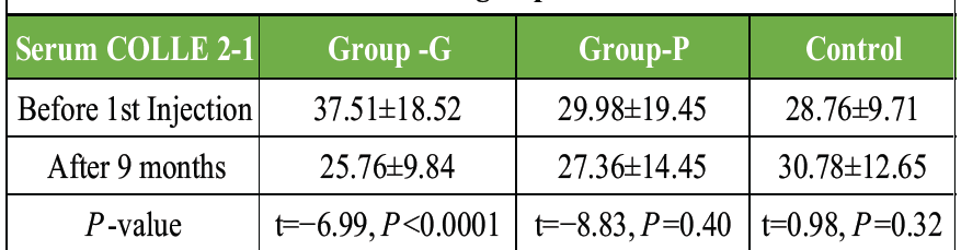
Table 3: Serum COLL 2-1 level before injection and after 9 months among three groups
There was no significant difference in K-L grade distribution between the groups. Group G had 43.33% in Grade II and 56.67% in Grade III, Group P had 40% in Grade II and 60% in Grade III, and the Control group had 36.67% in Grade II and 63.33% in Grade III, with P = 0.45 (Table 4).
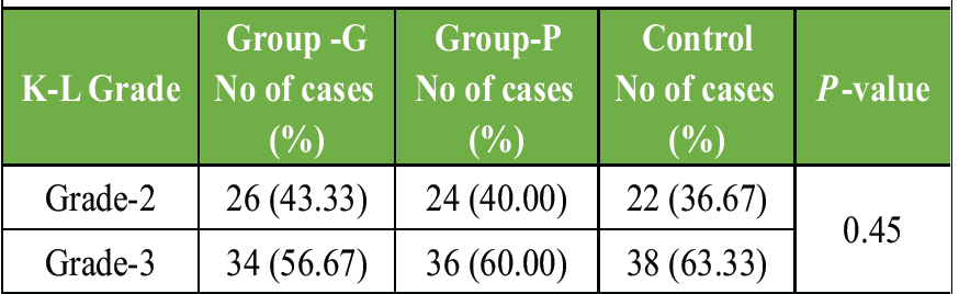
Table 4: Distribution according to K-L grade
Group G (GFC) showed the most significant improvement in VAS scores, decreasing from 5.70 ± 2.59 before the first injection to 1.68 ± 0.76 at 9 months (P < 0.0001). In comparison, Group P and the control group showed less reduction, with final scores of 5.76 ± 1.22 and 6.19 ± 1.86, respectively. This highlighted the superior efficacy of GFC in reducing pain (Table 5).
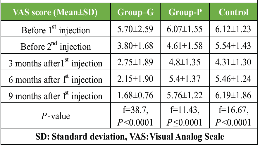
Table 5: VAS score at follow-up among three groups
Group G (GFC) showed the greatest improvement in WOMAC scores, decreasing from 58.88 ± 11.25 to 34.11 ± 9.98 over 9 months. In comparison, Group P (PRP) and the control group had higher final scores, indicating that GFC was more effective in reducing knee OA symptoms (Table 6).
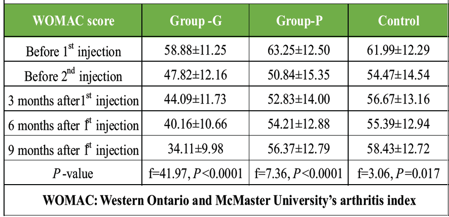
Table 6: WOMAC score at follow-up among the three groups
Group G (GFC) showed notable improvement in WORM scores, especially in the medial femorotibial joint and patellofemoral joint, with a decrease in the total score from 129.59 ± 12.87 to 125.09 ± 12.67 (P = 0.05). Group P (PRP) and the control group did not show significant changes. Hence, GFC proved to be the most effective treatment (Table 7).
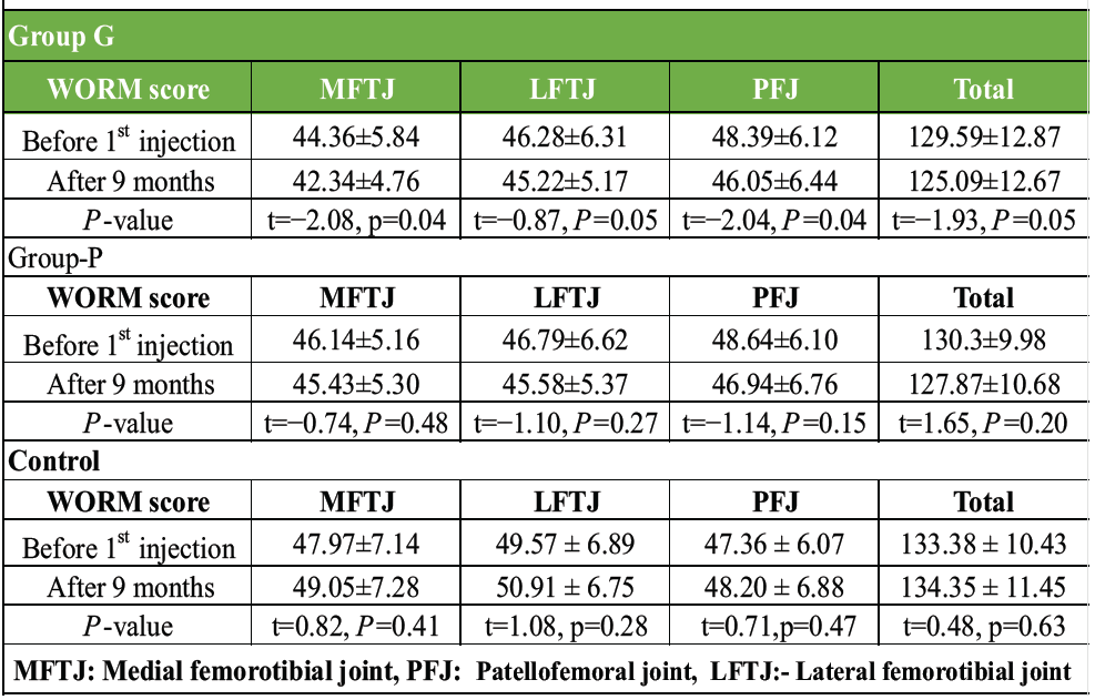
Table 7: WORM score before injection and after 9 months among the three groups
In the present study, we conducted a comprehensive comparison of the effects of PRP and CGF, both of which are commonly used treatments for KOA. Our findings highlight significant improvements in both VAS and WOMAC scores following CGF injections, suggesting its potential benefits over PRP. In addition, this trial provides valuable insights into the safety and efficacy of CGF therapy for KOA in comparison to PRP.
Despite recent advancements, achieving optimal outcomes in KOA remains a challenge, particularly when addressing long-term symptom relief and disease modification. This study offers a novel perspective by directly comparing intra-articular GFC and PRP injections, providing critical insights into their relative efficacy.
In this randomized investigation, 180 participants were evenly divided into three groups. The mean age and BMI were comparable across the groups (P = 0.29 and P = 0.65, respectively). Females were found to represent a higher percentage in all groups (P = 0.5). Similarly, Ozcan and Altun [11] and Raeissadat et al. [12] observed a female predominance in their study.
The present study findings revealed that GFC significantly reduced serum Coll2-1 levels, a biomarker of cartilage degradation, compared to PRP, which showed no significant changes (P = 0.40). A previous study performed by Saraf et al. [13] also has concordant results with our findings, showing a decrease in serum COLLE 2-1 levels 1 year after CGF therapy, with no significant change following NS injections, suggesting that CGF may exert a chondroprotective effect.
The most compelling evidence of GFC’s superiority emerged from the analysis of patient-reported outcomes. GFC injections produced substantial improvements in VAS scores, decreasing from 5.70 ± 2.59 pre-injection to 1.68 ± 0.76 at 9 months (P < 0.0001). Similarly, GFC demonstrated the greatest improvements in WOMAC scores, indicating a significant reduction in pain, stiffness, and physical limitations compared to PRP. These findings are consistent with prior studies by Ozcan and Altun [11], Vaquerizo et al. [14], and Anitua et al., [15] which highlighted the effectiveness of growth factor-rich therapies in symptom reduction.
Given the existing gap in the literature concerning direct comparisons of the efficacy of CGF and PRP in managing KOA, our study is the first to address this gap by evaluating and comparing the effectiveness of PRP and CGF in treating knee OA. By providing evidence-based insights into the relative effectiveness of these two biological treatments, this study not only enhances the understanding of their role in KOA management but also offers valuable guidance for clinicians. In addition, the present study findings will support more informed decision-making regarding treatment options, ultimately helping in the refinement of diagnostic methods and the optimization of therapeutic strategies for patients with knee OA.
Limitations of the study
The study has several limitations, including the relatively small sample size. In addition, since it was a single-center study, the results may not be applicable to other settings or populations. The 9-month follow-up period may also be insufficient to assess the long-term effects of the treatments. Unquantified dose–response and restricted biomarkers: Individual growth factor concentrations within each autologous injection were not quantified or statistically correlated with treatment clinical success, obscuring potential dose–response profiles. In addition, metabolic biochemical assessment was limited exclusively to serum Coll2-1NO2, lacking a comprehensive panel of overlapping inflammatory and catabolic joint markers. Imaging restrictions: Joint structural profiling was confined to semi-quantitative WORMS-based MRI scoring. Advanced quantitative imaging methodologies, such as T2 mapping, dGEMRIC, or cartilage volume analysis, were not implemented.
Confounding lifestyle factors and cost-effectiveness: Dynamic lifestyle confounders – including BMI, physical activity levels, occupational parameters, post-injection physiotherapy compliance, and dietary variables – were neither standardized nor statistically controlled. Finally, no economic or cost-effectiveness feasibility analysis was conducted to support real-world clinical translation.
GFC emerged as the most effective treatment modality for knee OA, demonstrating significant improvements in biochemical markers, pain reduction, and functional outcomes compared to PRP and the control group. These findings suggest that GFC is a superior option for managing knee OA symptoms. Further studies with larger sample sizes and longer follow-ups are needed for a better understanding.
* Efficacy: Intra-articular growth factor concentrate provides superior and longer-lasting pain relief compared to platelet-rich plasma, with improved functional outcomes.
* Indications: It is indicated for patients with early to moderate knee osteoarthritis as a minimally invasive, non-operative treatment option.
* Mechanism: It delivers concentrated autologous growth factors that reduce inflammation, enhance chondrocyte activity, and promote tissue repair.
* Clinical outcomes: It improves pain, mobility, and quality of life, supports cartilage preservation, and may delay disease progression and the need for knee arthroplasty.
References
- 1. Martel-Pelletier J, Barr AJ, Cicuttini FM, Conaghan PG, Cooper C, Goldring MB, et al. Osteoarthritis. Nat Rev Dis Primers 2016;2:16072. [Google Scholar] [PubMed]
- 2. Pal CP, Singh P, Chaturvedi S, Pruthi KK, Vij A. Epidemiology of knee osteoarthritis in India and related factors. Indian J Orthop 2016;50:518-22. [Google Scholar] [PubMed]
- 3. Huang Y, Liu X, Xu X, Liu J. Intra-articular injections of platelet-rich plasma, hyaluronic acid or corticosteroids for knee osteoarthritis: A prospective randomized controlled study. Orthopade 2019;48:239-47. [Google Scholar] [PubMed]
- 4. Patel S, Jindal K, Dhillon MS. The future of injectable orthobiologic substances for knee osteoarthritis: Options beyond platelet rich plasma. J Musculoskelet Surg Res 2020;4:173-81. [Google Scholar] [PubMed]
- 5. Ondresik M, Maia FR, Morais AD, Gertrudes AC, Dias Bacelar AH, Correia C, et al. Management of knee osteoarthritis. Current status and future trends. Biotechnol Bioeng 2017;114:717-39. [Google Scholar] [PubMed]
- 6. Ozer D, Eyigör S. New therapy methods in osteoarthritis. Ege J Med 2019;58:14-21. [Google Scholar] [PubMed]
- 7. Bhan K, Singh B. Efficacy of platelet-rich plasma injection in the management of rotator cuff tendinopathy: A review of the current literature. Cureus 2022;14:e26103. [Google Scholar] [PubMed]
- 8. Chen X, Wang J, Yu L, Zhou J, Zheng D, Zhang B. Effect of concentrated growth factor (CGF) on the promotion of osteogenesis in bone marrow stromal cells (BMSC) in vivo. Sci Rep 2018;8:5876. [Google Scholar] [PubMed]
- 9. KimTH, Kim SH, Sándor GK, Kim YD. Comparison of platelet-rich plasma (PRP), platelet-rich fibrin (PRF), and concentrated growth factor (CGF) in rabbit-skull defect healing. Arch Oral Biol 2014;59:550-8. [Google Scholar] [PubMed]
- 10. Zhang L, Ai H. Concentrated growth factor promotes proliferation, osteogenic differentiation, and angiogenic potential of rabbit periosteum-derived cells in vitro. J Orthop Surg Res 2019;14:146. [Google Scholar] [PubMed]
- 11. Ozcan S, Altun AY. Efficacy of intra-articular concentrated growth factor, platelet-rich plasma, and hyaluronic acid in knee osteoarthritis: A comparative analysis. Ann Med Res 2024;31(4):304-309. [Google Scholar] [PubMed]
- 12. Raeissadat SA, Ghazi Hosseini P, Bahrami MH, Salman Roghani R, Fathi M, Gharooee Ahangar A, et al. The comparison effects of intra-articular injection of platelet rich plasma (PRP), plasma rich in growth factor (PRGF), hyaluronic acid (HA), and ozone in knee osteoarthritis; a one year randomized clinical trial. BMC Musculoskelet Disord 2021;22:134. [Google Scholar] [PubMed]
- 13. Saraf A, Hussain A, Bishnoi S, Habib H, Garg A. Serial intraarticular injections of growth factor concentrate in knee osteoarthritis: A placebo controlled randomized study. J Orthop 2023;37:46-52. [Google Scholar] [PubMed]
- 14. Vaquerizo V, Plasencia M, Arribas I, Seijas R, Padilla S, Orive G, et al. Comparison of intra-articular injections of plasma rich in growth factors (PRGF-Endoret) versus Durolane hyaluronic acid in the treatment of patients with symptomatic osteoarthritis: A randomized controlled trial. Arthroscopy 2013;29:1635-43. [Google Scholar] [PubMed]
- 15. Anitua E, Sánchez M, Aguirre JJ, Prado R, Padilla S, Orive G. Efficacy and safety of plasma rich in growth factors intra-articular infiltrations in the treatment of knee osteoarthritis. Arthroscopy 2014;30:1006-17. [Google Scholar] [PubMed]






Related Articles in Journal of Orthopaedic Case Reports
 October 1, 2026 Comparative Efficacy of Leukocyte-Poor Platelet-Rich Plasma, Leukocyte-Rich Platelet-Rich Plasma, Injectable Platelet-Rich Fibrin, and Corticosteroid Injections in Kellgren–Lawrence Grade 2–3 Knee Osteoarthritis: A Prospective Comparative Study
 October 1, 2026 Efficacy of Intra-Articular Injection of Platelet-Rich Plasma and Growth Factor Concentrate in Degenerative Meniscal Lesions
 October 1, 2026 Arthrokinematic and Magnetic Resonance Imaging-Based Outcomes of Customized Lateral Wedge Insoles in Medial Compartment Knee Osteoarthritis: Six-Month Interim Findings of a Prospective Cohort Study
 August 1, 2026 The Effect of Intraarticular Platelet-Rich Plasma Injection in Knee Osteoarthritis