


Introduction
Osteoarthritis (OA) of the knee is the most common arthritis worldwide and is a major contributor to morbidity due to knee pain in adults aged 45 years and older [1]. According to the Global Burden of Disease Study, hip and knee OA impacts more than 250 million people worldwide, with most of the disease burden being for knee OA [2]. It is defined by progressive articular cartilage degeneration, subchondral remodeling, osteophyte formation, and low-grade synovial inflammation that collectively result in pain, stiffness, and functional decline [3]. The Kellgren–Lawrence (KL) system is used to grade radiographic severity, with Grades 2 and 3 defined as definite to moderate joint space narrowing and osteophytosis, for which intra-articular therapy is most commonly indicated prior to arthroplasty as a treatment option [4].
Intra-articular corticosteroid (iACR) injection has been a staple of conservative therapy for many years. Corticosteroids such as triamcinolone acetonide are potent anti-inflammatory agents of synovial inflammation and pro-inflammatory cytokines, and induce rapid but short-lived analgesia. Randomized evidence has raised concerns about accelerated cartilage volume loss with repeated use of corticosteroids, and no sustained benefit beyond 3 months [5,6]. This has led to an interest in the use of autologous biologic alternatives with potentially disease-modifying effects.
Platelet-rich plasma (PRP) is an autologous blood preparation with a supraphysiologic platelet concentration that serves as a source of growth factors such as platelet-derived growth factor (PDGF), transforming growth factor-beta (TGF-β), and vascular endothelial growth factor (VEGF), which can modulate the metabolism of chondrocytes and suppress catabolic cytokines, including interleukin-1 beta (IL-1β) and tumor necrosis factor-alpha (TNF-α) [7]. Based on the amount of leukocytes present, PRP formulations can be broadly divided into leukocyte-rich PRP (LR-PRP) and leukocyte-poor PRP (LP-PRP), and it is important to note that PRP contains both growth factors and pro-inflammatory mediators that can be released by the leukocytes, which is a distinction of mechanistic and clinical significance. Meta-analyses of these preparations show similar effectiveness, with LR-PRP having a greater rate of post-injection inflammatory reactions [8,9].
Injectable platelet-rich fibrin (iPRF) is a second-generation, anticoagulant-free PRP produced through low-speed centrifugation, resulting in a fibrin matrix that encapsulates and retains platelets and leukocytes for extended and gradual release of growth factors as opposed to a bolus effect. This scaffold-based delivery has been suggested to increase the local bioactivity of the product. It could lead to longer-lasting clinical effects, although there are limited clinical comparison studies with conventional PRP formulations and with corticosteroids in the treatment of knee OA [10,11].
Although the use of these types of biologic injections is increasing, there are very few prospective studies that compare more than two of the agents at a single time and even fewer studies that compare all four within one cohort. In current Osteoarthritis Research Society International/European Alliance of Associations for Rheumatology guidelines, intra-articular treatments have been mentioned as possible add-on therapies within a multilayered framework of non-pharmacological and pharmacological interventions, but have not been fully compared and ranked by the biologic subtypes [12,13,14]. In addition, standardization of PRP reporting, such as the inclusion of platelet count, leukocyte count, and preparation method, has been called for to enable meaningful comparison between studies, as there is significant heterogeneity in the published studies, which may be due to variation in preparation protocols [15,16,17].
Therefore, this study was designed to prospectively compare the clinical efficacy (as assessed through Visual Analog Scale [VAS] and Western Ontario and McMaster Universities OA Index [WOMAC] scores at 12-month follow-up) of LP-PRP, LR-PRP, iPRF, and corticosteroid injection in patients with KL Grade 2–3 knee OA and to clarify the relative short-term and long-term therapeutic profiles, and to guide evidence-based treatment choice in daily orthopedic practice.
Materials and Methods
A prospective, single-center, parallel-group comparative clinical study was conducted in the Department of Orthopaedics after approval by the Institutional Ethics Committee (IEC No. Dr. MGR-ERI/SLMCH/2025/016 dated March 05, 2025) in accordance with the Declaration of Helsinki. All participants gave written informed consent before being enrolled.
Study population
Patients aged 40–70 years, with primary knee OA of KL Grade 2 or 3, and who had persistent symptoms of knee OA for more than 3 months despite conservative treatment (analgesic, physiotherapy, and activity modification) were included in the study. The exclusion criteria were KL Grade 1 or 4 disease, intra-articular injection in the last 6 months, inflammatory or septic arthritis, coagulopathy or anticoagulant treatment, known malignancy, pregnancy, and body mass index (BMI) >35 kg/m2.
Group allocation
A total of 100 patients who met the eligibility criteria entered and were divided into four parallel groups: 25 patients were assigned to receive LP-PRP, 25 patients were assigned to receive LR-PRP, 25 were assigned to receive iPRF, and 25 patients were assigned to receive iACR (triamcinolone acetonide 40 mg). There were 23, 22, 24, and 21 patients in Groups 1, 2, 3, and 4, respectively, at the end of 12 months, who had completed the study.
Preparation of biologic products
In the case of LP-PRP and LR-PRP, 40 mL of peripheral venous blood was collected in acid-citrate-dextrose anticoagulated tubes and processed according to a standard double-spin centrifugation protocol, in which LP-PRP was prepared without buffy-coat leukocytes, whereas LR-PRP was prepared with buffy-coat leukocytes. The platelet concentration in the final product was aimed at 4–6 times the baseline whole-blood platelet concentration, in line with PAW classification quality standards. For iPRF, 20 mL of blood was collected into plain tubes without additives and spun for 8 min at low speed (700 rpm, or about 60 g), and the liquid fibrin-platelet concentrate was drawn off from the top layer of plasma and injected, following the protocol of Choukroun.
Injection technique
All injections were made by one experienced orthopedic surgeon under standard anterolateral or anteromedial patellar portal, without the use of image guidance, and after joint aspiration for effusion. Intra-articular injection (approximately 4–5 mL) of the assigned biologic product was given to all patients in groups 1–3, and 2 mL of 2% lignocaine mixed with triamcinolone acetonide 40 mg was given to each patient in group 4. A 48 h post-procedure rest period was advised, and rescue analgesia (paracetamol) was given as required (and its use was recorded).
Outcome measures and follow-up
Primary outcome measures included the 0–10 VAS for pain and the 0–96 WOMAC for pain, stiffness, and physical function, which were assessed by an assessor who was blind to group allocation; both were recorded at baseline (pre-injection) and at 1, 3, 6, and 12 months after injection. Any adverse event (pain, swelling, and effusion) was documented at every visit.
Statistical analysis
Values of continuous variables are presented as mean ± standard deviation. Each group’s changes from baseline were compared using repeated measures analysis of variance (ANOVA), and between-group comparisons at each time point were compared using one-way ANOVAs with Bonferroni correction. P < 0.05 was deemed to be statistically significant. To help define the clinical significance of changes observed, the minimal clinically important difference for WOMAC was taken from previous validation research.
Results
Patient demographics
Demographic and radiographic parameters such as proportion of KL grade, age and sex distribution, and BMI were comparable between groups, and no statistically significant between-group difference (P > 0.05) was found, indicating baseline homogeneity of treatment arms (Table 1).
Baseline demographic and radiographic characteristics
| Characteristic | Group 1 LP-PRP (n=23) | Group 2 LR-PRP (n=22) | Group 3 iPRF (n=24) | Group 4 Steroid (n=21) | P-value |
|---|---|---|---|---|---|
| Age, years (mean±SD) | 57.9±7.6 | 58.4±8.1 | 57.3±7.9 | 58.6±8.0 | 0.87 |
| Sex, female n (%) | 13 (56.5) | 13 (59.1) | 14 (58.3) | 12 (57.1) | 0.98 |
| BMI, kg/m2 (mean±SD) | 27.4±3.0 | 27.8±3.4 | 27.3±3.1 | 27.6±3.3 | 0.92 |
| KL Grade 2, n (%) | 13 (56.5) | 12 (54.5) | 13 (54.2) | 12 (57.1) | 0.97 |
| KL Grade 3, n (%) | 10 (43.5) | 10 (45.5) | 11 (45.8) | 9 (42.9) | 0.97 |
| Loss to follow-up, n | 2 | 3 | 1 | 4 | — |
LP-PRP: Leukocyte-poor platelet-rich plasma, LR-PRP: Leukocyte-rich platelet-rich plasma, iPRF: Injectable platelet-rich fibrin, SD: Standard deviation, BMI: Body mass index, KL: Kellgren–Lawrence
Platelet characterization
Platelet characteristics of the three platelet-based preparations were assessed to ensure comparability of the biologic products (Table 2). The baseline whole-blood platelet count was 258.6 ± 38.7 × 10³/µL in the LP-PRP group, 261.4 ± 40.2 × 10³/µL in the LR-PRP group, and 256.9 ± 37.5 × 10³/µL in the iPRF group. The final platelet concentration was 1.19 ± 0.16 × 10⁶/µL, 1.23 ± 0.18 × 10⁶/µL, and 1.34 ± 0.21 × 10⁶/µL, respectively, corresponding to platelet enrichment of approximately 4.60 ± 0.52-fold, 4.71 ± 0.56-fold, and 5.21 ± 0.73-fold over baseline. The leukocyte concentration was substantially lower in the LP-PRP preparation (0.78 ± 0.24 × 1063/µL) than in the LR-PRP preparation (5.82 ± 1.14 × 10³/µL), whereas the iPRF preparation demonstrated a leukocyte concentration of 3.96 ± 0.88 × 10³/µL, consistent with its fibrin-based cellular composition. Platelet recovery was 80.6 ± 7.2% for LP-PRP, 82.4 ± 7.6% for LR-PRP, and 76.8 ± 8.1% for iPRF. These findings demonstrated that LP-PRP and LR-PRP achieved the intended supraphysiological platelet concentration, whereas the iPRF preparation provided a concentrated platelet-fibrin product with retained cellular components, consistent with the respective preparation protocols described in the study.
Platelet characterization of three study groups
| Parameter | Group 1 LP- PRP (n =23) | Group 2 LR- PRP (n =22) | Group 3 iPRF (n =24) |
|---|---|---|---|
| Baseline platelet count (×103/µL) | 258.6±38.7 | 261.4±40.2 | 256.9±37.5 |
| Final platelet concentration (×106/µL) | 1.19±0.16 | 1.23±0.18 | 1.34±0.21 |
| Platelet enrichment (fold) | 4.60±0.52 | 4.71±0.56 | 5.21±0.73 |
| Leukocyte count (×103/µL) | 0.78±0.24 | 5.82±1.14 | 3.96±0.88 |
| Platelet recovery (%) | 80.6±7.2 | 82.4±7.6 | 76.8±8.1 |
LP-PRP: Leucocyte-poor platelet-rich plasma, LR-PRP: Leucocyte-rich platelet-rich plasma, iPRF: Injectable platelet-rich fibrin
Functional scores
Baseline VAS and WOMAC. Mean baseline VAS scores ranged from 7.02 ± 0.45 (Group 3) to 7.40 ± 0.74 (Group 2), and mean baseline WOMAC scores ranged from 60.77 ± 5.14 (Group 3) to 62.95 ± 5.76 (Group 2), with no significant between-group differences at baseline (Table 3).
VAS and WOMAC scores across follow-up (mean±SD)
| Time | LP-PRP VAS | LR-PRP VAS | iPRF VAS | Steroid VAS | LP-PRP WOMAC | LR-PRP WOMAC | iPRF WOMAC | Steroid WOMAC |
|---|---|---|---|---|---|---|---|---|
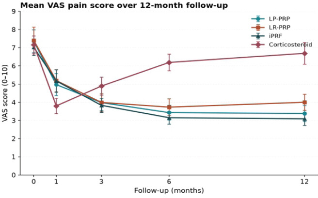
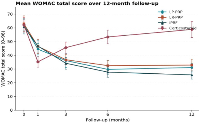
| Baseline | 7.36±0.63 | 7.40±0.74 | 7.02±0.45 | 7.16±0.49 | 62.22±4.40 | 62.95±5.76 | 60.77±5.14 | 62.14±5.24 |
| 1 month | 4.97±0.60 | 5.19±0.61 | 5.17±0.62 | 3.80±0.42 | 44.45±4.18 | 46.50±5.14 | 46.73±4.35 | 35.20±3.76 |
| 3 months | 3.98±0.43 | 3.99±0.49 | 3.83±0.39 | 4.89±0.49 | 36.12±4.90 | 36.77±3.75 | 34.19±4.30 | 45.59±4.06 |
| 6 months | 3.43±0.41 | 3.73±0.46 | 3.15±0.35 | 6.19±0.46 | 29.73±3.51 | 32.47±3.69 | 27.74±3.65 | 53.35±4.99 |
| 12 months | 3.38±0.44 | 4.00±0.44 | 3.09±0.36 | 6.69±0.59 | 31.07±3.12 | 32.86±4.37 | 25.72±2.94 | 58.79±5.83 |
LP-PRP: Leukocyte-poor platelet-rich plasma, LR-PRP: Leukocyte-rich platelet-rich plasma, iPRF: Injectable platelet-rich fibrin, SD: Standard deviation, BMI: Body mass index, KL: Kellgren–Lawrence, VAS: Visual Analog Scale, WOMAC: Western Ontario and McMaster Universities Osteoarthritis Index
Trajectory of VAS over follow-up
All four groups experienced a significant decrease in VAS score from baseline to 1 month (P < 0.001 in each group). This was highest in Group 4 (corticosteroid) (7.16 ± 0.49–3.80 ± 0.42 at 1 month), and showed the largest difference from all groups of biologic treatment at this time point. This effect was, however, transient, as VAS scores in Group 4 increased gradually to 4.89 ± 0.49 at 3 months, 6.19 ± 0.46 at 6 months, and 6.69 ± 0.59 at 12 months, becoming nearly at baseline level. Groups 1–3, on the other hand, still improved or at least did not worsen after 1 month. Group 3 (iPRF) achieved the lowest VAS score at both 6 months (3.15 ± 0.35) and 12 months (3.09 ± 0.36), followed closely by Group 1 (LP-PRP: 3.43 ± 0.41 and 3.38 ± 0.44) and Group 2 (LR-PRP: 3.73 ± 0.46 and 4.00 ± 0.44). The difference among the biologic groups and the corticosteroid group was statistically significant at 12 months (P < 0.001) (Fig. 1 and Table 3).
WOMAC recovery over time
The WOMAC total score was similar (Fig. 2 and Table 3). The improvement was largest for Group 4 (baseline 62.14 ± 5.24–35.20 ± 3.76 at 1 month). Still, the group deteriorated over time, with 53.35 ± 4.99 at 6 months and 58.79 ± 5.83 at 12 months, which approached baseline values and were significantly poorer than all biologic groups at 12 months (P < 0.001). There was the greatest maintenance of function in Group 3 (iPRF), with WOMAC scores of 27.74 ± 3.65 at 6 months and 25.72 ± 2.94 at 12 months. The intermediate scores of Groups 1 and 2 were similar (LP-PRP = 31.07 ± 3.12, LR-PRP = 32.86 ± 4.37), both being significantly lower than Group 4 (P < 0.001) but not between one another (P > 0.05).
Improvement in percentage terms from baseline
Near-complete regression to baseline symptoms was seen by 12 months, with the greatest percentage reduction in VAS (56%) and WOMAC (58%) seen for Group 3, followed by Group 1 (54% VAS, 50% WOMAC) and Group 2 (46% VAS, 48% WOMAC), with Group 4 showing only a 7% VAS and 5% WOMAC reduction (Fig. 3 and Table 4).
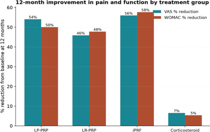
Percentage reduction from baseline at 12 months
| Group | VAS % reduction | WOMAC % reduction |
|---|---|---|
| Group 1 (LP-PRP) | 54 | 50 |
| Group 2 (LR-PRP) | 46 | 48 |
| Group 3 (iPRF) | 56 | 58 |
| Group 4 (Corticosteroid) | 7 | 5 |
VAS: Visual Analog Scale, WOMAC: Western Ontario and McMaster Universities Osteoarthritis Index, LP-PRP: Leukocyte-poor platelet-rich plasma, LR-PRP: Leukocyte-rich platelet-rich plasma
LP-PRP versus LR-PRP
There was no statistically significant difference in the VAS or WOMAC scores between Group 1 and Group 2 at any time point (P > 0.05 at all follow-up points). Still, a more favorable numerical trend was seen at 6 and 12 months for the VAS and WOMAC scores, respectively, in Group 1.
Adverse events
More frequently, post-injection pain and mild effusion (24–48 h) were reported in Group 2 (LR-PRP) than in Group 1 (LP-PRP) or Group 3 (iPRF), suggesting that the leukocyte content of LR-PRP was higher. There were no serious adverse events, joint infection, or systemic complications reported in any of the groups during the study period.
Discussion
In this prospective comparative study, an important difference in the time course of efficacy was found between corticosteroid and platelet-based biological injections for the treatment of KL Grade 2–3 knee OA. Corticosteroid was the most rapid to provide symptomatic relief at 1 month, as it is a very powerful anti-inflammatory drug with a quick onset of action. Still, this effect diminished by 3 months and was close to baseline at 12 months. The results of this pattern resemble those of the RESTORE trial and a number of systematic reviews that found that the effectiveness of corticosteroid treatment in knee OA is limited and does not compare favorably with PRP after 3 months [18,19]. It may also reflect the temporary effect on synovial inflammation, without providing a substrate for chondral matrix support, and the possible catabolic effect of repeated or prolonged corticosteroid use on chondrocytes, as suggested by McAlindon et al. in a randomized trial, which showed that there was more cartilage volume loss with intra-articular triamcinolone than with saline [20].
All three platelet-based groups had a more gradual onset. Yet, more lasting improvement, as the biological mechanism of PRP and platelet-rich fibrin (PRF) suggests: Prolonged delivery of growth factors (PDGF, TGF-β, and VEGF), which affect chondrocyte anabolism, and suppression of catabolic cytokines (IL-1β and TNF-α), at the local level [21,22]. This is consistent with the meta-analytic findings that PRP is more effective than corticosteroid and hyaluronic acid at 6 and 12 months, despite similar or poorer short-term results [23,24].
In our cohort, the similar effect of both preparations (LP-PRP and LR-PRP) with knee OA fits with recent double blind randomized studies, including the study by Di Martino et al., which showed comparable clinical outcomes between the two preparations, with a higher incidence of transient inflammatory reactions after LR-PRP [25]. This safety signal is confirmed by our observation of more frequent, but self-limited, effusion and pain in the LR-PRP group, possibly because of the release of pro-inflammatory mediators in addition to growth factors, mediated by leukocytes [26].
The superior 12-month results seen with iPRF, though it has a slower onset of action, make sense from a biological standpoint, as the unique preparation method maintained a three-dimensional fibrin scaffold that is instrumental in attracting platelets and leukocytes, allowing the gradual release of growth factors over days to weeks as opposed to the immediate bolus release of liquid PRP [27,28]. This sustained release feature, well documented in the oral and maxillofacial literature for the soft tissue and bone regeneration environment, may prove favorable in the intra-articular space, where a sustained low-level anabolic activity may be more beneficial to maintaining chondrocyte and synovial homeostasis within a therapeutic window appropriate for OA progression [29]. Comparative clinical trials of iPRF in knee OA are relatively scarce; however, our results are promising and need to be replicated using larger, multicenter, randomized trials with standardized iPRF preparation protocols.
These results have translational implications in relation to how shared decision-making is applied in day-to-day clinical activities. However, if the patient needs rapid relief for an event such as a rise in activity or travel or a milestone that demands mobility for the short term, then corticosteroid, despite its transient effect, might be the most effective choice for this purpose, provided that they are warned of the likely reduction in effect after 3 months. Other patients, however, who want to delay surgery, want to have a long symptom-free interval, or cannot attend clinics frequently for re-injection of the biologic, may benefit more from a single injection, especially of iPRF, given its impressive 12-month results in this group of patients [10]. Despite the promising efficacy results, some practical factors may affect the actual use of these products, including cost, availability of point-of-care centrifugation equipment, and operator experience with biologic preparation techniques.
This study has several limitations that should be considered when interpreting the findings. First, only 100 patients were enrolled, with approximately 21–24 completing follow-up in each group; this limits statistical power and the ability to detect smaller between-group differences. Second, the study was conducted at a single institution, which limits generalizability of the findings to other populations, healthcare settings, and clinical practices. Third, all injections were administered by one experienced surgeon, which improves procedural consistency but may limit external validity, as outcomes can vary with operator expertise. Fourth, the 12-month follow-up is insufficient to determine the long-term durability of benefit or whether these treatments alter the natural progression of OA. Fifth, no MRI, radiographic progression, cartilage thickness, or other imaging-based outcomes were assessed; therefore, the possibility of a disease-modifying effect cannot be established from the present study. Sixth, although the outcome assessor was blinded, true patient blinding was difficult to achieve because the injections differed in preparation, volume, appearance, and post-injection reactions, introducing potential placebo and expectation bias. Seventh, the absence of a saline or sham injection control group makes it difficult to distinguish the specific treatment effect from the natural course of symptoms, placebo response, or the effect of the injection procedure itself. Eighth, patients with KL Grade 2 and Grade 3 OA were pooled; these represent different degrees of structural disease, and treatment response may differ between them, and the sample size did not permit meaningful subgroup analyses according to age, sex, BMI, KL grade, baseline severity, or other prognostic factors. Ninth, although platelet and leukocyte concentrations were reported, other important characteristics of PRP/iPRF, including growth-factor concentrations, red blood cell contamination, fibrin architecture, and cellular composition, were not comprehensively characterized, and LP-PRP, LR-PRP, and iPRF were prepared using fundamentally different techniques and blood volumes, making it difficult to determine whether the observed differences were attributable specifically to leukocyte content, fibrin architecture, platelet concentration, or other preparation-related factors. Tenth, only a single injection was evaluated; different PRP/iPRF regimens, including repeated injections, may produce different clinical outcomes and could alter the comparative efficacy observed. Eleventh, outcome assessment relied mainly on VAS and WOMAC scores, and objective functional performance, activity levels, patient satisfaction, quality of life, and return to desired activities were not evaluated. Finally, details regarding standardized physiotherapy, weight management, exercise, analgesic use, and activity modification during follow-up are limited, and differences in these co-interventions could have influenced the clinical outcomes observed. In addition, some of the comparative literature does not report the platelet and leukocyte counts, making it hard to compare PRP/PRF across studies, which underscores the importance of a standardized reporting system for PRP/PRF such as the PAW and MIBO systems in future comparative studies.
Notwithstanding these limitations, our findings suggest a clinically meaningful difference in risk–benefit profile between corticosteroid and platelet-based biologics: corticosteroid is a reasonable option for rapid symptom relief in the short term, while LP-PRP, LR-PRP, and especially iPRF hold promise of more durable improvement in pain and function. However, the present study demonstrates improvement in pain and function rather than superiority in disease modification or cartilage preservation, and the possibility of a disease-modifying effect of iPRF is not supported without structural or biological evidence. In keeping with this, a growing body of literature supports the use of biologic injections for intermediate-to-long-term knee OA management [23,30,31,32]. Larger, multicenter, adequately blinded randomized controlled trials with a placebo/saline control arm, standardized biologic characterization, and longer follow-up are required to confirm these findings.
Conclusion
A corticosteroid injection is the most rapid but least persistent source of symptom relief in patients with knee OA, Grade 2–3, with almost complete return to baseline at 12 months. Both LR-PRP and iPRF produced a slower-onset but significantly more lasting improvement in pain and function, and in this cohort, the greatest improvement at 12 months was with iPRF. The results support the use of platelet-based biologic injections, especially iPRF, when treatment is aimed at obtaining durable symptomatic and functional improvement, and corticosteroids may be suitable if a quick short-term improvement is the desired treatment goal. These findings demonstrate improvement in pain and function but do not establish superiority in terms of disease modification or cartilage preservation. Further, larger, multicenter, adequately blinded randomized controlled trials with standardized biologic characterization and longer follow-up are warranted to confirm these findings and to elucidate their disease-modifying potential.
Clinical Message
Corticosteroids provide the fastest pain relief in KL Grade 2–3 knee OA, but their effect diminishes within 3 months and nearly returns to baseline by 12 months.
Platelet-based biologics (LP-PRP, LR-PRP, and iPRF) offer slower onset but more durable improvement, with iPRF showing the greatest sustained benefit at 12 months.
Clinical benefit is symptomatic, not disease-modifying – larger, multicenter trials with standardized biologic characterization and longer follow-up are needed to confirm efficacy and explore structural outcomes.
Conflict of Interest:
Nil
Source of Support:
Nil
Consent:
The authors confirm that informed consent was obtained from the patient for publication of this article
How to Cite this Article
Muruganandam A, Mohan KC, Muruganandam P, Praveen K, Jeyaraman N, Jeyaraman M. Comparative Efficacy of Leukocyte-Poor Platelet-Rich Plasma, Leukocyte-Rich Platelet-Rich Plasma, Injectable Platelet-Rich Fibrin, and Corticosteroid Injections in Kellgren–Lawrence Grade 2–3 Knee Osteoarthritis: A Prospective Comparative Study. Journal of Orthopaedic Case Reports 2026 October;16(10): 330-338.
References
- Cui A, Li H, Wang D, Zhong J, Chen Y, Lu H. Global, regional prevalence, incidence and risk factors of knee osteoarthritis in population-based studies. EClinicalMedicine 2020. 29-30:100587. [Google Scholar] | [PubMed]
- Xu J, Lei M, Xu D. Global, regional, and national burden of knee osteoarthritis attributable to high BMI: A systematic analysis from 1990 to 2021 and projections to 2050. Front Public Health 2025;13:1668257. [Google Scholar] | [PubMed]
- Zhang X, Huang C, Hu Z, Tan Y, Wang P, Zhu L. Global, regional, and country-specific lifetime risks of osteoarthritis, 1990-2021: A systematic analysis for the global burden of disease study 2021. Glob Health Res Policy 2025;10:29. [Google Scholar] | [PubMed]
- Kohn MD, Sassoon AA, Fernando ND. Classifications in brief: Kellgren-Lawrence classification of osteoarthritis. Clin Orthop 2016;474:1886-93. [Google Scholar] | [PubMed]
- Felson DT. Evidence suggests that intraarticular corticosteroids are effective (short term) and safe (long term). Osteoarthritis Cartilage 2023;31:140-1. [Google Scholar] | [PubMed]
- Si-Hyeong Park S, Li B, Kim C. Efficacy of intra-articular injections for the treatment of osteoarthritis: A narrative review. Osteoarthr Cartil Open 2025;7:100596. [Google Scholar] | [PubMed]
- Moretti L, Maccagnano G, Coviello M, Cassano GD, Franchini A, Laneve A. Platelet rich plasma injections for knee osteoarthritis treatment: A prospective clinical study. J Clin Med 2022;11:2640. [Google Scholar] | [PubMed]
- Rodríguez-Merchán EC. Intra-articular platelet-rich plasma injections in knee osteoarthritis: A review of their current molecular mechanisms of action and their degree of efficacy. Int J Mol Sci 2022;23:1301. [Google Scholar] | [PubMed]
- Peng YN, Huang D, Chou IL, Lew HL. Comparative clinical effectiveness of intra-articular leukocyte-rich platelet-rich plasma and hyaluronic acid in treating knee osteoarthritis pain: An updated systematic review and meta-analysis of randomized controlled trials. Arch Rehabil Res Clin Transl 2026;8:100573. [Google Scholar] | [PubMed]
- Costa FR, De Souza SA, Martins RA, Costa BR, Pires L, De Macedo AP. The role of injectable platelet-rich fibrin in orthopedics: Where do we stand?. Curr Issues Mol Biol 2025;47:239. [Google Scholar] | [PubMed]
- Miron RJ, Gruber R, Farshidfar N, Sculean A, Zhang Y. Ten years of injectable platelet-rich fibrin. Periodontol 2000 2024;94:92-113. [Google Scholar] | [PubMed]
- Uson J, Rodriguez-García SC, Castellanos-Moreira R, O'Neill TW, Doherty M, Boesen M. EULAR recommendations for intra-articular therapies. Ann Rheum Dis 2021;80:1299-305. [Google Scholar] | [PubMed]
- Moseng T, Vliet Vlieland TP, Battista S, Beckwée D, Boyadzhieva V, Conaghan PG. EULAR recommendations for the non-pharmacological core management of hip and knee osteoarthritis: 2023 Update. Ann Rheum Dis 2024;83:730-40. [Google Scholar] | [PubMed]
- Bannuru RR, Osani MC, Vaysbrot EE, Arden NK, Bennell K, Bierma-Zeinstra SM. OARSI guidelines for the non-surgical management of knee, hip, and polyarticular osteoarthritis. Osteoarthritis Cartilage 2019;27:1578-89. [Google Scholar] | [PubMed]
- Berdini M, Clementi G, Torcianti M, Del Bianco D, Cantori I, Procaccini R. Standardization of platelet-rich plasma preparation in orthopedics: A review of the literature and proposal for a reproducible protocol. LabMed 2026;3:8. [Google Scholar] | [PubMed]
- Anitua E, Padilla S, Prado R, Tierno R, Hamdan Alkhraisat M. Influence of platelet-rich plasma composition on pain and functional performance in knee osteoarthritis: A systematic review and network meta-analysis. Knee Surg Relat Res 2026;38:17. [Google Scholar] | [PubMed]
- Chahla J, Cinque ME, Piuzzi NS, Mannava S, Geeslin AG, Murray IR. A call for standardization in platelet-rich plasma preparation protocols and composition reporting: A systematic review of the clinical orthopaedic literature. J Bone Joint Surg Am 2017;99:1769-79. [Google Scholar] | [PubMed]
- Sundman EA, Cole BJ, Fortier LA. Growth factor and catabolic cytokine concentrations are influenced by the cellular composition of platelet-rich plasma. Am J Sports Med 2011;39:2135-40. [Google Scholar] | [PubMed]
- Bennell KL, Paterson KL, Metcalf BR, Duong V, Eyles J, Kasza J. Effect of intra-articular platelet-rich plasma vs placebo injection on pain and medial tibial cartilage volume in patients with knee osteoarthritis: The RESTORE randomized clinical trial. JAMA 2021;326:2021-30. [Google Scholar] | [PubMed]
- McAlindon TE, LaValley MP, Harvey WF, Price LL, Driban JB, Zhang M. Effect of intra-articular triamcinolone vs saline on knee cartilage volume and pain in patients with knee osteoarthritis: A randomized clinical trial. JAMA 2017;317:1967-75. [Google Scholar] | [PubMed]
- Wu WS, Chen LR, Chen KH. Platelet-rich plasma (PRP): Molecular mechanisms, actions and clinical applications in human body. Int J Mol Sci 2025;26:10804. [Google Scholar] | [PubMed]
- Zhang JY, Xiang XN, Yu X, Liu Y, Jiang HY, Peng JL. Mechanisms and applications of the regenerative capacity of platelets-based therapy in knee osteoarthritis. Biomed Pharmacother 2024;178:117226. [Google Scholar] | [PubMed]
- Bensa A, Previtali D, Sangiorgio A, Boffa A, Salerno M, Filardo G. PRP injections for the treatment of knee osteoarthritis: The improvement is clinically significant and influenced by platelet concentration: A meta-analysis of randomized controlled trials. Am J Sports Med 2025;53:745-54. [Google Scholar] | [PubMed]
- Wang R, Xie Y, Xie L, Liu J, Jia J, Chen X. Platelet-rich plasma versus corticosteroid in the treatment of knee osteoarthritis: A systematic review and meta-analysis of randomized controlled trials. Georgian Med News 2024;349:169-82. [Google Scholar] | [PubMed]
- Di Martino A, Boffa A, Andriolo L, Romandini I, Altamura SA, Cenacchi A. Leukocyte-rich versus leukocyte-poor platelet-rich plasma for the treatment of knee osteoarthritis: A double-blind randomized trial. Am J Sports Med 2022;50:609-17. [Google Scholar] | [PubMed]
- Xiong Y, Gong C, Peng X, Liu X, Su X, Tao X. Efficacy and safety of platelet-rich plasma injections for the treatment of osteoarthritis: A systematic review and meta-analysis of randomized controlled trials. Front Med 2023;10:1204144. [Google Scholar] | [PubMed]
- Narayanaswamy R, Patro BP, Jeyaraman N, Gangadaran P, Rajendran RL, Nallakumarasamy A. Evolution and clinical advances of platelet-rich fibrin in musculoskeletal regeneration. Bioengineering (Basel) 2023;10:58. [Google Scholar] | [PubMed]
- Iozon S, Caracostea GV, Páll E, Şoriţău O, Mănăloiu ID, Bulboacă AE. Injectable platelet-rich fibrin influences the behavior of gingival mesenchymal stem cells. Rom J Morphol Embryol 2020;61:189-98. [Google Scholar] | [PubMed]
- Miron RJ, Fujioka-Kobayashi M, Bishara M, Zhang Y, Hernandez M, Choukroun J. Platelet-rich fibrin and soft tissue wound healing: A systematic review. Tissue Eng Part B Rev 2017;23:83-99. [Google Scholar] | [PubMed]
- Khalilizad M, Emadian ST, Marzban Abbas Abadi M. Comparative efficacy of different doses of platelet-rich plasma injection in the treatment of knee osteoarthritis: A systematic review and network meta-analysis. J Orthop Surg 2025;20:221. [Google Scholar] | [PubMed]
- Qiao X, Yan L, Feng Y, Li X, Zhang K, Lv Z. Efficacy and safety of corticosteroids, hyaluronic acid, and PRP and combination therapy for knee osteoarthritis: A systematic review and network meta-analysis. BMC Musculoskelet Disord 2023;24:926. [Google Scholar] | [PubMed]
- Li B, Zhang Y, Bi L. Comparative efficacy of treatments for patients with knee osteoarthritis: A network meta-analysis. Eur J Med Res 2020;25:27. [Google Scholar] | [PubMed]
© 2026 Journal of Orthopaedic Case Reports - Published by Indian Orthopaedic Research Group
About the Authors
-

Anandanarayan Muruganandam
-

K C Mohan
-

Parimalam Muruganandam
-

K Praveen
-

Naveen Jeyaraman
-

Madhan Jeyaraman
Related Articles in Journal of Orthopaedic Case Reports
 October 1, 2026 Efficacy of Intra-Articular Injection of Platelet-Rich Plasma and Growth Factor Concentrate in Degenerative Meniscal Lesions
 August 1, 2026 The Effect of Intraarticular Platelet-Rich Plasma Injection in Knee Osteoarthritis
 August 1, 2026 Outcome Analysis of Leukocyte-Rich Platelet-Rich Plasma, Leukocyte-Poor Platelet-Rich Plasma, and Injectable Platelet-Rich Fibrin in the Management of Chronic Plantar Fasciitis: A Prospective Comparative Study
 July 1, 2026 Efficacy of Intra-articular Growth Factor Concentrate in Knee Osteoarthritis: A Randomized Controlled Trial