

The mini-open posterior approach provides a safe, reproducible, and technically straightforward means of achieving anatomical reduction and stable internal fixation of displaced PCL tibial avulsion fractures, yielding excellent functional and radiological outcomes and offering a practical alternative to arthroscopic fixation in resource-limited settings
Dr. Priyank Sundriyal, Department of Orthopedics, Government Medical College, Chhatrapati Sambhajinagar, Maharashtra, India. E-mail: priyank.sl17@gmail.com
Abstract
Introduction: The posterior cruciate ligament (PCL) is a key posterior stabilizer of the knee, and its injuries commonly present as bony avulsion fractures at the tibial insertion rather than midsubstance tears. Displaced fractures (Meyers–McKeever type II–IV) generally require surgical fixation to restore posterior stability and prevent chronic laxity, pain, and premature osteoarthritis. The mini-open posterior approach allows direct visualization and anatomical reduction of the avulsed fragment while minimizing neurovascular risk.
Case Report: We present a prospective series of patients (age range 20–65 years) with displaced PCL tibial avulsion fractures treated with the mini-open posterior approach and internal fixation using cannulated cancellous screws. Injury mechanisms included road traffic accidents and sports injuries. All patients were followed for a minimum of 12 months and assessed clinically and radiologically, with functional outcomes recorded using the Lysholm and International Knee Documentation Committee (IKDC) scores. Fracture union was achieved in nearly all patients by approximately 12 weeks. Mean Lysholm score improved from 52.3 ± 10.5 preoperatively to 93.6 ± 4.2 postoperatively, and mean IKDC score improved from 48.7 ± 9.8 to 90.1 ± 5.3. Mean flexion improved from 95.2° ± 15.4 to 128.6° ± 8.7. Tegner Active score increased from 2.0 ± 0.8 to 5.5 ± 0.9. KT arthrometer improved from 8.1 ± 2.0 to 1.6 ± 0.7. Complications were infrequent and included mild residual laxity in a small subset of patients, with no neurovascular injuries.
Conclusion: The mini-open posterior approach with internal fixation is a safe, reproducible, and clinically effective technique for displaced PCL tibial avulsion fractures, providing excellent restoration of knee stability, range of motion, and functional outcomes and remains a practical alternative to arthroscopic fixation, particularly where arthroscopic resources are limited.
Keywords: Posterior cruciate ligament, tibial avulsion fracture, mini-open posterior approach, internal fixation, Lysholm score, International Knee Documentation Committee score.

The posterior cruciate ligament (PCL) is the primary restraint against posterior translation of the tibia relative to the femur [1]. It originates from the lateral surface of the medial femoral condyle and inserts on the posterior tibial plateau, approximately 1–1.5 cm below the joint line, through two functional bundles: The anterolateral bundle, which is taut in flexion, and the posteromedial bundle, which is taut in extension.
Unlike midsubstance ligament tears seen more often in adults with high-energy trauma, PCL injuries frequently occur as bony avulsion fractures at the tibial insertion, particularly in younger patients whose ligament is relatively stronger than the underlying bone [2]. The most common mechanism is a dashboard injury sustained during road traffic accidents, in which a posteriorly directed force is applied to the proximal tibia with the knee flexed. Other mechanisms include hyperflexion injuries during sporting activity, hyperextension injuries, and falls onto a flexed knee with direct impact on the tibia.
If left untreated, displaced PCL tibial avulsion fractures can result in chronic posterior instability, anterior knee pain, and accelerated degenerative change [3]. Surgical fixation is generally indicated for displaced fractures corresponding to Meyers–McKeever type II and III injuries [2]. Open reduction and internal fixation through a posterior approach allows direct visualization of the fragment and stable anatomical fixation, and the Burks and Schaffer mini-open approach in particular minimizes neurovascular risk while providing adequate surgical exposure [4]. Although arthroscopic-assisted fixation techniques have been described, open posterior approaches continue to be widely used because of their technical simplicity, cost-effectiveness, and reproducibility, especially in resource-limited settings [5].
This study was undertaken to evaluate the clinical, functional, and radiological outcomes of patients with PCL tibial avulsion fractures treated with the mini-open posterior approach and internal fixation.
• To evaluate the clinical outcomes of patients with PCL tibial avulsion fractures treated using a mini-open posterior approach and internal fixation
• To assess functional recovery using validated scoring systems – the Lysholm Knee Score and the International Knee Documentation Committee (IKDC) score
• To determine the effectiveness of the procedure in restoring knee stability (posterior drawer test) and range of motion (ROM)
• To analyze radiological outcomes, including time to fracture union and quality of reduction
• To identify and evaluate postoperative complications, including infection, knee stiffness, residual laxity, and hardware-related issues.
Study design and setting
This was a prospective observational study conducted in the Department of Orthopaedics of a tertiary care teaching hospital, over a period of 18–24 months, with a minimum follow-up of 12 months for each patient.
Inclusion criteria
- Patients aged 18–65 years
- Diagnosed with PCL tibial avulsion fracture
- Meyers–McKeever type II, III, or IV fractures
- Duration of injury <12 weeks.
Exclusion criteria
- Type I (undisplaced) fractures
- Chronic injuries of more than 12-week duration
- Previous knee surgery
- Open fractures or associated neurovascular injury
- Patients medically unfit for surgery.
Surgical technique: The mini-open posterior approach
All patients were operated using the mini-open posterior approach, which utilizes the interval between the medial head of the gastrocnemius and the semimembranosus muscle [4,6].
- Patient positioning: Prone or lateral decubitus position
- Incision: A simplified “L”-shaped or longitudinal incision was made over the popliteal fossa
- Dissection: The neurovascular bundle (popliteal artery and tibial nerve) was carefully protected by retracting the medial head of the gastrocnemius laterally, exposing the posterior capsule and the avulsed PCL fragment
- Fixation: Anatomical reduction of the fragment was achieved and fixation performed using 4.0 mm or 4.5 mm cannulated cancellous screws with washers; pull-out sutures were used occasionally for smaller, comminuted fragments.
Postoperatively, patients were mobilized in a hinged knee brace with a graduated, protected range-of-motion protocol, and progressed to full weight-bearing and active rehabilitation based on radiological evidence of healing.
Cases
Case 1
A 25-year-old male with a sports injury presented with acute posterior knee instability. Pre-operative workup confirmed a Type III PCL tibial avulsion. He underwent mini-open fixation; at 8-week follow-up, excellent ROM was demonstrated, and radiological union was achieved at 10 weeks (Fig. 1).
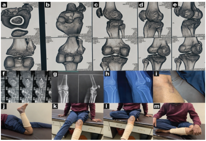
Figure 1: (a, b, c, d, e, f) pre-op workup; (g and h) post-op X-rays; (i) mini-incision; (j, k, l, m) showing range of motion on 8-week follow-up.
Case 2
A 20-year-old male with a renal tubular acidosis (RTA) mechanism had a displaced PCL avulsion with shaft femur fracture confirmed on plain radiographs. Post-operative radiographs demonstrated anatomical reduction; mini-incision scar was cosmetically acceptable. ROM was near-normal at 8 weeks (Fig. 2).
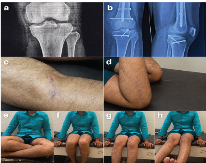
Figure 2: (a) pre-op X-ray; (b) post-op X-ray; (c) mini-incision; (d, e, f, g, h) Showing range of motion at post-op 8 weeks.
Case 3
A 20-year-old male with RTA presented with PCL avulsion combined with a lateral condyle fracture. Both injuries were addressed surgically; post-operative radiographs confirmed satisfactory reduction of both fragments (Fig. 3).
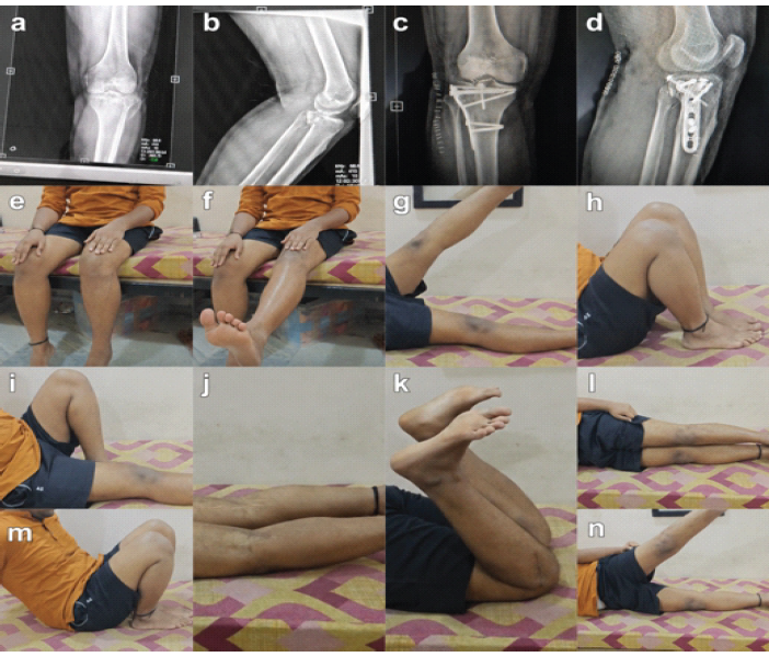
Figure 3: (a and b) pre-op X-rays; (c and d) post-op X-rays; (e, f, g, h, i, j, k, l, m, n) showing range of motion on 3-month follow-up.
Case 4
A 40-year-old male with RTA presented with osseous avulsion of PCL confirmed on radiographs and magnetic resonance imaging managed with open posterior approach; post-operative radiographs confirmed satisfactory reduction with good ROM at 12 weeks of follow-up (Fig. 4).
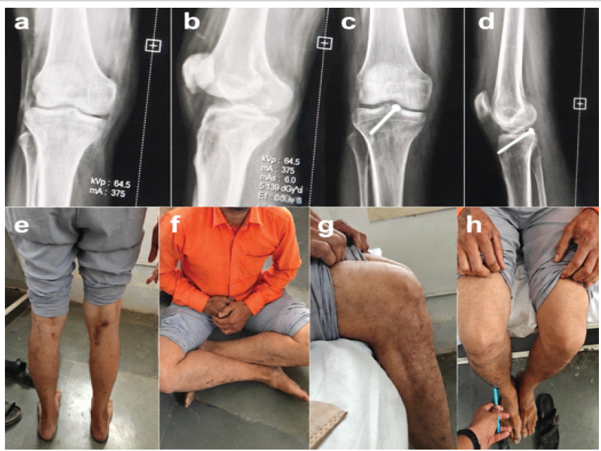
Figure 4: (a and b) pre-op X-rays; (c and d) post-op X-rays; (e) incision mark; (f) sitting cross-legged; (g and h) sitting with legs hanging down the table.
All patients achieved fracture union, with union confirmed radiologically in nearly all patients by approximately 12 weeks postoperatively. ROM improved consistently, with an average terminal flexion of approximately 120°–128° and full extension achieved in all patients by final follow-up. Knee stability, assessed clinically using the posterior drawer test, was well restored, with only minimal residual laxity noted in a small number of patients.
Functional outcomes, summarized in Table 1, showed marked improvement in both Lysholm and IKDC scores from the pre-operative to the 12-month post-operative assessment, consistent with previously reported outcomes for this technique [7]. The majority of patients achieved an “excellent” Lysholm grade (score range approximately 90–95) at final follow-up.
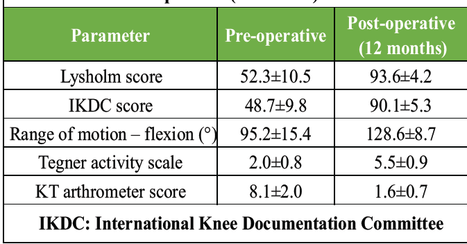
Table 1: Functional outcomes – pre-operative versus post-operative (12 months)
Postoperative complications were rare and are summarized in Table 2. No patient in this series developed a deep infection, hardware failure, or neurovascular injury.
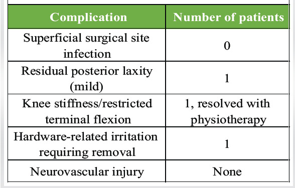
Table 2: Post-operative complications
Displaced PCL tibial avulsion fractures require anatomical reduction and stable fixation to restore the native tension and function of the ligament. The mini-open posterior approach described by Burks and Schaffer [4] remains a widely used technique for this purpose, offering several advantages over both closed management and arthroscopic-assisted fixation.
The principal advantages of the open posterior approach include direct visualization of the fracture fragment, the ability to achieve precise anatomical reduction under direct vision, strong and reliable fixation with cannulated screws, and a comparatively short learning curve for the operating surgeon. These features make it an attractive option in settings where arthroscopic expertise or equipment may be limited.
Compared with arthroscopic techniques, which are minimally invasive but technically demanding and equipment-dependent, the open posterior approach is technically simpler, cost-effective, and reproducible across a range of surgical settings, without compromising functional outcomes [5,8]. The results of the present series with mean Lysholm scores improving from 52.3 to 93.6 and IKDC scores from 48.7 to 90.1 are comparable to outcomes reported in the existing literature for both open and arthroscopic fixation of PCL tibial avulsion fractures [7,9,10,11].
The principal limitation of the posterior approach is the theoretical risk of injury to the popliteal neurovascular bundle during dissection; however, this risk is minimized with careful technique and thorough knowledge of the regional anatomy [12]. Occasional need for subsequent hardware removal, due to local irritation from prominent screws or washers, was also noted as a limitation of the technique.
This series, while demonstrating consistently favorable outcomes, is limited by its small sample size and the absence of a comparative arthroscopic cohort. Larger, prospective, randomized comparative studies with longer-term follow-up are required to further define the relative roles of open and arthroscopic fixation for this injury pattern.
The mini-open posterior approach for fixation of PCL tibial avulsion fractures is a reliable and effective surgical technique that allows direct visualization and accurate anatomical reduction of the avulsed fragment. It consistently results in excellent clinical and functional outcomes, with high Lysholm and IKDC scores, restoration of near-normal knee kinematics, and high patient satisfaction. The technique demonstrates high rates of fracture union and restoration of posterior knee stability, with minimal residual laxity on follow-up clinical examination.
Compared to traditional open procedures, it offers the advantages of a smaller incision, reduced soft-tissue dissection, shorter operative time, and faster rehabilitation, and it serves as a practical alternative to arthroscopic fixation, particularly in settings with limited arthroscopic resources, without compromising outcomes. The procedure is associated with a low complication rate, with rare instances of infection, neurovascular injury, or fixation failure when performed with proper technique.
Early diagnosis and timely surgical intervention remain critical to achieving optimal outcomes and preventing long-term complications such as chronic instability and degenerative joint change. Overall, the mini-open posterior approach with internal fixation is a safe, reproducible, and clinically successful method for managing displaced PCL tibial avulsion fractures, although larger randomized controlled trials with long-term follow-up are warranted to further strengthen this evidence base.
Limitations
The present study is a prospective case series without any comparative control group which demonstrates that the mini-open posterior approach provides reliable fracture union, restoration of knee stability, and excellent functional outcomes in patients with displaced PCL tibial avulsion fractures. Nevertheless, the findings should be interpreted in light of several limitations, including the small sample size, case series design without a comparative cohort, single-center setting, relatively short follow-up, and the absence of objective instrumented stability assessment and comprehensive patient-reported outcome measures. Larger multicenter prospective comparative studies with longer follow-up are required to validate these findings, compare different fixation techniques, and determine long-term functional and radiological outcomes.
The mini-open posterior approach provides a safe, reproducible, and technically straightforward means of achieving anatomical reduction and stable internal fixation of displaced PCL tibial avulsion fractures, yielding excellent functional and radiological outcomes and offering a practical alternative to arthroscopic fixation in resource-limited settings.
References
- 1. LaPrade CM, Civitarese DM, Rasmussen MT, LaPrade RF. Emerging updates on the posterior cruciate ligament: A review of the current literature. Am J Sports Med 2015;43:3077-92. [Google Scholar] [PubMed]
- 2. Meyers MH, McKeever FM. Fracture of the intercondylar eminence of the tibia. J Bone Joint Surg Am 1959;41:209-22. [Google Scholar] [PubMed]
- 3. Katsman A, Strauss EJ, Campbell KA, Alaia MJ. Posterior cruciate ligament avulsion fractures. Curr Rev Musculoskelet Med 2018;11:503-9. [Google Scholar] [PubMed]
- 4. Burks RT, Schaffer JJ. A simplified approach to the tibial attachment of the posterior cruciate ligament. Clin Orthop Relat Res 1990;254:216-9. [Google Scholar] [PubMed]
- 5. Hooper PO 3rd, Silko C, Malcolm TL, Farrow LD. Management of posterior cruciate ligament tibial avulsion injuries: A systematic review. Am J Sports Med 2018;46:734-42. [Google Scholar] [PubMed]
- 6. Sharma A, Gandhi N. Management of posterior cruciate ligament tibial avulsion fractures using a mini-open screw fixation technique. J Clin Orthop Trauma 2026;76:103410. [Google Scholar] [PubMed]
- 7. Joshi S, Bhatia C, Gondane A, Rai A, Singh S, Gupta S. Open reduction and internal fixation of isolated posterior cruciate ligament avulsion fractures: Clinical and functional outcome. Knee Surg Sports Traumatol Arthrosc 2017;25:210-6. [Google Scholar] [PubMed]
- 8. Song JG, Nha KW, Lee SW. Open posterior approach versus arthroscopic suture fixation for displaced posterior cruciate ligament avulsion fractures: systematic review. Knee Surg Relat Res 2018;30:275-83. [Google Scholar] [PubMed]
- 9. Khatri K, et al. Posterior cruciate ligament tibial avulsion treated with open reduction and internal fixation through Burks and Schaffer approach. Malays Orthop J 2015;9:35-9. [Google Scholar] [PubMed]
- 10. Mondal A, et al. Surgical fixation of posterior cruciate ligament tibial avulsion fracture using cannulated cancellous screw: clinical and functional outcome evaluation. Int J Res Orthop 2021;7:456-62. [Google Scholar] [PubMed]
- 11. Pu JS, et al. Suture bridge versus screw fixation for posterior cruciate ligament tibial avulsion fractures: A comparative study. BMC Surg 2025;25:58. [Google Scholar] [PubMed]
- 12. Nicandri GT, Klineberg EO, Wahl CJ, Mills WJ. Treatment of posterior cruciate ligament tibial avulsion fractures through a modified open posterior approach: Operative technique and 12- to 48-month outcomes. J Orthop Trauma 2008;22:317-24. [Google Scholar] [PubMed]






Related Articles in Journal of Orthopaedic Case Reports
 August 1, 2026 Comparative Outcomes of Hamstring Tendon versus Peroneus Longus Tendon Autograft in Arthroscopic Single-Bundle Anterior Cruciate Ligament Reconstruction: A Prospective Comparative Study
 October 1, 2026 Outcomes of Blood Flow Restriction Therapy as an Adjunct to Standard Rehabilitation Following Anterior Cruciate Ligament Reconstruction: A Prospective Observational Cohort Study
 October 1, 2026 Arthroscopic Posterior Cruciate Ligament Tibial Spine Avulsion Fixation Using a Suture Pull-Through Single-Tunnel Technique
 September 1, 2026 Serial Magnetic Resonance Imaging Graft Signal Intensity and its Correlation with Functional Recovery Following Primary Anterior Cruciate Ligament Reconstruction