

Early Tibial resection during robot-assisted total knee arthroplasty was associated with improved flexion and extension gap symmetry and may reduce the need for extensive soft-tissue releases within a tibia-first workflow.
Dr. Anurag H Daxini, Department of Orthopaedics and Joint Replacement, Mahajan Ortho and Surgical Hospital, Nagpur, India. E-mail: dranuragdaxini@gmail.com
Abstract
Introduction: The essence of total knee arthroplasty (TKA) is achieving optimal soft tissue balance. While robotic systems provide real-time gap feedback, the isolated impact of tibial resection on gap symmetry remains underexplored.
Objective: To evaluate the association between tibial resection and coronal and sagittal gap balancing in patients undergoing robot-assisted TKA (RATKA).
Materials and Methods: A retrospective study of 1137 knees using the VELYS system was conducted. Medial-lateral gap differences in extension and flexion were compared before and after tibial resection. Mechanical alignment correction and insert size distribution were also analyzed.
Results: Tibial resection was associated with improved extension gap symmetry in 88.4% of cases and flexion symmetry in 63.9%. Mean hip-knee-ankle correction was 5.2°, with 99.8% achieving neutral alignment. No meaningful correlation was found between post-tibial gaps and insert size.
Conclusion: Tibial resection was associated with improved gap symmetry and mechanical axis correction during RATKA using a tibia-first workflow. These findings support the potential role of early tibial resection in optimizing gap balance; however, prospective multicenter studies incorporating long-term clinical and functional outcomes are needed to validate these observations.
Keywords: Tibial resection, gap balancing, robot-assisted total knee arthroplasty, VELYS

Total knee arthroplasty (TKA) is one of the most successful surgical treatments for end-stage knee osteoarthritis, with over one million procedures performed globally each year [1]. Achieving accurate coronal and sagittal balance is essential for both patient satisfaction and long-term implant survival [2,3]. In the Indian population, patients frequently present late with advanced deformities and higher body mass index (BMI), with over 60% of patients exhibiting more than 10° of angular deformity at the time of surgery [4].
Conventional gap balancing relies on measured resections and soft tissue releases, which are highly surgeon-dependent and variable. Robotic-assisted systems, such as the VELYS robotic-assisted solution, offer improved reproducibility in component alignment and soft tissue balance without the need for pre-operative computed tomography imaging [5]. Robot-assisted TKA (RATKA) has been shown to enhance alignment accuracy and reduce outliers in mechanical axis restoration [6,7,8].
Among various robotic workflows, the tibia-first approach has gained popularity for its logical sequencing – where the tibial cut serves as the foundation for femoral planning. Tibial resection influences soft tissue tension and gap symmetry in both extension and flexion [9,10]. However, most literature evaluates balance only after both femoral and tibial resections, failing to isolate the specific impact of the tibial step [6]. Given the prevalence of severe deformity and soft tissue imbalance in Indian TKA patients, understanding the independent contribution of tibial resection during a tibia-first robotic workflow is clinically relevant. Accordingly, this retrospective study aimed to evaluate the association of tibial resection with intraoperative gap symmetry and mechanical alignment correction during RATKA using the VELYS system.
This was a retrospective, single-surgeon, single-center observational study conducted at a high-volume joint replacement center in a second-tier Indian city. The study included consecutive patients who underwent RATKA between January 2024 and December 2024. All data were collected from a prospectively maintained robotic joint replacement registry using the VELYS Robotic-Assisted Solution (DePuy Synthes).
Exclusion criteria included incomplete intraoperative data, revision TKA cases, and severe bone loss or extra-articular deformities requiring augmentation.
All surgeries followed a tibia-first workflow using the VELYS robotic-assisted solution. Pre-resection gaps were recorded, followed by tibial resection and re-measurement of extension and flexion gaps. Final femoral resections and implant sizing were based on remaining gap asymmetries. Importantly, no soft tissue releases were performed before tibial resection or during the gap measurements analyzed in this study, allowing assessment of the immediate association between tibial resection and intraoperative gap balance. Any soft-tissue releases performed later in the procedure, when required, were not included in the present analysis.
Parameters collected included pre- and post-tibial resection medial and lateral gaps in both flexion and extension, pre- and post-operative hip-knee-ankle (HKA), and final tibial insert size. Gap symmetry was defined as the absolute difference between medial and lateral gaps.
To ensure consistency across all cases, intraoperative gap measurements were obtained using the standardized tensioning protocol integrated into the VELYS system. Balanced gaps were defined as ≤2 mm difference between medial and lateral compartments at a given flexion angle. Pre-tibial resection flexion and extension gaps were measured (Fig. 1). Followed by post-tibial resection, flexion and extension gaps were measured (Fig. 2), and lastly, final flexion and extension gaps were measured (Fig. 3).
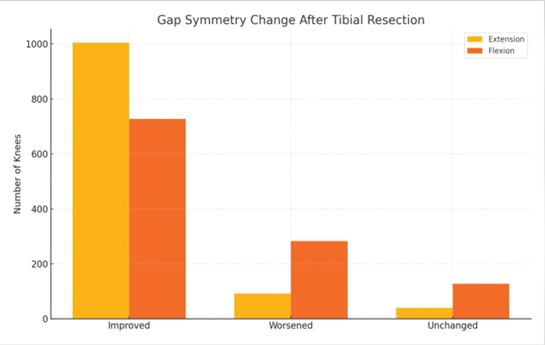
Figure 1: Unbalanced pre-tibial resection gaps: The graph on the right side of the figure shows a flexion gap of 6.5 mm and 10 mm on the medial and lateral sides, respectively, and an extension gap of 6 mm and 11 mm on the medial and lateral sides, respectively. Both the flexion and extension gaps have a difference of >2mm between the medial and lateral sides, denoting unbalanced gaps.
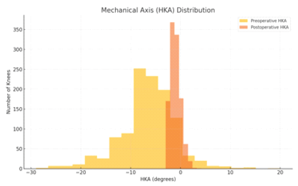
Figure 2: Balanced post-tibial resection gaps: The graph on the right side shows flexion gaps of 7.5 mm and 9.5 mm, respectively, and extension gaps of 7 mm and 8.5 mm on the medial and lateral sides, respectively. Both the flexion and extension gaps have a difference of <2 mm between the medial and lateral sides, denoting balanced gaps.
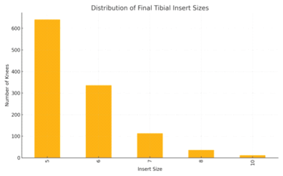
Figure 3: Balanced gaps. Showing final gap balance post-femur cuts showing balanced medial and lateral gaps on flexion and extension.
Pre-operative full leg films were compared with post-operative full leg films for HKA axis. All extension gaps were measured with the knee in full extension possible, and flexion gaps were assessed at 90° of flexion under standardized manual valgus and varus stress applied by the surgeon. The robotic system recorded and displayed dynamic gap readings in real time using a standardized tensioning protocol. Although manual stress application may introduce some operator-dependent variability, the use of a single experienced surgeon and a uniform measurement protocol helped minimize variability. Data were digitally extracted from the VELYS database immediately following surgery and cross-verified with intraoperative reports for accuracy.
All procedures were performed using the same posterior-stabilized implant design (Attune, DePuy Synthes) to maintain prosthetic consistency. Intraoperative gap assessments were conducted at full extension and 90° flexion using the VELYS system’s dynamic tensioning interface, under standardized manual varus-valgus stress. Balanced gaps were defined as medial-lateral differences ≤2 mm. Simultaneous bilateral knees were analyzed independently, with each knee treated as a separate unit of analysis. This approach was adopted because gap balancing and implant planning are performed independently for each knee during surgery. Intraoperative data were extracted from the robotic system and verified against surgical logs for completeness. Individual consent was waived off due to retrospective, de-identified data analysis. No formal power analysis was performed, as the sample included all eligible cases over the study period.
Descriptive statistics were reported as means with standard deviations or medians with interquartile ranges, depending on the distribution of the data. Continuous variables were analyzed using paired or unpaired Student’s t-tests, while categorical variables were expressed as frequencies and percentages and compared using chi-square tests. Paired tests were used for intra-subject comparisons, such as pre- versus post-tibial resection gap differences and HKA correction. Correlation between continuous variables, such as post-tibial gaps and insert size, was evaluated using Pearson correlation coefficients. A P < 0.05 was considered statistically significant. All statistical analyses were performed using the Statistical Package for the Social Sciences software (version 20.0; IBM Corp., Armonk, NY, USA) and Microsoft Excel (version 2019; Microsoft Corp., Redmond, WA, USA).
Extension gaps
Before tibial resection, the mean medial-lateral extension gap difference was 2.26 ± 2.04 mm.
Following tibial resection, this reduced to 0.77 ± 1.15 mm, indicating a marked improvement in gap symmetry. Gap balance improved in 1005 knees (88.4%), worsened in 92 knees (8.1%), and remained unchanged in 40 knees (3.5%).
Flexion gaps
Before tibial resection, the mean medial-lateral flexion gap difference was 1.98 ± 1.91 mm.
After tibial resection, this improved to 1.28 ± 1.47 mm, showing a positive effect on flexion gap symmetry. Flexion gap balance improved in 727 knees (63.9%), worsened in 283 knees (24.9%), and remained unchanged in 127 knees (11.2%) (Fig. 4).
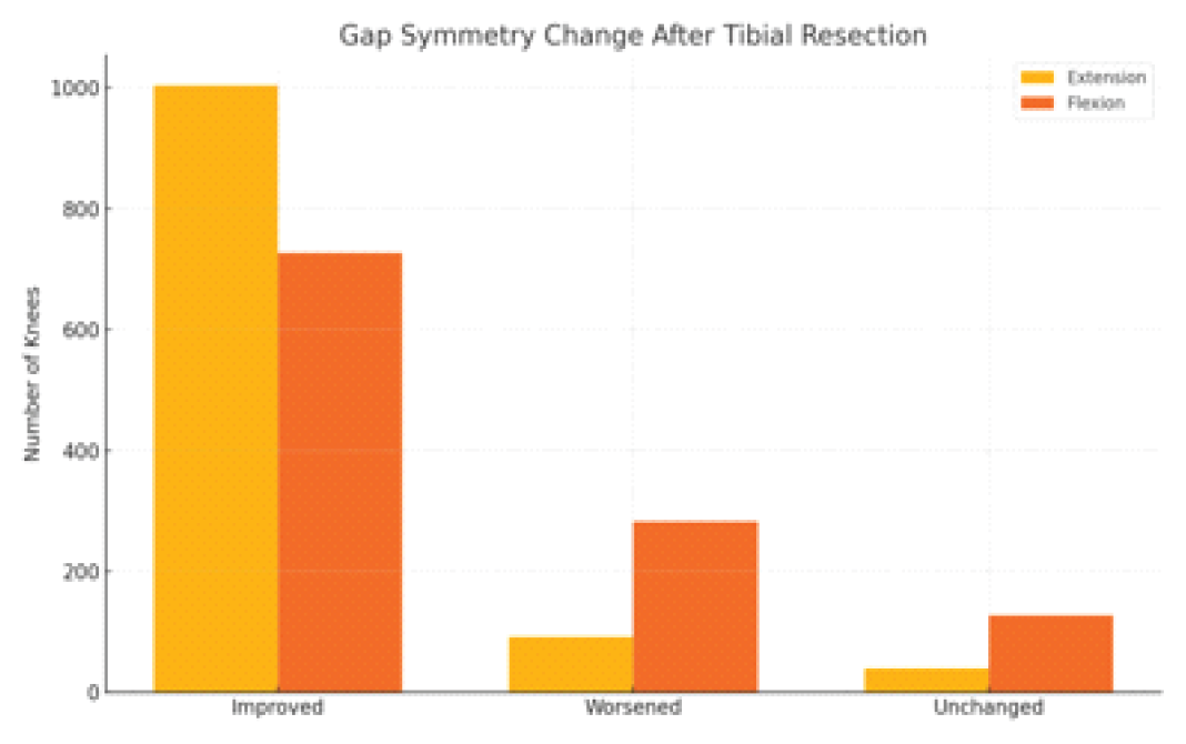
Figure 4: Gap symmetry change after tibial resection: The bar diagram showing the number of knees where gap symmetry improved, worsened, and was unchanged.
HKA correction
The mean pre-operative HKA angle was –6.51° ± 5.48°, indicating a predominant varus alignment in the study cohort. Following surgery, the mean post-operative HKA improved to –1.28° ± 2.47°, reflecting an average correction of +5.23° ± 3.76° toward mechanical neutrality.
Neutral alignment, defined as an HKA within ±3°, was achieved in 1135 knees (99.8%), while only 2 knees (0.2%) remained outside the desired alignment range post-operatively (Fig. 5).
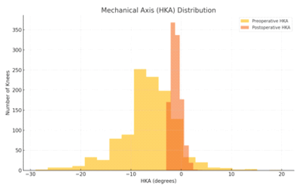
Figure 5: Mechanical axis distribution: The graph showing pre-operative hip knee ankle axis distribution as against post-operative hip knee ankle axis distribution. The post-operative alignment was within ±3° in 1135 knees (99.8%).
Insert size distribution
The most commonly used tibial insert thickness was 5 mm, implanted in 650 knees (57.2%).
An insert size of 6 mm was used in 308 knees (27.1%), while 7 mm inserts were selected in 101 knees (8.9%). Larger inserts (≥8 mm) were required in only 78 knees (6.8%), suggesting that the majority of cases achieved acceptable gap balancing with minimal polyethylene thickness (Fig. 6).
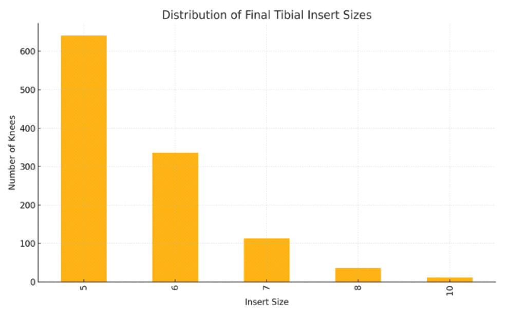
Figure 6: Final insert sizes: Shows the maximum number of tibial inserts being used is 5 and 6.
Correlation between post-tibial gaps and insert size
Pearson correlation analysis was performed to evaluate the relationship between post-tibial resection gap measurements and final tibial insert thickness. There was no meaningful correlation between the post-resection extension gap and insert size (r = 0.014), or between the post-resection flexion gap and insert size (r = 0.094). These findings suggest that final insert selection may be influenced by multiple factors beyond static gap values alone, including dynamic balancing and intraoperative femoral adjustments
This study evaluated the association between tibial resection and medial-lateral gap balance in RATKA using the VELYS Robotic-Assisted Solution. Our findings showed that tibial resection was associated with improved extension gap symmetry in the vast majority of cases, with a comparatively more variable but still meaningful impact on flexion gap symmetry. These results are particularly relevant in the context of a tibia-first workflow, where early gap balance directly influences downstream femoral planning and implant positioning [9,10].
The observed improvement in extension gap symmetry (88.4% of knees) reinforces the concept that the coronal plane alignment and soft tissue tension can be substantially normalized with precise tibial resection alone [2,3,10]. This is consistent with the principle that the tibia acts as the foundation of gap balancing, especially in robotic workflows, and supports the rationale for minimizing soft tissue releases when using robotic planning and execution systems [6]. In flexion, gap symmetry improved in nearly two-thirds of cases, though a notable proportion (24.9%) experienced worsening – possibly due to posterior femoral geometry, changes in slope, or unaddressed soft tissue imbalances, which are typically managed during femoral preparation [7].
Our study adds to the existing literature by specifically evaluating the association between tibial resection and intraoperative gap symmetry during a tibia-first robotic workflow, an area that has received relatively limited attention. Few studies, if any, have attempted to isolate the contribution of tibial resection in robotic workflows [6,8,10]. In this regard, the present study contributes a novel and data-driven insight, particularly relevant to surgeons adopting sensor-based, gap-first or adjustable planning models using imageless robotic platforms, such as VELYS.
Interestingly, we found no strong correlation between post-tibial gaps and insert size, suggesting that final implant thickness is influenced by a constellation of factors, including rotational alignment, dynamic trailing, and surgeon preference rather than static medial-lateral gaps alone. This underscores the complexity of intraoperative decision-making, even in highly digitized systems, and reflects the adaptive nature of RATKA, where femoral component planning evolves based on intraoperative balance goals.
From a clinical perspective, the ability to achieve symmetrical gaps early in the procedure without performing soft tissue releases may help reduce operative time, limit morbidity, and streamline training in robotic workflows [9,11]. In high-volume centres or resource-limited settings common in India, these efficiency gains have practical significance. Although post-operative mechanical axis is a surrogate marker rather than a direct measure of clinical function, the high rate (99.8%) of neutral alignment demonstrates the precision and reliability of RATKA [7,8].
This study has a few limitations. First, its retrospective observational design allows demonstration of an association rather than a causal relationship between tibial resection and improved gap balance. Second, the study represents the experience of a single high-volume surgeon at a single centre using only the VELYS Robotic-Assisted Solution, which may limit generalizability of the findings to other surgeons, institutions and robotic platforms. Third, the study did not include a conventional TKA control group or comparison with alternate robotic workflows, such as femur-first or gap-first techniques. Fourth, patient-reported outcome measures and long-term follow-up data were not available; therefore, the relationship between improved intraoperative gap symmetry and post-operative function, patient satisfaction, implant survival, polyethylene wear, or revision rates could not be assessed. Fifth, although gap measurements were obtained using a standardized robotic tensioning protocol by a single experienced surgeon, manual application of varus-valgus stress may have introduced some operator-dependent variability. In addition, soft tissue releases performed after tibial resection were not quantitatively analyzed; simultaneous bilateral knees were analyzed as independent observations, formal assessment of interobserver or intraobserver reliability was not performed, and subgroup analyses based on deformity severity, BMI, and arthritis grade were beyond the scope of this study.
Nonetheless, this study offers strong preliminary evidence supporting the potential role of tibial resection in improving intraoperative gap symmetry during RATKA. Future prospective multicentre studies comparing different robotic platforms and surgical workflows, while incorporating patient-reported outcomes and long-term clinical follow-up, are required to validate these findings and determine their clinical significance [11].
This retrospective study found that tibial resection was associated with improved coronal and sagittal gap symmetry during RATKA using a tibia-first workflow. The majority of knees, particularly in extension, demonstrated improved medial-lateral gap symmetry following tibial resection, while restoration of mechanical alignment was achieved in nearly all cases. These findings support the potential role of a structured tibia-first robotic workflow in optimizing intraoperative gap balance. However, prospective multicenter studies with long-term clinical and functional outcomes are required to confirm these observations.
Within a Tibia-first robotic-assisted total knee arthroplasty workflow, tibial resection was consistently associated with improved intraoperative coronal and sagittal gap symmetry, potentially reducing the need for extensive soft tissue releases.
References
- 1. Carr AJ, Robertsson O, Graves S, Price AJ, Arden NK, Judge A, et al. Knee replacement. Lancet 2012;379:1331-40. [Google Scholar] [PubMed]
- 2. Bellemans J, D’Hooghe P, Vandenneucker H, Van Damme G, Victor J. Soft tissue balance in total knee arthroplasty: does stress relaxation occur perioperatively? Clin Orthop Relat Res. 2006 Nov;452:49-52. doi: 10.1097/01.blo.0000238790.29102.95. PMID: 16936582. [Google Scholar] [PubMed] [CrossRef]
- 3. Mihalko WM, Saleh KJ, Krackow KA, Whiteside LA. Soft-tissue balancing during total knee arthroplasty in the varus knee. J Am Acad Orthop Surg 2009;17:766-74. [Google Scholar] [PubMed]
- 4. Pal CP, Singh P, Chaturvedi S, Pruthi KK, Vij A. Epidemiology of knee osteoarthritis in India and related factors. Indian J Orthop 2016;50:518-22. [Google Scholar] [PubMed]
- 5. VELYS Robotic-Assisted Solution. DePuy Synthes. Product Literature. 2021. [Google Scholar] [PubMed]
- 6. Hampp EL, Chughtai M, Scholl LY, Sodhi N, Bhowmik-Stoker M, et al. Robotic-arm assisted total knee arthroplasty demonstrated greater accuracy and precision to plan compared with manual techniques. J Knee Surg 2019;32:239-50. [Google Scholar] [PubMed]
- 7. Deckey DG, Rosenow CS, Verhey JT, Brinkman JC, Mayfield CK, Clarke HD, et al. Robotic-assisted total knee arthroplasty improves accuracy and precision compared to conventional techniques. Bone Joint J 2021;103-B:74-80. [Google Scholar] [PubMed]
- 8. Alrajeb R, Zarti M, Shuia Z, Alzobi O, Ahmed G, Elmhiregh A. Robotic-assisted versus conventional total knee arthroplasty: A systematic review and meta-analysis of randomized controlled trials. Eur J Orthop Surg Traumatol 2024;34:1333-43. [Google Scholar] [PubMed]
- 9. St Mart JP, Goh EL. The current state of robotics in total knee arthroplasty. EFORT Open Rev 2021;6:270-9. [Google Scholar] [PubMed]
- 10. Daines BK, Dennis DA. Gap balancing vs. measured resection technique in total knee arthroplasty. Clin Orthop Surg 2014;6:1-8. [Google Scholar] [PubMed]
- 11. Marchand RC, Sodhi N, Anis HK, Ehiorobo J, Newman JM, Taylor K, et al. One-year patient outcomes for robotic-arm-assisted versus manual total knee arthroplasty. J Knee Surg 2019;32:1063-8. [Google Scholar] [PubMed]




Related Articles in Journal of Orthopaedic Case Reports
 June 10, 2024 Precise Calibration of Femoral Component Rotation Using the Posterior Condylar Axis as a Reference during Image-free Robot-assisted Total Knee Arthroplasty: A Technical Note
 January 1, 2026 Anteromedial Knee Pain Unmasked: Schwannoma of the Infrapatellar Branch of the Saphenous Nerve – A Rare Case Report
 June 10, 2024 Impending Atypical Femoral Fracture on the Contralateral Side Progressed to a Complete Fracture Early after Surgery for an Atypical Femoral Fracture: A Case Report
 December 10, 2021 Clinical and Surgical Insights on Bilateral Total Knee Arthroplasty in Ochronotic Arthropathy: A Case-based Review