

Successful knee arthrodesis with the Ilizarov ring fixator depends on complete infection eradication, meticulous debridement, and stable circular fixation, allowing reliable fusion even in the presence of severe bone loss and compromised soft tissues.
Dr. Rohit Kumar Yadav, Department of Orthopaedics, All India Institute of Medical Sciences, Deoghar - 814152, Jharkhand, India. E-mail: rk169816@gmail.com
Abstract
Introduction: In the present study, we describe clinical outcomes and observations of Ilizarov ring fixation used for a knee joint infection with pronounced joint destruction, soft tissue and bone involvement, which are serious diseases in which not only the joint but also the entire extremity is directly endangered. Salvage procedure in infected knees resulting from failed total knee arthroplasty, osteomyelitis, or septic arthritis.
Materials and Methods: We report a case series of four patients (two males and two females), including patients presenting with periarticular knee joint infection characterized by extensive soft-tissue involvement and significant osseous destruction, often complicated by chronic discharging sinuses and profound functional impairment. All patients underwent detailed clinical assessment and standard radiographic evaluation and comprehensive evaluations including C-reactive protein, erythrocyte sedimentation rate, limb-length discrepancy (LLD), knee injury and osteoarthritis outcome score, knee society score, Visual Analog Scale (VAS) score and Lysholm score. Intraoperative tissue samples were collected for post-operative microbiological analysis to identify the causative organisms and to guide targeted antimicrobial therapy, and an Ilizarov ring fixator was applied in compression mode.
Results: Union was achieved in all patients at both the distal femur and proximal tibia at a mean duration of 7 months (29.5 ± 4 weeks). The mean residual LLD was 2.1 ± 0.6 cm. Pain relief was substantial, with the VAS score decreasing from 7.5 ± 0.6 preoperatively to 2.0 ± 0.8 postoperatively. Similarly, the Lysholm score improved from 45.3 ± 2.5 preoperatively to 76.5 ± 4.3 postoperatively.
Conclusion: The Ilizarov technique is an effective salvage procedure for complex knee joint pathologies. Its advantages include cost-effectiveness, avoidance of secondary surgical interventions, allowance for immediate weight-bearing, a high rate of arthrodesis, and a low incidence of complications.
Keywords: Arthrodesis, Ilizarov fixator, Lysholm score, limb-length discrepancy, C-reactive protein, erythrocyte sedimentation rate, knee injury and osteoarthritis outcome score, knee society score, Visual Analog Scale score

Knee arthrodesis is a widely performed surgical procedure with well-defined indications [1,2,3], and this procedure represents the best choice for patients with significant bone loss and recurrent knee infections. The main goal of applying a procedure is to achieve a stable and pain-free lower limb with a low risk of reinfection and a better functional outcome compared with above-knee amputation. Knee arthrodesis remains a method of limb salvage [4]. In the last decades, many surgical techniques have been proposed for performing knee arthrodesis: internal osteosynthesis, intramedullary (IM) nailing and external fixation [5,6,7]. The existing literature on femoral-tibial fusion with an Ilizarov circular external fixator reports success rates that vary in different studies from 64% to 100% [8,9].
Four patients with complex infected knee conditions underwent knee arthrodesis using an Ilizarov circular external fixator. Indications included chronic periprosthetic joint infection (PJI) following failed total knee arthroplasty (Fig. 1), post-traumatic septic knee after a high-grade open fracture (Fig. 2), and chronic infection of the native knee with persistent sinus tracts (Fig. 3 and 4). All patients presented with pain, periarticular swelling, inability to bear weight, and elevated inflammatory markers.
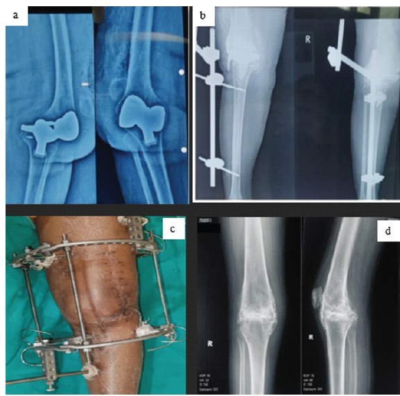
Figure 1: Case of prosthetic joint infections post (a) failed total knee arthroplasty done elsewhere, (b) post-implant removal with cement spacer and external fixator application, (c) post-external fixator removal and Ilizarov application, (d) radiograph post-arthrodesis.
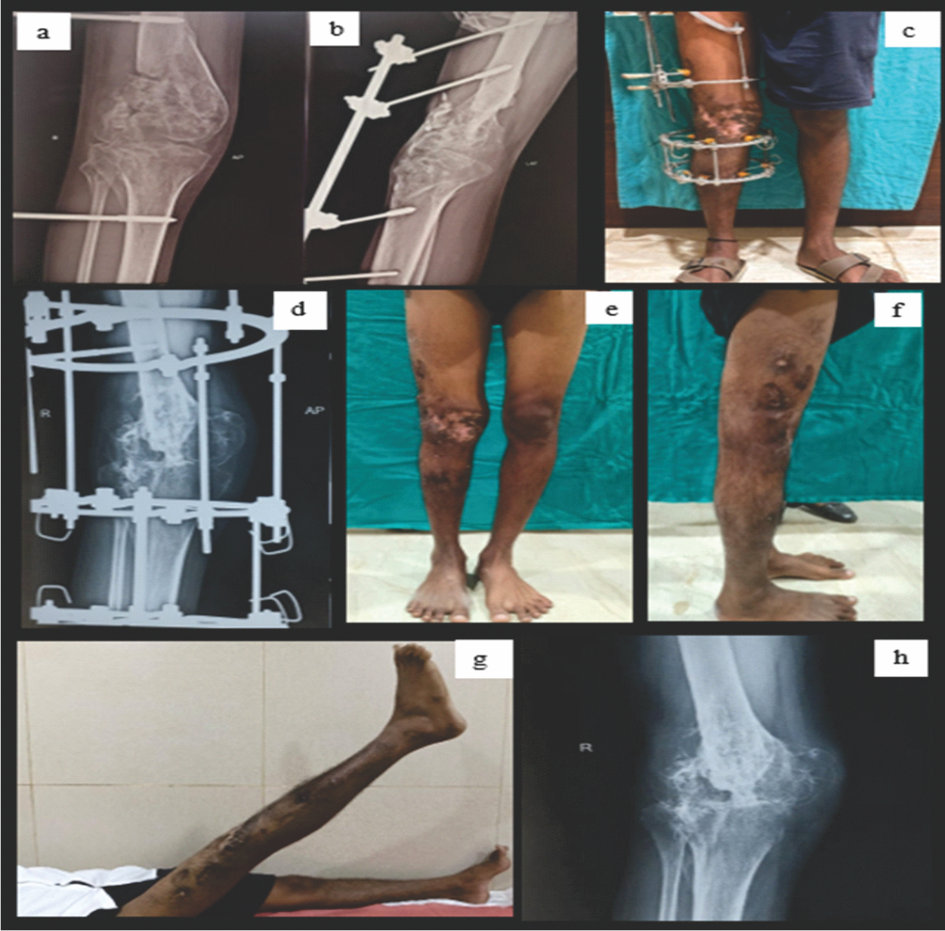
Figure 2: Case of post traumatic septic arthritis (a) & (b) knee spanning external fixation done elsewhere for GA IIIB distal femur fracture (c) & (d) after external fixator removal and Ilizarov application (e), (f), (g) & (h) after removal of Ilizarov frame.
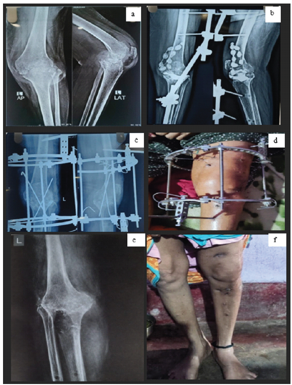
Figure 3: Case of septic arthritis (a) initially managed with external fixator and antibiotic beads, (b) after external fixator removal, (c) after Ilizarov fixator application, (d) after removal of frame. (e) Post arthrodesis radiograph (f) post arthrodesis clinical picture
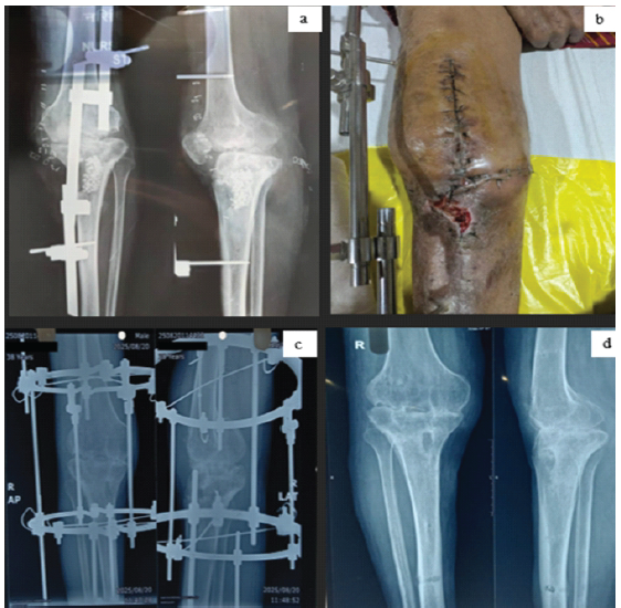
Figure 4: Case of septic arthritis (a and b) debridement, stimulant application and external fixation (c) post-Ilizarov frame application (d) post-Ilizarov frame removal.
A standardized staged treatment protocol was used in all cases. Stage 1 consisted of radical debridement with complete excision of sinus tracts and infected tissue, combined with local antibiotic delivery (polymethylmethacrylate spacers or antibiotic-loaded calcium sulfate beads) and culture-directed systemic antibiotics. Temporary linear external fixation was used when required to maintain limb alignment and length. Stage 2 was performed after clinical resolution of infection and normalization of inflammatory markers and involved definitive knee arthrodesis using an Ilizarov circular fixator applied in acute compression across the femorotibial junction. Adjunctive procedures, including autologous bone grafting, transarticular Kirschner-wire fixation, femoral corticotomy, and distraction osteogenesis, were performed selectively to address bone loss and limb-length discrepancy (LLD).
Outcome assessment
Serial radiographs were reviewed to evaluate clinical and radiographic outcomes following arthrodesis. In the absence of clinical signs of infection (pain, fever, swelling, or effusion), inflammatory markers (erythrocyte sedimentation rate and C-reactive protein) were not routinely rechecked. Radiographic union was defined as continuous trabecular bridging across the arthrodesis site on both anteroposterior and lateral radiographs. Clinical union was defined as a stable, pain-free limb without the need for external support.
LLD was assessed during routine follow-up by measuring the distance from the anterior superior iliac spine to the medial malleolus on the operated limb and comparing it with the contralateral side.
Functional and pain outcomes were assessed using validated scoring systems, including the Western Ontario and McMaster Universities Osteoarthritis Index (WOMAC), knee society score (KSS), knee injury and osteoarthritis outcome score (KOOS), Visual Analog Scale (VAS), and Lysholm score.
Outcomes and follow-up
Radiographic union was achieved in all four patients, with time to union ranging from 26 to 36 weeks. Despite severe bone loss and active or quiescent infection at presentation, stable arthrodesis was consistently obtained. Residual LLD was modest (mean, approximately 2 cm) and was well-tolerated with shoe lifts.
Functional outcomes improved substantially across all measures. The mean WOMAC score improved from 40.5 ± 1.3 preoperatively to 63.5 ± 1.3 postoperatively (Table 1). The mean KSS improved from 32.8 ± 2.2 to 69.0 ± 2.6 (Table 2), indicating restoration of a stable, painless limb capable of independent ambulation. KOOS improved from 39.0 ± 2.6 to 66.5 ± 2.1, reflecting improved knee-related quality of life.
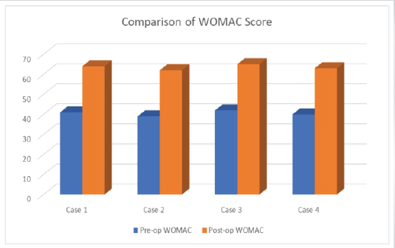
Table 1: Western Ontario and McMaster Universities Osteoarthritis Index scores in pre-operative and post-operative periods of individual cases and mean score mentioned in outcome section
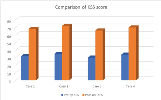
Table 2: Knee Society Score in pre-operative and post-operative periods of individual cases and mean score mentioned in outcome section
Pain relief was notable, with mean VAS scores decreasing from 7.5 ± 0.6 preoperatively to 2.0 ± 0.8 postoperatively. Correspondingly, Lysholm scores improved from 45.3 ± 2.5 to 76.5 ± 4.3, indicating good functional recovery and patient satisfaction following knee arthrodesis (Table 3). All cases are summarized in Table 4.
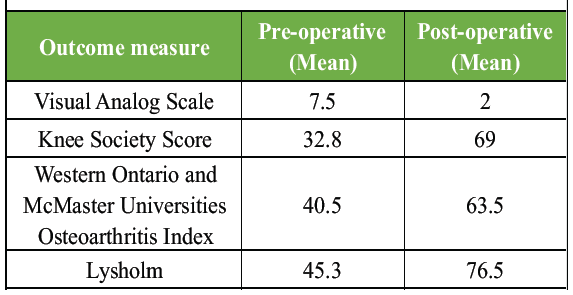
Table 3: Key clinical outcomes before and after Ilizarov knee arthrodesis
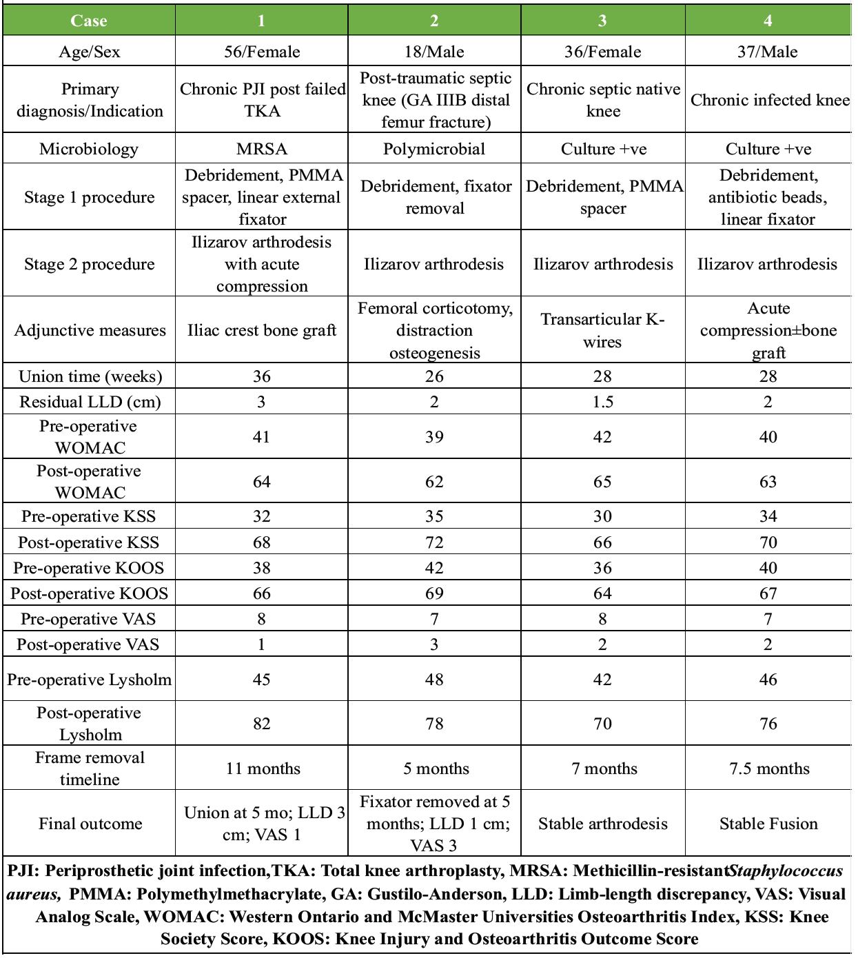
Table 4: Summary of all cases in the series
Knee arthrodesis remains a well-established salvage procedure for the management of persistent and complex knee pathologies, particularly PJI. In patients with multiple failed surgical interventions – including prior use of static or articulating antibiotic-loaded cement spacers – arthrodesis provides a stable, pain-free limb while effectively eradicating infection. Various surgical options have been described, including internal fixation with IM nailing, external fixation, and tibiofemoral screw-plate constructs [10].
Screw plate constructs provide rigidity but seldom achieve adequate compression for successful fusion at weight-bearing joints [11]. IM introduction of infection due to reaming is a major complication related to arthrodesis using nails. Furthermore, nail migration, poor rotational control and tibial fractures compound the need for an alternative to nails and plates [12].
In the present case series, all four patients treated with knee arthrodesis using the Ilizarov ring fixator achieved successful union, with a mean time to union of 29.5 ± 4.1 weeks (range: 26–36 weeks) [13]. These findings are consistent with previously published studies by Watanabe et al. [14] and Leroux et al. [15], supporting the reliability of circular external fixation in achieving union in complex infected scenarios.
Residual LLD remains a concern following arthrodesis. In our series, the mean LLD was 2.1 ± 0.6 cm, comparable to reports by Bargiotas et al. [16] and Incavo et al. [17]. Importantly, this degree of shortening was clinically well tolerated and could be effectively managed with simple measures such as shoe lifts, without significantly compromising patient satisfaction or function.
Functional outcomes following knee arthrodesis are inherently limited compared to joint-preserving procedures; however, they remain acceptable in the context of limb salvage. In our study, post-operative evaluation using validated scoring systems – including the KSS, Knee Society Functional Score, KOOS, VAS for pain, and Lysholm score – demonstrated that most patients achieved satisfactory pain relief, although some functional limitations persisted.
Knee arthrodesis continues to serve as an important alternative to above-knee amputation in complex and refractory cases. The outcomes observed in this series support its role as a reliable salvage option in carefully selected patients. Our findings align with Conway et al. [18], who reported that successful arthrodesis offers superior overall outcomes compared to amputation or failed revision arthroplasty. Arthrodesis can therefore provide a stable, painless, and functionally acceptable limb, particularly in patients who are physically and psychologically compromised after multiple failed procedures.
A key factor contributing to the favorable outcomes in our series was meticulous surgical technique. Complete eradication of infection prior to definitive fixation, combined with preparation of large, congruent contact surfaces of viable bleeding cancellous bone, was critical in achieving a high union rate and satisfactory functional results.
In our case series, Arthrodesis appears to be a valuable option in cases of complex knee pathologies, particularly in patients with multiple prior surgeries or significant comorbidities. However, given the small sample size and retrospective design without a control group, larger prospective studies are needed to generate more robust and generalizable evidence.
Knee arthrodesis using the Ilizarov ring fixator is a reliable and effective salvage procedure for complex knee pathologies, particularly in the setting of infection. It provides predictable union, satisfactory pain relief, and acceptable functional outcomes, making it a valuable alternative to amputation in carefully selected patients. The existing literature on femoral-tibial fusion with an Ilizarov circular external fixator reports success rates that vary in different studies from 64% to 100%. In our series, the rate of complete healing was 76.5% at the first surgical attempt.
The Ilizarov ring fixator remains an effective and cost-efficient salvage technique for complex infected knees, providing durable limb preservation, early functional rehabilitation, and a viable alternative to amputation in carefully selected patients.
References
- 1. Bruno AA, Kirienko A, Peccati A, Dupplicato P, De Donato M, Arnaldi E, et al. Knee arthrodesis by the Ilizarov method in the treatment of total knee arthroplasty failure. Knee 2017;24:91-9. [Google Scholar] [PubMed]
- 2. Ngona NS, Bouyou AS, Ikounga YP, Nkoua MF, Ellah MR, Diao S, et al. Impact of the mixed and simple methods on bone fusion at average follow-up at the Idrissa Pouye General Hospital (HOGIP) in Dakar. Int J Orthop Sci 2024;10:374-8. [Google Scholar] [PubMed]
- 3. Waszczykowski M, Niedzielski K, Radek M, Fabis J. Arthroscopic-assisted arthrodesis of the knee joint with the ilizarov technique: A case report and literature review. Med (Baltimore) 2016;95:e2540. [Google Scholar] [PubMed]
- 4. Mvili Gampio Ngona NS, Bouhelo-Pam KP, Bilongo-Bouyou AS, Nzengui ZF, Diao S, Kasse AN. Arthrodesis impact on the functional outcome of operated knees at the last follow-up. Int J Orthop Sci 2024;10:357-60. [Google Scholar] [PubMed]
- 5. Bengtson S, Knutson K. The infected knee arthroplasty. A 6-year follow-up of 357 cases. Acta Orthop Scand 1991;62:301-11. [Google Scholar] [PubMed]
- 6. Oostenbroek HJ, Van Roermund PM. Arthrodesis of the knee after an infected arthroplasty using the Ilizarov method. J Bone Joint Surg Br 2001;83:50-4. [Google Scholar] [PubMed]
- 7. Rand JA. Alternatives to reimplantation for salvage of the total knee arthroplasty complicated by infection. J Bone Joint Surg Am 1993;75:282-9. [Google Scholar] [PubMed]
- 8. Garberina MJ, Fitch RD, Hoffmann ED, Hardaker WT, Vail TP, Scully SP. Knee arthrodesis with circular external fixation. Clin Orthop 2001;382:168-78. [Google Scholar] [PubMed]
- 9. David R, Shtarker H, Horesh Z, Tsur A, Soudry M. Arthrodesis with the Ilizarov device after failed knee arthroplasty. Orthopedics 2001;24:33-6. [Google Scholar] [PubMed]
- 10. Ramazzini-Castro R, Pons-Cabrafiga M. Knee arthrodesis in rescue surgery: A study of 18 cases. Rev Esp Cir Ortop Traumatol 2013;57:45-52. [Google Scholar] [PubMed]
- 11. Matthews AC, Mallin BA, Pritchett JW. Knee arthrodesis with a tension-band plate. J Bone Joint Surg Am 1988;70:285-8. [Google Scholar] [PubMed]
- 12. Benson ER, Resine ST, Lewis CG. Functional outcome of arthrodesis for failed total knee arthroplasty. Orthopedics 1998;21:875-9. [Google Scholar] [PubMed]
- 13. Emerson RH Jr., Muncie M, Tarbox TR, Higgins LL. Comparison of a static with a mobile spacer in total knee infection. Clin Orthop Relat Res 2002;404:132-8. [Google Scholar] [PubMed]
- 14. Watanabe K, Minowa T, Takeda S, Otsubo H, Kobayashi T, Kura H, et al. Outcomes of knee arthrodesis following infected total knee arthroplasty: A retrospective analysis of 8 cases. Mod Rheumatol 2014;24:243-9. [Google Scholar] [PubMed]
- 15. Leroux B, Aparicio G, Fontanin N, Ohl X, Madi K, Dehoux E, et al. Arthrodesis in septic knees using a long intramedullary nail: 17 Consecutive cases. Orthop Traumatol Surg Res 2013;99:399-404. [Google Scholar] [PubMed]
- 16. Bargiotas K, Wohlrab D, Sewecke JJ, Lavinge G, Demeo PJ, Sotereanos NG. Arthrodesis of the knee with a long intramedullary nail following the failure of a total knee arthroplasty as the result of infection. J Bone Joint Surg Am 2006;88:553. [Google Scholar] [PubMed]
- 17. Incavo SJ, Lilly JW, Bartlett CS, Churchill DL. Arthrodesis of the knee: Experience with intramedullary nailing. J Arthroplasty 2000;15:871-6. [Google Scholar] [PubMed]
- 18. Conway JD, Mont MA, Bezwada HP. Arthrodesis of the knee. J Bone Joint Surg Am 2004;86:835-48. [Google Scholar] [PubMed]




Related Articles in Journal of Orthopaedic Case Reports
 March 1, 2026 Comparative Analysis of Suprapatellar and Infrapatellar Nailing in Tibial Fracture: A Randomized Clinical Study
 March 1, 2026 Evaluation of Post-operative Serum Biomarkers (D-Dimer, ESR, and CRP) in Patients Following Total Joint Arthroplasty: A Prospective Observational Study
 February 1, 2026 Clinico-Laboratory Correlates of Inflammatory Markers in Elderly Knee Osteoarthritis
 April 1, 2025 Effect of DMARD Therapy on Inflammatory Biomarkers and Disease Activity in Rheumatoid Arthritis Patients