


Introduction
Glomus tumor is a benign neoplasm of the neuromyoarterial tissue that is commonly seen in the subungual region of the digit’s most commonly of the upper extremity. The normal glomus is a myoarterial apparatus located on the surface of the body at the reticular stratum level in the deepest layer of the corium. These cells play a role in regulating physiologic processes, such as temperature, through changes in the blood flow. They are most prevalent in the subungual area of the extremities and in the pre-coccygeal soft tissue [1]. It accounts for only 1.6% of all soft-tissue tumors. It occurs most commonly in women aged between 30 and 50 years and is frequently associated with delayed diagnosis. The peripheries are most affected, with up to 75% of cases involving the hand. Less than 30% of tumors are found in extra-digital locations, including head, limbs, tongue, trachea, lungs, mediastinum, stomach, and rectum, but a predominance is noted in the thigh, leg, and forearm. Extradigital Glomus tumors are more common in men [2].
Glomus tumor of the toe is an uncommon presentation, and the diagnosis may not be readily apparent. Differential diagnoses for Glomus tumors include Cysts, Hidradenomas, Angiomas, Neuromas, Melanomas, Kaposi sarcomas, Blue Naevi, Foreign Bodies, and various venous malformations. Further differential diagnoses include the 6 painful tumors of the skin, which are Eccrine Spiradenoma, Neurilemmoma, Leiomyoma, Angiolipoma, Neuroma, and Dermatofibroma.
Case Report
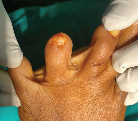
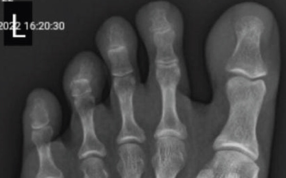
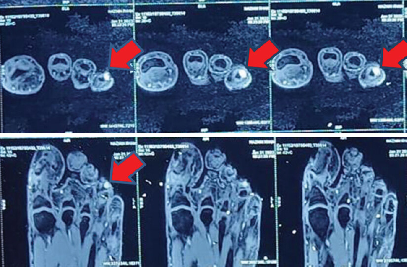
A 34-year-old lady presented to our institution with complaints of pain overlying the apex of her left 4th toe since approximately 2 years, for which she had received treatment from multiple physicians with intermittent symptomatic relief; after which she presented to us, she did not give any history of trauma. She complained of persistent pain, exaggeration while wearing footwear and during the winter season. On clinical examination (Fig. 1), there was no obvious swelling. Severe tenderness was present over the nail bed of 4th toe, and sensations were decreased over 4th toe compared to the other toes. Pinpoint pressure using the tip of a pen elicited painful symptoms that were not present when applied to the adjacent nail beds of the toe; consistent with a positive Love test. As a preliminary diagnostic test, we requested for plain radiograph (Fig. 2) of the involved foot, which was essentially normal, showing no bony involvement of the 4th toe distal phalanx. Magnetic resonance imaging (MRI) was done, which showed approximately 6.5 × 5.8 × 5.4 mm moderately sized and homogenously enhancing lesion on the dorsal aspect of the distal part of 4th toe, in the subungual region with smooth scalloping of the adjacent bony cortex and no evidence of cortical erosion or marrow edema which was suggestive of Glomus tumor/Glomangioma (Fig. 3).
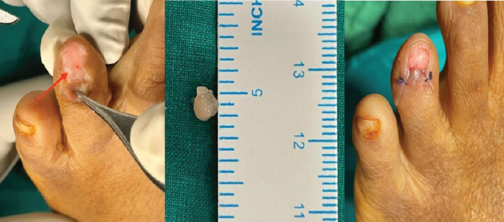
After investigating and proper counseling, the patient was taken up for excisional biopsy of the tumor. The lesion was approached via square flap technique, toe nail was removed, under which showed white colored mass (Fig. 4). The tumor was excised in toto and sent for histopathology examination.
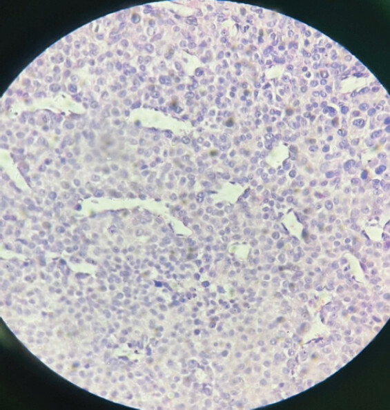
Histopathology examination showed tissue lined by stratified squamous epithelium. Subepithelium showed a tumor arranged in solid sheets surrounding thin-walled blood vessels. Individual cells were round, uniform, with round, monotonous vesicular nuclei and a moderate amount of eosinophilic cytoplasm. Many nuclei showed prominent nucleoli. Myxoid changes were seen in large areas. There was no evidence of atypia or malignancy. These findings were reported as consistent with Glomus tumor (Fig. 5).
Fourteen days postoperatively, suture removal was done, and the patient did not complain of any pain, 4 months postoperatively the patient returned; there was no recurrence of pain or mass, and she was able to wear closed shoe and was completely asymptomatic.
Discussion
Glomus tumor is a rare mesenchymal neoplasm composed of cells that resemble the modified perivascular smooth muscle cells (glomus cells) [1,3]. Glomus bodies are arteriovenous shunts, surrounded by glomus cells, present in the Sucquet-Hoyer canal, which contract via sympathetic activity, thereby regulating blood flow to the extremities. Hence, they play a role in thermoregulation, decreasing blood flow when the body is exposed to cold and increasing it when it is hot. Glomus bodies are located in the dermis throughout the body but more densely concentrated in apical skin areas, primarily the fingertips, followed by the base of the foot and the rest of the body [3]. Although these tumors arise from glomus cells, these tumors can also arise in the extracutaneous locations not known to have glomus cells [4]. The initiating, or causative, event associated with glomus cell proliferation and eventual Glomus tumor formation is unknown. Once established, Glomus tumors are typically composed of 3 components: Glomus cells, vessels, and smooth muscle cells [5]. In its commonest location – namely, the subungual tissue of the fingers – the Glomus tumor generally presents with a characteristic triad of intense paroxysmal pain, pinpoint tenderness, and hypersensitivity to cold. Pain is the most common symptom. Presentation of a painful lesion in the toe turning out to be a Glomus tumor is an uncommon event. A clinical differential diagnosis for painful toe soft-tissue lesion includes hidradenoma, ganglion cyst, neuroma, melanoma, blue nevi, neurilemmoma, and foreign body [6].
Toes being an unusual location for a Glomus tumor, it is likely to be missed for a while. On physical examination, the most common symptom is pain due to a change in temperature, cold being the trigger. It is common to see the classic clinical triad of intense paroxysmal pain, positive love test, and hypersensitivity to cold [6]. The pain precedes any other symptom, even before any structural change in the nail, which is why initial diagnosis is difficult. The exact mechanism of the pain is not known; however, it involves the P substance released by the tumor, and this could be connected with the paroxysmal pain and pain when exposed to cold [7]. In the initial stage of the tumor, it is difficult to diagnose due to its small size, and the diagnosis is aided with the use of MRI [8]. Around 1% of all the Glomus tumors are considered to be malignant, which, on histopathological examination, shows a size greater than 2cm, deep location, and mild-to-high degree of nuclear mitosis, and presence of atypical mitotic figures; in such cases, the risk of metastasis is more than 25% [9]. The treatment of a Glomus tumor should be surgical, and multiple ways of accessing a tumor have been described [10,11].
Conclusion
The purpose of this case report is to make the physicians aware of the occurrence of a Glomus tumor in the toes. Excision of the tumor is completely curative and relieves the patient of all the symptoms.
Clinical Message
Being a rare presentation in the foot, the Glomus tumor often goes undiagnosed or is misdiagnosed. A high level of suspicion is required to correctly diagnose Glomus tumor in the foot.
Conflict of Interest:
Source of Support:
Nil
Consent:
The authors confirm that informed consent was obtained from the patient for publication of this article
References
- Lui TH, Mak SM.Glomus tumor of the great toe. J Foot Ankle Surg 2014;53:360-3. [Google Scholar] | [PubMed]
- Workman M, Saragas NP, Ferrao P.Glomus tumors in the foot. J Foot Ankle 2020;14:183-6. [Google Scholar] | [PubMed]
- Mohindra M, Sambandam B, Gautam VK, Maini L.A rare case of glomus tumor of the great toe: An analysis of behavior at this rare site. Foot Ankle Spec 2016;9:83-7. [Google Scholar] | [PubMed]
- Gombos Z, Fogt F, Zhang PJ.Intraosseous glomus tumor of the great toe: A case report with review of the literature. J Foot Ankle Surg 2008;47:299-301. [Google Scholar] | [PubMed]
- Polo C, Borda D, Poggio D, Asunción J, Peidro L.Glomus tumor of the hallux. Review of the literature and report of two cases. Foot Ankle Surg 2012;18:89-93. [Google Scholar] | [PubMed]
- Sprinkle RL 3rd, Sangueza OP, Schwartz GA.Glomus tumor of the toe. J Am Podiatr Med Assoc 2017;107:257-60. [Google Scholar] | [PubMed]
- Assmus H, Dombert T.Glomus tumours of the extremities: Localisation and operative treatment in 36 cases. Handchir Mikrochir Plast Chir 2002;34:103-7. [Google Scholar] | [PubMed]
- Verma A, Parmar R, Pawar I, Agarwal S.Diagnostic dilemma of subungual glomus tumor of great toe: A Case report and review of the literature. J Foot Ankle Surg (Asia Pacific) 2021;9:35-9. [Google Scholar] | [PubMed]
- Abou Jaoude JF, Farah AR, Sargi Z, Khairallah S, Fakih C.Glomus tumors: Report on eleven cases and a review of the literature. Chir Main 2000;19:243-52. [Google Scholar] | [PubMed]
- Iglesias de la Torre L.Consideraciones clinicas, anatómicas, radiológicas y quirúrgicas del glomus tumoral de masson. Cir Ortoped Traumatol (Habana) 1939;7:11-7.[TPS1.1]. [Google Scholar] | [PubMed]
- Takata H, Ikuta Y, Ishida O, Kimori K.Treatment of subungual glomus tumour. Hand Surg 2001;6:25-7. [Google Scholar] | [PubMed]
© 2026 Journal of Orthopaedic Case Reports - Published by Indian Orthopaedic Research Group
About the Authors
-

Dr. Pratik Mankar
-

Dr. Madhav Khadilkar
-

Dr. Amit Mahajan
-

Dr. A Kannan
Related Articles in Journal of Orthopaedic Case Reports
 October 1, 2026 Early Recognition of Glomus Tumor of Hand Using Clinical Tests: Experience from an Indian Tertiary Center
 September 1, 2026 The Tiny Tumor with Disproportionate Pain: Subungual Glomus Tumor of the Thumb Managed with Nail-Sparing Excision: A Case Report
 August 1, 2026 Intramuscular Glomus Tumor of Foot Presenting as Neuroma of Sural Nerve: A Case Report
 July 1, 2026 Extradigital Glomus Tumor of the Knee Mimicking Osteoarthritis: A Case Report