

Myxofibrosarcoma may present as a pathological fracture with MRI features that mimic a benign vascular lesion, so mandatory pre-operative tissue biopsy and prompt referral to a specialist sarcoma center are essential to prevent diagnostic delay, unplanned excision, and tumor progression.
Dr. Amit Bhardwaj, Department of Orthopaedic Surgery, Sengkang General Hospital, Sengkang, Singapore. E-mail: dramitortho@gmail.com
Abstract
Introduction: Myxofibrosarcoma (MFS) is among the most common soft-tissue sarcomas of the extremities in elderly patients; however, presentation as a pathological fracture is exceptionally rare. The tumor’s fluid-rich myxoid matrix produces magnetic resonance imaging signal characteristics that closely mimic benign vascular lesions, creating a significant diagnostic challenge when encountered outside a tertiary setting. We report a case of low-grade myxofibrosarcoma presenting as a pathological pilon fracture.
Case Report: A 60-year-old male presented with a 2-week history of ankle pain following an inversion injury that was refractory to conservative management, accompanied by a 6 kg weight loss over 4 months. Examination revealed cachexia, diffuse lower-limb swelling, and skin induration. Plain radiographs demonstrated a sagittally angulated pathological pilon fracture with diffuse moth-eaten osteolysis of the tibia and fibula. Magnetic resonance imaging revealed a large 35 × 6.5 × 6.1 cm inter- and intramuscular lesion initially reported as cavernous lymphangioma. Open core needle biopsy established the diagnosis of low-grade myxofibrosarcoma. The patient declined the recommended high transfemoral amputation; 3 weeks later, cutaneous fungation developed. Final histopathology of the amputation specimen confirmed progression to high-grade myxofibrosarcoma (350 mm) with multifocal bone invasion and clear resection margins. At 48 months postoperatively, the patient remains disease-free.
Conclusion: MFS may present as a pathological fracture with imaging features indistinguishable from benign pathology. Mandatory pre-operative tissue biopsy and prompt referral to a specialist sarcoma center are essential to prevent diagnostic delay, unplanned excision, and the associated risk of tumor progression.
Keywords: Myxofibrosarcoma, pathological pilon fracture, soft-tissue sarcoma, cavernous lymphangioma, limb amputation, bone invasion, magnetic resonance imaging misdiagnosis.

Myxofibrosarcoma (MFS) is a malignant fibroblastic neoplasm characterized by a variably myxoid stroma, nuclear pleomorphism, and a distinctive curvilinear vascular pattern. The term was introduced by Angervall, Kindblom, and Merck to reflect the tumor’s dual myxoid and fibroblastic differentiation. Recognized by the World Health Organization (WHO) as one of the most common soft-tissue sarcomas of the extremities in elderly patients, MFS has a mean age at diagnosis of approximately 67 years.
Its clinical behavior is characterized by a local recurrence rate of 14–30% and a relatively low metastatic rate of approximately 6%, with the lung and bone being the most common sites of distant spread. A key biological feature is the tendency of low-grade lesions to recur at progressively higher histological grades, thereby acquiring metastatic potential through stepwise dedifferentiation. This natural history underscores the importance of early, definitive surgical management at a specialist sarcoma center.
Presentation as a pathological fracture is exceptionally rare and diagnostically challenging. The tumor’s myxoid, fluid-rich matrix produces magnetic resonance imaging (MRI) signal characteristics that can closely mimic benign lesions, including cavernous lymphangioma, myxoid liposarcoma, and low-grade fibromyxoid sarcoma. Unplanned excision – performed without prior histological diagnosis by a surgeon without specialist sarcoma expertise – is associated with a threefold increase in 5-year cumulative local recurrence (45% vs. 15%). We present this case to highlight this rare presentation and the diagnostic and surgical pitfalls it entails.
A 60-year-old male presented with a 2-week history of ankle pain following an inversion injury that was refractory to conservative treatment. Systemic enquiry revealed multiple longstanding swellings of the left leg and an unintentional 6 kg weight loss over 4 months. Examination demonstrated cachexia, diffuse firm swelling of the entire left lower limb, and overlying skin induration (Fig. 1).
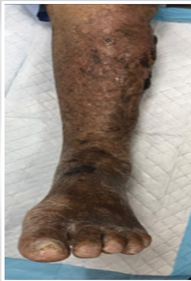
Figure 1: Clinical photograph of the left lower limb at presentation. Diffuse swelling, skin induration, and hyperpigmentation extend from the mid-leg to the foot. Multiple subcutaneous nodular deposits are evident over the anterolateral leg.
Plain radiographs demonstrated a sagittally angulated pathological pilon fracture of the distal tibia and fibula with diffuse moth-eaten osteolysis, cortical scalloping, and cortical thinning along both shafts, findings inconsistent with simple trauma (Fig. 2).
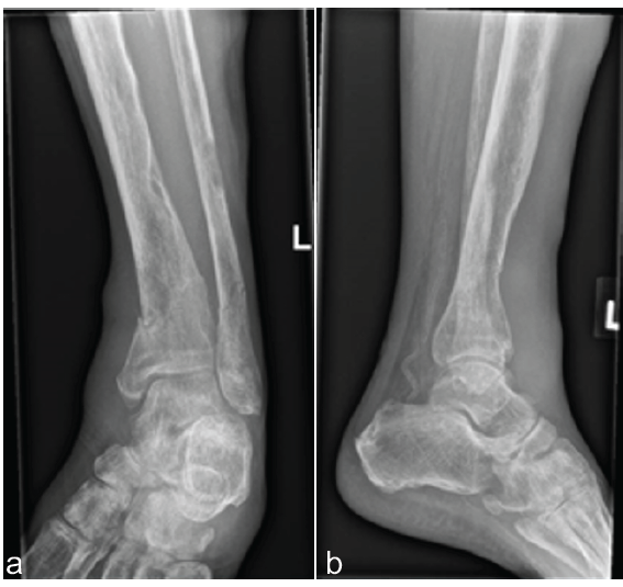
Figure 2: Plain radiographs of the left ankle. (a) Anteroposterior and (b) lateral views demonstrating a sagittally angulated pathological pilon fracture of the distal tibia with an associated non-displaced fibular fracture, together with diffuse moth-eaten osteolysis and cortical scalloping of the distal tibia and fibula, indicating chronic osseous invasion by the underlying neoplasm.
MRI revealed a lobulated, septated T2-hyperintense lesion measuring 35.0 × 6.5 × 6.1 cm occupying the anterior and peroneal compartments from the infrapatellar region to the ankle, with near-complete replacement of muscle architecture and transgression of the intermuscular septum (Fig. 3).
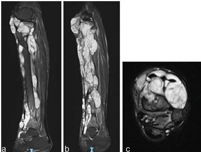
Figure 3: Magnetic resonance imaging of the left lower limb. (a and b) Coronal T2-weighted sequences demonstrate an extensive lobulated hyperintense lesion with fine internal septations occupying the anterior and peroneal compartments from the infrapatellar region to the ankle. (c) Axial T2-weighted image at the mid-leg demonstrating compartment filling, transgression of the intermuscular septum, and near-complete replacement of normal muscle architecture.
The initial radiological impression was a benign cavernous lymphangioma; however, the clinical features and the degree of osseous erosion raised concern for malignancy. Staging computed tomography of the thorax, abdomen, and pelvis demonstrated no lymphadenopathy or distant metastases. Whole-body bone scintigraphy showed uptake confined to the left lower limb (Fig. 4), and MRI of the thigh demonstrated no skip lesions.
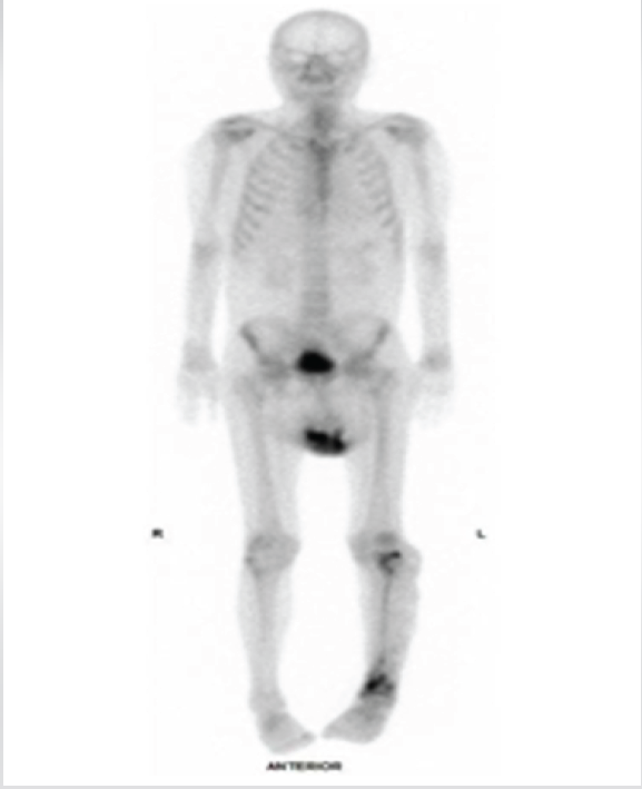
Figure 4: Whole-body bone scintigraphy (anterior projection). Focally increased tracer uptake is confined to the left lower extremity. No skip lesions, contralateral involvement, or distant osseous metastases are identified.
An open core needle biopsy was performed with an anterolateral approach. Histopathology revealed a myxoid neoplasm composed of atypical pleomorphic spindle cells, thin-walled curvilinear vessels with adherent tumor cells, pseudolipoblasts, and a Ki-67 index <5%, consistent with low-grade myxofibrosarcoma (Fig. 5).
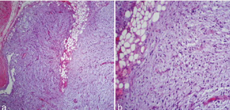
Figure 5: Core needle biopsy, hematoxylin and eosin stain. (a) Low-power view (×10) demonstrating a myxoid neoplasm with pleomorphic spindle cells, incomplete fibrous septa, and scattered thin-walled curvilinear vessels – the hallmark vascular architecture of myxofibrosarcoma. (b) High-power view (×40) showing atypical hyperchromatic mononuclear cells, pseudolipoblasts with cytoplasmic myxoid vacuoles, and abundant myxoid stroma. Mitotic figures are rare, consistent with low-grade designation.
Following multidisciplinary discussion, high transfemoral amputation was recommended; however, the patient initially declined. Over the subsequent 3 weeks, progressive cutaneous ulceration with extrusion of gelatinous myxoid material developed, suggesting rapid local progression (Fig. 6). The patient subsequently consented to surgery, and high-transfemoral amputation was performed without complication.
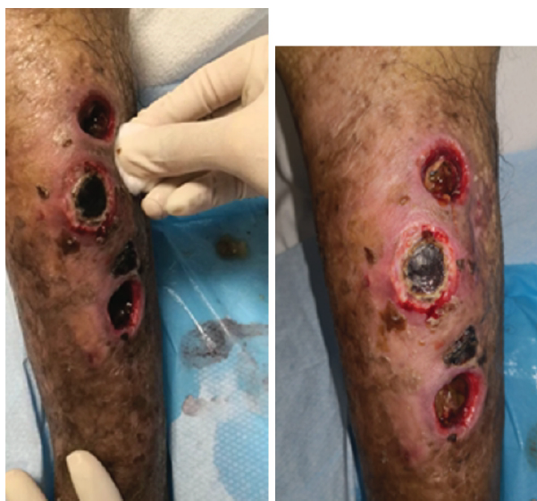
Figure 6: Clinical photograph of the left lower leg following the patient’s initial refusal of amputation. Multiple ulcerated nodules with discharge of gelatinous myxoid material are visible over the anterolateral skin, consistent with cutaneous tumor fungation and indicative of accelerating local grade progression.
Histopathological examination of the above-knee amputation specimen confirmed a high-grade myxofibrosarcoma measuring 350 mm within the anterior extensor compartment, with multifocal cortical invasion of the proximal tibia, anterior tibial shaft, and proximal fibula, extending to the subarticular region and associated with a non-displaced distal tibial fracture. All resection margins were clear (bony margin 16 cm; soft-tissue and skin margin 10.5 cm), and no vascular tumor emboli were identified. At 48 months postoperatively, the patient remains disease-free with no evidence of local recurrence or distant metastasis (Table 1).
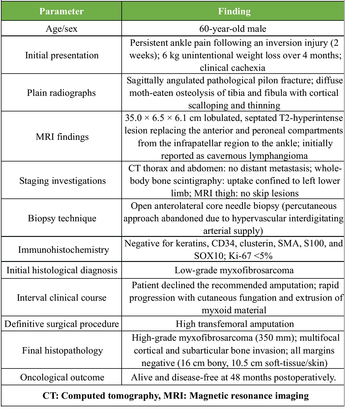
Table 1: Case summary
The most diagnostically consequential feature of this case was the misidentification of a high-grade sarcoma as a benign cavernous lymphangioma on MRI. The 35 cm lobulated, septated, T2-hyperintense mass replacing the anterior and peroneal compartments demonstrated homogeneous myxoid signal without overt solid nodularity, favoring a vascular malformation. However, the coexistence of diffuse tibial and fibular cortical scalloping, moth-eaten osteolysis, and a pathological pilon fracture represented radiological red flags inconsistent with a benign lesion. The T2-hyperintense morphology of MFS, attributable to its fluid-rich myxoid extracellular matrix, closely replicates the signal characteristics of lymphangioma, myxoid liposarcoma, cellular myxoma, and low-grade fibromyxoid sarcoma on conventional MRI sequences [1]. Kaya et al. demonstrated that infiltrative tail-like projections along fascial planes represent the most reliable MRI indicator of malignancy in myxoid lesions. However, this feature may be absent in early or low-grade tumors [1]. No single MRI sequence reliably distinguishes low-grade MFS from benign myxoid entities; histological sampling therefore remains the definitive diagnostic modality [2]. In the present case, imaging alone was insufficient to resolve the differential diagnosis, and the initial radiological impression created a genuine risk of inadvertent excision.
Pathological fracture as the index presentation of soft-tissue sarcoma is uncommon, with published reports largely limited to isolated case series [3]. In MFS, fracture typically results from progressive cortical erosion caused by periosteal infiltration and pressure-mediated bone destruction from the extraosseous mass, rather than the hematogenous intramedullary spread characteristic of primary bone sarcomas [3]. Final histopathology in our patient confirmed multifocal cortical invasion of both tibia and fibula with scalloped borders and subperiosteal infiltration, supporting this mechanism. The distal tibial plafond is biomechanically vulnerable, and even focal cortical thinning contiguous with a soft-tissue mass may precipitate articular fracture under axial loading [3]. Any lytic bone lesion in an elderly patient contiguous with a myxoid soft-tissue mass should therefore prompt urgent oncological evaluation and must not be attributed to trauma without investigation.
Core biopsy demonstrated low-grade MFS with a Ki-67 proliferative index <5%. However, the definitive resection specimen revealed high-grade disease with marked pleomorphism and extensive cortical invasion, a discordance consistent with intratumoral heterogeneity and the recognized natural history of MFS. Mentzel et al., in a landmark clinicopathological analysis of 75 cases, described the propensity of low-grade MFS to accumulate progressive pleomorphism, mitotic activity, and necrosis with each recurrence, thereby acquiring metastatic potential through stepwise dedifferentiation [4]. Sambri et al. subsequently identified delay to definitive surgery as an independent predictor of grade progression, with intervals exceeding 8 weeks associated with significantly higher rates of upward grade shift [5]. Haglund et al. further reported a 5-year disease-specific survival of 60% for high-grade MFS compared with 90% for low-grade disease, underscoring the clinical consequences of tumor progression [6]. In this case, the 3-month interval during which the patient declined amputation was accompanied by cutaneous ulceration and extrusion of gelatinous myxoid material, reflecting rapid local progression, and the final specimen confirmed high-grade transformation. These findings emphasize the importance of timely definitive management once MFS is diagnosed, particularly in large, compartment-spanning tumors with osseous involvement.
Contemporary series report amputation rates for MFS of approximately 4%, with wide local excision constituting the standard treatment in most cases [7,8]. In this patient, limb salvage was oncologically untenable because of tumor extension from the infrapatellar region to the distal tibia (35 cm), complete compartmental replacement, multifocal cortical destruction with pathological fracture, proximity (6 mm) to the popliteal vessels, and severe muscle atrophy precluding functional reconstruction. Negative resection margins remain the most important determinant of local control in MFS [7,9]. Gronchi et al. demonstrated that margin-positive resection independently predicted local recurrence, with a hazard ratio of 3.8 on multivariate analysis [9]. High transfemoral amputation achieved clear margins of 16 cm (bony) and 10.5 cm (soft-tissue and skin), consistent with ESMO sarcoma guideline principles [10]. At 48 months, the patient remains free of local recurrence and distant metastasis.
The initial radiological misdiagnosis created conditions highly favorable for limb salvage. Fiore et al., in a series of 172 patients with MFS, reported that the 5-year cumulative local recurrence rate increased from 15% in primarily treated cases to 45% following unplanned excision, with a hazard ratio of 5.0 on multivariate analysis [11]. Patients at greatest risk are those with superficial, smaller, and apparently lower-grade tumors – precisely the subgroup in which MFS most convincingly mimics benign pathology [11]. This case reinforces the principle that biopsy prior to excision of any indeterminate soft-tissue mass is mandatory [12], irrespective of reassuring imaging features, and that such lesions should be referred to a specialist sarcoma center before operative intervention.
We report a 35 cm myxofibrosarcoma of the left lower limb presenting as a pathological distal tibial pilon fracture and initially misinterpreted on MRI as a benign cavernous lymphangioma. This case highlights the inability of conventional MRI to reliably distinguish benign from malignant myxoid lesions, the potential for pathological fracture due to direct cortical invasion by extraosseous sarcoma, the risk of biopsy-resection grade discordance with rapid progression during surgical delay, and the importance of margin-negative resection as the cornerstone of durable local control. High transfemoral amputation achieved oncologically adequate margins, and the patient remains disease-free at 48 months. Early biopsy of indeterminate myxoid masses and timely definitive surgery are essential to minimize progression and optimize oncological outcomes.
MFS is a rare but aggressive soft-tissue sarcoma that may mimic benign vascular lesions on MRI and occasionally present as a pathological fracture. A high index of suspicion, mandatory pre-operative tissue biopsy, and prompt referral to a specialist sarcoma center are essential to prevent diagnostic delay, unplanned excision, and tumor progression. Timely definitive surgical management with clear resection margins remains the cornerstone of durable local control and favorable oncological outcomes.
References
- 1. Kaya M, Wada T, Nagoya S, Sasaki M, Matsumura T, Yamaguchi T, et al. MRI and histological evaluation of the infiltrative growth pattern of myxofibrosarcoma. Skeletal Radiol 2008;37:1085-90. [Google Scholar] [PubMed]
- 2. Murphey MD, McRae GA, Fanburg-Smith JC, Temple HT, Levine AM, Aboulafia AJ. Imaging of soft-tissue myxoma with emphasis on CT and MR and comparison of radiologic and pathologic findings. Radiology 2002;225:215-24. [Google Scholar] [PubMed]
- 3. Manoso MW, Frassica DA, Deune EG, Frassica FJ. Outcomes of surgical resection of soft-tissue sarcomas presenting with pathologic fractures. J Surg Oncol 2005;91:9-14. [Google Scholar] [PubMed]
- 4. Mentzel T, Calonje E, Wadden C, Camplejohn RS, Beham A, Smith MA, et al. Myxofibrosarcoma. Clinicopathologic analysis of 75 cases with emphasis on the low-grade variant. Am J Surg Pathol 1996;20:391-405. [Google Scholar] [PubMed]
- 5. Sambri A, Bianchi G, Righi A, Dei Tos AP, Picci P, Donati DM. Surgical margins and local recurrence in myxofibrosarcoma. Eur J Surg Oncol 2019;45:1076-82. [Google Scholar] [PubMed]
- 6. Haglund KE, Raut CP, Nascimento AF, Wang Q, George S, Baldini EH. Recurrence patterns and survival for patients with intermediate- and high-grade myxofibrosarcoma. Int J Radiat Oncol Biol Phys 2012;82:361-7. [Google Scholar] [PubMed]
- 7. Look Hong NJ, Hornicek FJ, Raskin KA, Yoon SS, Szymonifka J, Yeap B, et al. Prognostic factors and outcomes of patients with myxofibrosarcoma. Ann Surg Oncol 2013;20:80-6. [Google Scholar] [PubMed]
- 8. Sanfilippo R, Miceli R, Grosso F, Fiore M, Puma E, Pennacchioli E, et al. Myxofibrosarcoma: Prognostic factors and survival in a series of patients treated at a single institution. Ann Surg Oncol 2011;18:720-5. [Google Scholar] [PubMed]
- 9. Gronchi A, Lo Vullo S, Colombo C, Collini P, Stacchiotti S, Mariani L, et al. Extremity soft tissue sarcoma in a series of patients treated at a single institution: Local control directly impacts survival. Ann Surg 2010;251:506-11. [Google Scholar] [PubMed]
- 10. Casali PG, Abecassis N, Bauer S, Biagini R, Bielack S, Bonvalot S, et al. Soft tissue and visceral sarcomas: ESMO-EURACAN-clinical practice guidelines for diagnosis, treatment and follow-up. Ann Oncol 2018;29 Suppl 4:iv51-67. [Google Scholar] [PubMed]
- 11. Fiore M, Casali PG, Miceli R, Mariani L, Bertulli R, Lozza L, et al. Prognostic effect of re-excision in adult soft tissue sarcoma of the extremity. Ann Surg Oncol 2006;13:110-7. [Google Scholar] [PubMed]
- 12. Mankin HJ, Mankin CJ, Simon MA. The hazards of the biopsy, revisited. Members of the musculoskeletal tumor society. J Bone Joint Surg Am 1996;78:656-63. [Google Scholar] [PubMed]


Related Articles in Journal of Orthopaedic Case Reports
 August 1, 2026 Not All Red Flags Indicate Malignancy: A Case Report of a Benign Lipoma Mimicking Soft-tissue Sarcoma
 July 1, 2026 Radiation-Associated Femoral Nonunion and Fixation Failure Following Limb-Salvage Surgery for Thigh Soft-tissue Sarcoma: Two Case Reports and a Review of the Literature
 December 10, 2023 Successful Outcome Following Limb Conservation Surgery for a Recurrent Malignant Peripheral Nerve Sheath Tumour: A Case Report
 July 7, 2017 Articular Reconstruction using Subchondral Cementation and Threaded Kirschner-wires in Giant Cell Tumor: A Novel Technique