

Ulnar neuropathy in individuals with Split-Hand/Split-Foot Malformation may have more acute deficits due to cleft reliance on the ulnar nerve for cleft-based prehension.
Mr. Seth Stidham, Saint Louis University School of Medicine, St. Louis, Missouri. E-mail: seth.stidham@health.slu.edu
Abstract
Introduction: Split-hand/split-foot malformation (SHFM) is a congenital limb defect characterized by central ray deficiency, resulting in deep median clefts of the hands and/or feet. Due to the nature of the malformation, the outer digits – those predominantly innervated by the ulnar nerve – are typically spared. In patients who rely on central cleft adduction and abduction as a substitute for thumb-index opposition, the ulnar nerve may assume a disproportionately critical role in hand function.
Case Report: We report a case of bilateral cubital tunnel syndrome (CubTS) in a 23-year-old patient with familial SHFM. We review the variable phenotypes of SHFM, the Manske and Halikis classification system, the clinical presentation and management of CubTS, and the functional anatomy of cleft closure.
Conclusion: We argue that the prioritization of neuropathy treatment in patients with congenital hand malformations should be advanced, given their overreliance on different nerve–distributed musculature for essential hand function.
Keywords: Split-hand/split-foot malformation, cubital tunnel syndrome, ulnar neuropathy, cleft-based prehension.

Split-hand/split-foot malformation (SHFM) is a congenital limb defect that results in deep median clefts of the hands and/or feet, and effects approximately 1 in 18,000 newborns [1]. Phenotypic variation is highly prevalent, with milder forms of aplasia solely affecting distal central phalanges, to more severe malformations that result in clefts of the metacarpal space and carpals [2]. SHFM may be non-syndromic (isolated) or may appear as part of a broader syndrome.
Surgical correction varies with severity. The Manske and Halikis classification, based on first web space preservation, is conventionally used to evaluate hand function. In Types I, II, and III, where an index ray is present, surgical goals include creation of a stable oppositional thumb, widening of the first web space using available index and thumb structures, and cleft closure [3]. However, some authors note that the cleft itself serves as a critical pinch mechanism – a “crab-like” motion relying on intrinsic hand muscles for abduction and adduction of the bordering rays, making the second web space functionally important [4]. Regardless of cleft type or age, patients with cleft hands demonstrate impaired function and lower outcome scores compared with age-matched controls [5].
Cubital tunnel syndrome (CubTS) is one of the most common compression neuropathies, entrapping the ulnar nerve at the elbow within the medial elbow, affecting 2–6% of the population [6]. Clinical presentation of CubTS includes numbness and tingling in the ulnar fourth and fifth digits and corresponding parts of the dorsal and palmar hand. These symptoms can be worsened by activities in elbow flexion, including phone use, desk work, and sleeping [7]. Difficulty or weakness with thumb and finger abduction, pinching, grip strength, and fine motor control, such as writing with a pencil or playing an instrument, are motor deficits that may be appreciated in moderate CubTS [8]. If CubTS is left untreated, 50% of patients do not report improvement, and approximately one in five cases progressively worsens [9].
There is extremely limited literature discussing the management of compression mononeuropathies in individuals with congenital hand deformities and malformations. Specifically, there is no literature traversing how to differently manage acquired mono-neuropathies in individuals with cleft hands.
A 23-year-old man with SHFM, having undergone corrective surgeries at ages 1, 7, and 15, for the creation of a merged cleft on his right hand, was referred to the orthopedic surgery department. He had a 4-year history of bilateral hand weakness, paresthesia, and numbness in an ulnar nerve distribution, decreased fine manipulation, noticed in writing and typing due to fasciculations, as well as decreased sensation when using fine manipulations of tools, resulting in occasional deep tissue pressure injuries in the 4th and 5th digits. Throughout much of this time, he had not experienced any grossly decreased cleft-based prehension strength; however, he noted emerging prehension changes prompting re-evaluation. A prior nerve conduction study and evaluation by an orthopedic hand specialist at the time of symptom onset were grossly normal, and the patient was recommended to continue conservative management, as per guideline standards for someone with normal morphology, consisting of bracing and nerve glide exercises. This recommendation was not made by his previous hand surgeon, a congenital malformation specialist.
On physical examination, the hands demonstrated clear aplasia of the central rays consistent with ectrodactyly (Fig. 1).
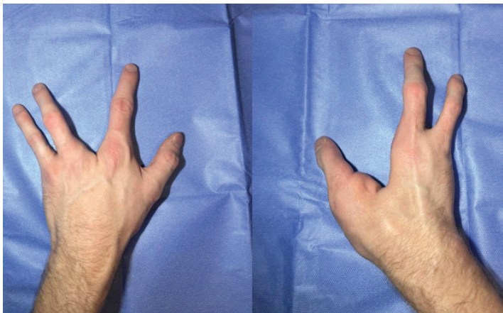
Figure 1: Extensor view of hands, demonstrating Type I/unmerged cleft (Left) and Type IV/merged cleft (Right).
There was noted thenar and first web space atrophy on the left; merged first web space on the right complicated evaluation. Special testing was notable for positive Tinel’s sign at the wrist and elbow, Phalen test, Wartenburg sign, and Froment sign. There was also mild bilateral ulnar clawing, more pronounced on the left than the right. electromyography /nerve conduction studies demonstrated bilateral demyelinating ulnar mononeuropathies, greater on the left, confirming bilateral CubTS, and review of previous hand X-rays demonstrated depth of cleft (Fig. 2).
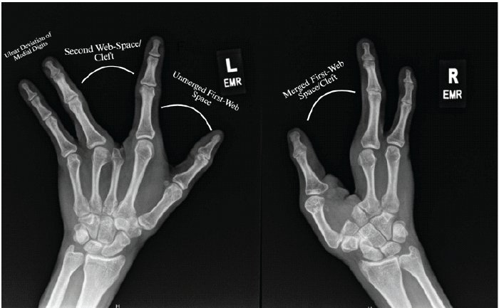
Figure 2: Bilateral merged hand X-rays demonstrating different cleft depth and difference in unmerged and merged phenotype.
Left cubital tunnel release
Surgery was scheduled and performed 4 months after the pre-operative evaluation. A left ulnar nerve in situ decompression was performed under general anesthesia with a sterile arm tourniquet. Through a 5 cm longitudinal incision between the medial epicondyle and anteromedial olecranon, the ulnar nerve was identified. The fascial roof overlying the nerve was released 8 cm proximal to the elbow, and the Osborne ligament and flexor carpi ulnaris fascia were released 8 cm distally, constituting the site of compression. The preservation of traversing cutaneous nerves was ensured throughout. The ulnar nerve remained stable with elbow flexion and extension; transposition was not required. The wound was irrigated and closed in layers. The patient tolerated the procedure without complications.
At 8 months post-operative, the patient reports less frequent symptoms, including paresthesia and fasciculations; however, notes that these symptoms still occur. Ulnar clawing, thenar atrophy, and webspace atrophy are approximately the same as before the operation.
Functional anatomy in SHFM
This case illustrates the variable phenotypes of SHFM and highlights the critical importance of the ulnar nerve in patients who rely on cleft-based hand function. The Manske and Halikis classification system stratifies cleft hands based on the integrity of the first web space. In Types I-II, the presence of an index ray allows for the persistence of a merged or unmerged cleft-based pinch. In our patient, whose left hand was a Type I, the mild cleft results in the formation of a functional cleft, with a preserved 1st web space, thus allowing for the use of the cleft as an alternative and preferred lateral prehension mechanism. This is furthered by the structural deviation of the retained 5th and 4th digits, which have ulnar deviation, allowing for large-degrees of cleft abduction and adduction (Fig. 3).
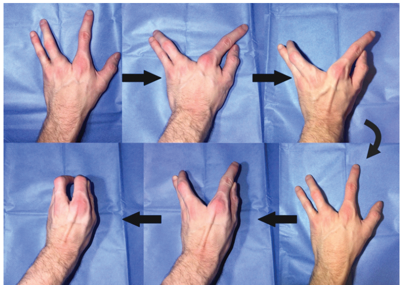
Figure 3: Abduction of the left unmerged central cleft into adduction functioning as lateral (key) prehension.
In patients who use the central cleft as a substitute for the pinching mechanism normally performed by thumb opposition, the ulnar nerve assumes a disproportionately critical role. The “crab-like” cleft closure motion – bringing the bordering rays together across the cleft – depends primarily on the palmar interossei for adduction and the dorsal interossei for abduction. These structures are normally innervated by the deep branch of the ulnar nerve (Tables 1 and 2) [10].
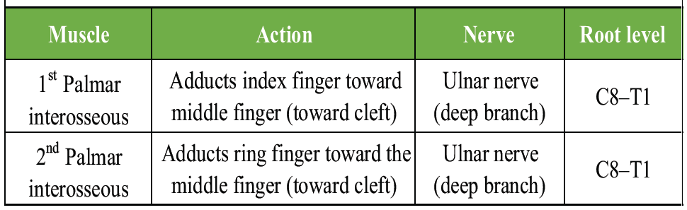
Table 1: Muscles involved in cleft closure: Adduction toward the cleft
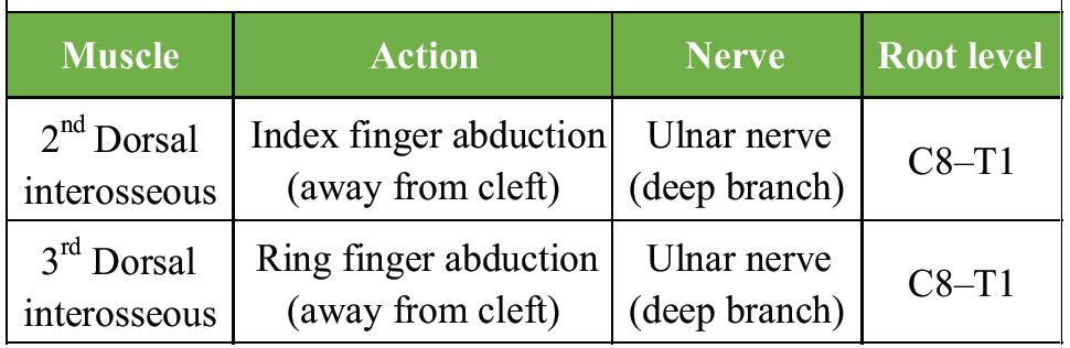
Table 2: Muscles involved in cleft opening: Abduction away from the cleft
In total, the ulnar nerve innervates all seven interossei, the medial two lumbricals, the three hypothenar muscles, the adductor pollicis, and partially the flexor pollicis brevis – constituting the majority of intrinsic hand musculature [10]. In a normal hand, the functional consequences of ulnar neuropathy are distributed across a full complement of digits and can be partially compensated by median nerve–innervated muscles.
Functional consequences of ulnar neuropathy in SHFM
Studies of ulnar nerve injury in anatomically normal hands demonstrate that grasping strength decreases by approximately 59–62%, with normal index-thumb-based key pinch by 51–61%, and pinch-to-zoom strength by 75–76% compared with the unaffected hand [11]. Grip strength receives greater contributions from the ulnar nerve than the median nerve, and ulnar nerve palsy results in reductions across all pinch and grip strength measures. The primary intrinsic muscles responsible for lateral and tip pinch – the adductor pollicis and first dorsal interosseous – are both ulnar-innervated [12].
In the context of SHFM, these deficits are amplified. Loss of interosseous function due to CubTS would effectively eliminate the cleft-based pinch mechanism, rendering the hand functionally inert for fine manipulation. This stands in contrast to the anatomically normal hand, where ulnar neuropathy impairs but does not eliminate prehension, as thumb opposition (primarily median-innervated) remains intact.
Furthermore, peripheral nerve anatomy in cleft hand syndrome has been shown to reflect the presence and absence of corresponding muscle and skin innervation areas. In a case of severe bilateral clefts, high-resolution ultrasound demonstrated severe bilateral reduction in median nerve size, with relative sparing of the ulnar nerve, consistent with the preservation of ulnar-innervated structures [13]. This finding underscores that in SHFM, the ulnar nerve may be the dominant – and in some cases, the sole – functional motor nerve to the remaining hand musculature.
Implications for neuropathy management
The disproportionate reliance on ulnar nerve–innervated musculature in SHFM has direct implications for the prioritization of neuropathy treatment. In the general population, CubTS is managed along a stepwise algorithm: conservative measures for mild disease, with surgical intervention reserved for severe or refractory cases [14]. In patients with SHFM—or others reliant on cleft-based pinch, even mild ulnar neuropathy may produce functionally significant deficits. Conservative management should be initiated promptly and monitored closely, and surgical evaluation should be considered earlier in the disease course than would be typical for the general population.
This case highlights the intersection of congenital hand malformation and acquired compression neuropathy. This is the only reported case discussing the management of CuTS in SHFM. In patients with SHFM who do not have a merged cleft-and-first webspace (Types I-II), the cleft may still act and may even be used more favorably than the first webspace. In these individuals, the ulnar nerve is the primary motor supply to the muscles that enable cleft closure-the dorsal and palmar interossei. CubTS in this population threatens to eliminate the primary grasping mechanism of the hand. Clinicians should recognize that the functional impact of ulnar neuropathy may be amplified in patients with congenital hand anomalies and should prioritize early diagnosis and treatment accordingly. Further study is warranted to characterize the prevalence of compression neuropathies in patients with congenital limb differences and to develop tailored management algorithms for this population.
Those with Split-hand/split-foot malformation with compression mononeuropathies should be monitored closely, and treatment expedited due to their functional anatomy’s potential over-reliance on certain nerves
References
- 1. Sowińska-Seidler A, Socha M, Jamsheer A. Split-hand/foot malformation – molecular cause and implications in genetic counseling. J Appl Genet 2014;55:105-15. [Google Scholar] [PubMed]
- 2. Gurrieri F, Everman DB. Clinical, genetic, and molecular aspects of split-hand/foot malformation: An update. Am J Med Genet Part A 2013;161A:2860-72. [Google Scholar] [PubMed]
- 3. Manske PR, Halikis MN. Surgical classification of central deficiency according to the thumb web. J Hand Surg Am 1995;20:687-97. [Google Scholar] [PubMed]
- 4. Aleem AW, Wall LB, Manske MC, Calhoun V, Goldfarb CA. The transverse bone in cleft hand: A case cohort analysis of outcome after surgical reconstruction. J Hand Surg Am 2014;39:226-36. [Google Scholar] [PubMed]
- 5. Rosa MF, Do Monte TM, Raposo-Amaral CE, Raposo-Amaral CA, De Abreu MF. Functionality assessment of patients with cleft hands. J Craniofac Surg 2022;33:104-7. [Google Scholar] [PubMed]
- 6. An TW, Evanoff BA, Boyer MI, Osei DA. The prevalence of cubital tunnel syndrome: A cross-sectional study in a U.S. metropolitan cohort. J Bone Joint Surg Am 2017;99:408-16. [Google Scholar] [PubMed]
- 7. Burahee AS, Sanders AD, Shirley C, Power DM. Cubital tunnel syndrome. EFORT Open Rev 2021;6:743-50. [Google Scholar] [PubMed]
- 8. Dawson DM. Entrapment neuropathies of the upper extremities. N Engl J Med 1993;329:2013-8. [Google Scholar] [PubMed]
- 9. Padua L, Aprile I, Caliandro P, Foschini M, Mazza S, Tonali P. Natural history of ulnar entrapment at elbow. Clin Neurophysiol 2002;113:1980-4. [Google Scholar] [PubMed]
- 10. Stewart JD. Peripheral nerve fascicles: Anatomy and clinical relevance. Muscle Nerve 2003;28:525-41. [Google Scholar] [PubMed]
- 11. Bertelli JA. Prior to repair functional deficits in above- and below-elbow ulnar nerve injury. J Hand Surg Am 2020;45:552.e1-10. [Google Scholar] [PubMed]
- 12. Misirlioglu TO, Palamar D, Taskiran OO, Terlemez R, Akgun K. Relationship between ultrasonographic hand muscle thickness measurements and muscle strength following median or ulnar nerve reconstruction. Muscle Nerve 2021;63:351-6. [Google Scholar] [PubMed]
- 13. Falco P, Hovius S, Van Alfen N. Peripheral nerve innervation in bilateral cleft hand syndrome elucidated by ultrasound. Front Neurol 2022;13:857363. [Google Scholar] [PubMed]
- 14. Wade RG, Griffiths TT, Flather R, Burr NE, Teo M, Bourke G. Safety and outcomes of different surgical techniques for cubital tunnel decompression: A systematic review and network meta-analysis. JAMA Netw Open 2020;3:e2024352. [Google Scholar] [PubMed]




Related Articles in Journal of Orthopaedic Case Reports
 July 10, 2024 Intramuscular Myxoma of the Flexor Carpi Ulnaris Causing Cubital Tunnel Syndrome: A Case Report
 June 10, 2023 Transscaphoid Transcapitate Perilunate Fracture-dislocation with Inferior Arc Injury and Acute Ulnar Nerve Compression: A Case Report
 December 10, 2022 Intra-articular Fracture at the Base of the Proximal Phalanx in the Index Finger Following a Punching Mechanism in a Patient with an Ipsilateral Ulnar Claw Deformity: A Case Report
 August 1, 2026 Intra-articular Ganglionic Cysts of Knee: A Cause for Anterior Knee Pain