

This case highlights the possibility that retained orthopedic implants may contribute to chronic mechanical irritation and fibrosis of adjacent neural structures, resulting in very late neurological manifestations, and emphasizes the importance of comprehensive clinical assessment, electrodiagnostic evaluation, and advanced imaging in establishing the diagnosis and guiding management.
Dr. Grace Hui Zhen Chua, Department of Orthopaedic Surgery, Singapore General Hospital, Outram, Singapore. E-mail: gracechua@outlook.com
Abstract
Introduction: Causes of foot drop are varied, one of which is sciatic nerve neuropathy. This report demonstrates a delayed presentation of foot drop due to sciatic nerve neuropathy on a background of ipsilateral femur intramedullary nailing 50 years ago.
Case Report: A 77-year-old female with a background of previous left femur intramedullary nail presented with a sudden onset of left foot drop for 1-day duration following minimal trauma. Magnetic resonance imaging and electrodiagnostic studies were conducted, with overall findings suggestive of a left sciatic neuropathy. The femur nail was eventually removed in the operating theater. Intraoperatively, the nail was found to be proud and extending past the bony surface of the femoral greater trochanter, with the left sciatic nerve noted to have significant scarring. Sciatic nerve neurolysis was done in the same setting, but the foot drop did not recover.
Discussion: Ipsilateral foot drop after intramedullary nailing of the femur is a rare but known complication, usually due to sciatic nerve palsy. In this case, the delayed sciatic neuropathy was likely due to the chronic mechanical irritation from the retained femoral nail causing scarring and fibrosis.
Conclusion: While it is rare, it remains possible that retained metal implants may contribute to chronic nerve irritation or compression decades after the index surgery. Careful clinical evaluation, electrodiagnostic studies, and relevant imaging are important for accurate diagnosis and management of the foot drop.
Keywords: Foot drop, sciatic nerve neuropathy, femur intramedullary nail.

Foot drop is characterized by the inability to dorsiflex the forefoot due to weakness in the dorsiflexor muscles [1]. Underlying causes of foot drop are varied and could be due to muscular, neurological, spinal, autoimmune, or musculoskeletal disorders. Common causes include L5 radiculopathy, peroneal or sciatic nerve injury, stroke, or motor neuron diseases.
This report demonstrates a delayed presentation of foot drop due to sciatic nerve neuropathy following minimal trauma, on a background of ipsilateral femur intramedullary nailing 50 years ago.
A 77-year-old female with a body mass index of 27.9 kg/m2 presented to the Emergency Department with a sudden onset of left foot drop for 1-day duration. This occurred after bending forward to pick up an item from the ground. She experienced sharp lower back pain and noted left foot weakness in dorsiflexion, with numbness up to the left mid shin. There was no associated lower limb radicular pain or bladder or bowel dysfunction. This was on a background of a previous left femoral intramedullary Kuntscher nail inserted exactly 50 years ago for a left femoral shaft fracture, with the implant remaining in situ since the index surgery. Postoperatively, the patient was functionally independent and ambulating well with no aids. She was noted to have a history of lumbar spinal stenosis which was managed conservatively.
On clinical examination, her right lower limb power was full, but the left lower limb power was zero on the Medical Research Council (MRC) scale at levels L4 (ankle dorsiflexion), L5 (big toe dorsiflexion) and S1 (ankle plantarflexion). Ankle inversion and eversion were also very weak, with sensation reduced in the L4 and L5 dermatomes, but spared in the sole (S1). Anal tone and perianal sensation were intact. A photograph of the patient’s bilateral ankles on dorsiflexion is seen in Fig. 1, demonstrating the left foot drop.
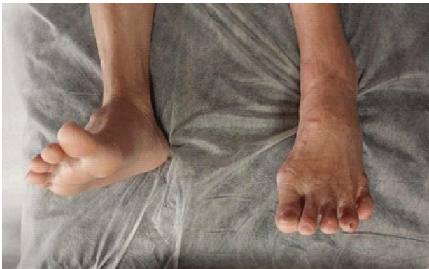
Figure 1: Photograph of bilateral ankle dorsiflexion, demonstrating patient’s left foot drop.
Radiographs of the patient’s full-length lower limbs, pelvis, femur, and lumbar spine were taken as follows in Figs. 2, 3, 4. There were no prior radiographs to compare with as the patient was no longer on follow-up for her femoral nail.
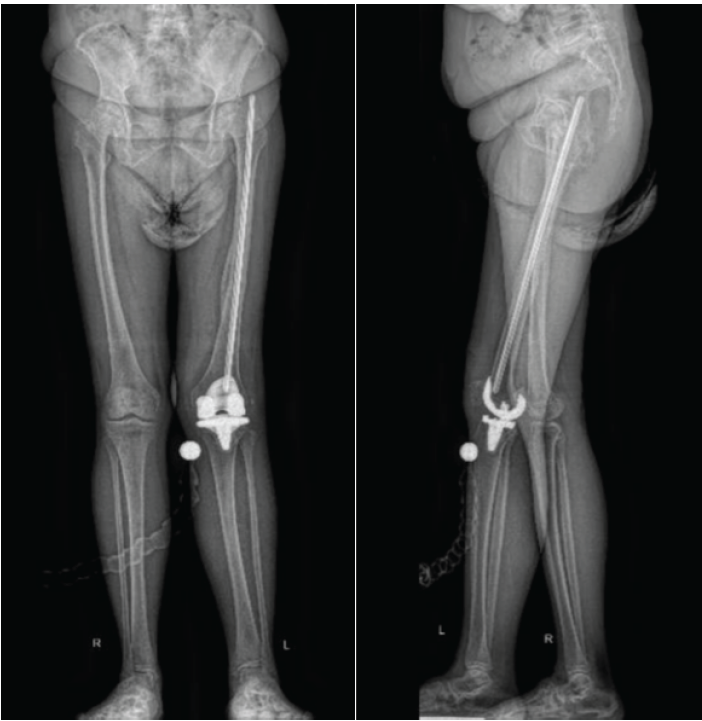
Figure 2: Radiographs of full-length lower limbs anteroposterior and lateral.
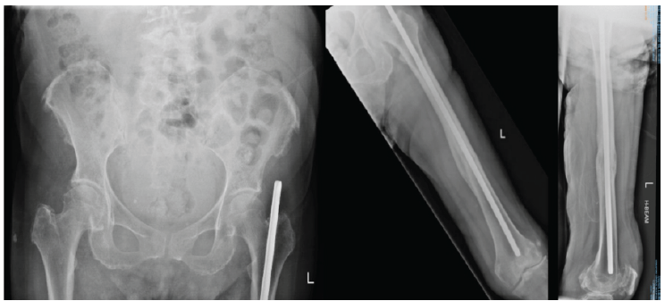
Figure 3: Radiographs of pelvis anteroposterior and left femur anteroposterior and lateral.
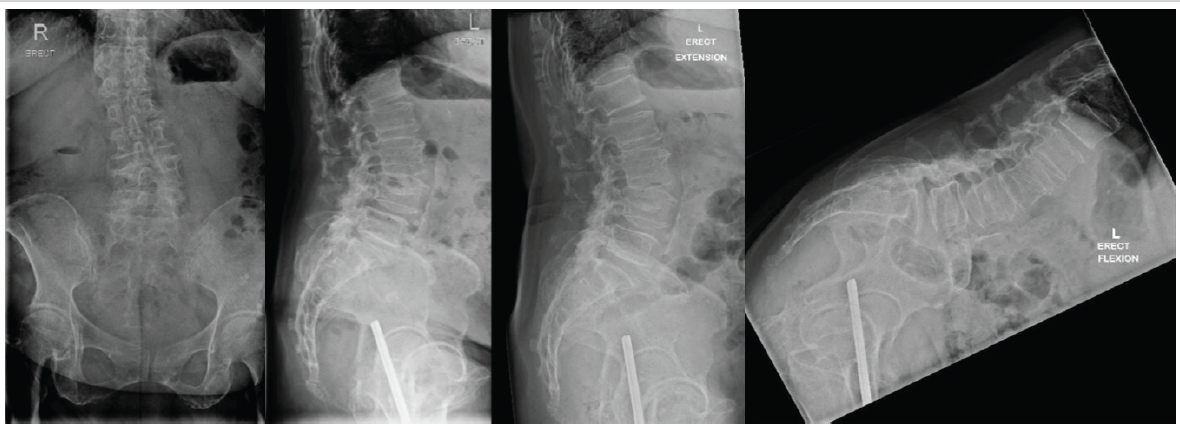
Figure 4: Erect radiographs of lumbar spine anteroposterior, lateral, extension, and flexion views.
Magnetic resonance imaging (MRI) of the lumbar spine was carried out the next day, but no significant left L4/L5 neural foraminal narrowing or nerve root compression was demonstrated to account for the left foot drop. An MRI of her left thigh was then carried out 5 days later to trace the route of the sciatic nerve. It showed a chronic tear of the gluteus medius tendon with surrounding scarring, with the sciatic nerve running very closely to the tendon stump. The sciatic nerve showed scarring to the level of the ischiofemoral space and appeared slightly hyperintense on turbo inversion recovery magnitude sequence, as seen in Fig. 5.
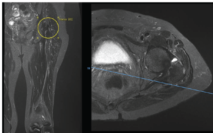
Figure 5: Magnetic resonance imaging of left thigh turbo inversion recovery magnitude sequence coronal and transverse views.
This was followed up with left lower limb electrodiagnostic studies conducted 9 days after the onset of foot drop. Nerve conduction study (NCS) and electromyography (EMG) demonstrated:
- Absent left peroneal sensory nerve response and reduced left sural nerve compound muscle action potential amplitude.
- Near-absent nerve response in left peroneal nerve at extensor digitorum brevis and absent nerve response in left peroneal at tibialis anterior (TA).
- Left TA muscle elicited abundant spontaneous discharges of active denervation; no voluntary motor unit action potentials could be elicited.
The overall findings were suggestive of a left sciatic neuropathy, on a background of underlying lumbosacral radiculopathy in the lower limbs. Findings were discussed with the patient and family. The option of removing the femoral nail was offered, but in view of the chronic irritation of the sciatic nerve, it was unsure if the foot drop would eventually recover.
The patient was keen to proceed with the nail removal due to the persistent foot drop, and the femoral nail was removed in the operating theater, 12 days after the onset of foot drop. During the operation, the Kuntscher femoral nail was noted to be proud, extending past the bony surface of the femoral greater trochanter, and the whole femoral nail was removed intact. The left sciatic nerve was noted to have significant scarring, and neurolysis was carried out, with the sciatic nerve decompressed both proximally (to the level of the sciatic notch) and distally (to the level of the gluteus maximus tendon insertion).
Postoperatively, the patient’s foot drop did not recover, with only a slight improvement of S1 power to MRC grade one from grade zero, while the rest of the power remained similar to preoperatively. NCS and EMG were carried out again 6 weeks postoperatively, which showed similar findings of a persistent left sciatic neuropathy, with no motor unit action potentials elicited from the left TA muscle, and electrophysiological evidence of active denervation in the left TA, biceps femoris (short head) and medial gastrocnemius muscles. The patient had been prescribed an ankle-foot orthosis for her foot drop, and after a period of rehabilitation, she was able to return to being functionally independent.
Ipsilateral foot drop after intramedullary nailing of the femur is a rare but known complication, usually due to sciatic nerve palsy. This is more commonly an acute foot drop, which may occur due to various causes reported such as direct pressure from the misplacement of clamps [2], nerve entrapment at the fracture site [3], post-operative hematoma formation [4,5], or traction-related when using the orthopedic traction table [6,7]. While there have been previous reports of delayed presentation of foot drop due to other metal implants such as hip hemiarthroplasty and total hip arthroplasty [8,9], to our knowledge, there are none reported from femur intramedullary nails.
In this case of foot drop, a lumbar etiology was excluded by radiological studies, and thereafter electrodiagnostic tests localized to a sciatic nerve neuropathy. While there are various causes to explain a delayed sciatic neuropathy, in this patient, it was likely due to the chronic mechanical irritation from the retained femoral nail causing scarring and fibrosis. Another differential to consider would be sciatic nerve neuropathy from myositis ossificans traumatica which has been reported in the literature [10], although scan findings were not suggestive of it.
The case highlights the possibility that retained orthopedic implants may contribute to chronic mechanical irritation and fibrosis of adjacent neural structures, resulting in very late neurological manifestations. It also emphasizes the importance of comprehensive clinical assessment, electrodiagnostic evaluation, and advanced imaging in establishing the diagnosis and guiding management.
Management options include conservative versus surgical removal of the nail with neurolysis, with the latter option being preferred as it allows exploration and direct visualization of the nerve, although restoration of motor function is not guaranteed. In this case, even though the onset of foot drop was only for 1-day duration, it is likely that the irritation to the sciatic nerve was a chronic process over the past five decades; hence, recovery was limited.
This report highlights a rare case of delayed presentation of foot drop due to sciatic nerve neuropathy on a background of previous femur intramedullary nailing carried out 50 years ago. While it is rare, it remains possible that retained metal implants may contribute to chronic nerve irritation or compression even many decades after the index surgery. Careful clinical evaluation, electrodiagnostic studies, and relevant imaging are important for accurate diagnosis and management of the foot drop.
The case highlights the possibility that retained orthopedic implants may contribute to chronic mechanical irritation and fibrosis of adjacent neural structures, resulting in very late neurological manifestations. It also emphasizes the importance of comprehensive clinical assessment, electrodiagnostic evaluation, and advanced imaging in establishing the diagnosis and guiding management. We believe that this case will be of interest to orthopedic surgeons and clinicians managing peripheral nerve disorders and late complications of retained implants.
References
- 1. Nori SL, Stretanski MF. Foot Drop. In: StatPearls. Treasure Island, FL: StatPearls Publishing; 2026. Available from: https://www.ncbi.nlm.nih.gov/books/nbk554393 [Last accessed on 2026 May 15]. [Google Scholar] [PubMed]
- 2. Chloros GD, Giannoudis VP, Giannoudis PV. Iatrogenic sciatic nerve injury secondary to clamp placement around the femoral neck during proximal femoral fracture intramedullary nailing. Trauma Case Rep 2021;35:100532. [Google Scholar] [PubMed]
- 3. Britton JM, Dunkerley DR. Closed nailing of a femoral fracture followed by sciatic nerve palsy. J Bone Joint Surg Br 1990;72:318. [Google Scholar] [PubMed]
- 4. Pedri TG, Treme GP. Sciatic nerve palsy after operative treatment of subtrochanteric femur fracture resulting from postoperative hematoma: A case report. UNM Orthop Res J 2017;6:148-150. [Google Scholar] [PubMed]
- 5. Kim YM, Joo YB, Song SH. Delayed sciatic nerve palsy due to hematoma related with anticoagulants prophylaxis in the femur intramedullary nailing: A case report. J Korean Fract Soc 2017;30:198-202. [Google Scholar] [PubMed]
- 6. Alzahrani JA, Alabdali AA, Albariqi MO. Ipsilateral foot drop after leg traction on fracture table for mid-shaft femur fracture nailing: A rare case report. Cureus 2023;15:e43826. [Google Scholar] [PubMed]
- 7. Runu R, Kumar M, Kumar I, Subhash A, Kumar S. Sciatic nerve palsy following femoral interlock nailing. J Indira Gandhi Inst Med Sci 2015;1:48-9. [Google Scholar] [PubMed]
- 8. Xu LW, Veeravagu A, Azad TD, Harraher C, Ratliff JK. Delayed presentation of sciatic nerve injury after total hip arthroplasty: Neurosurgical considerations, diagnosis, and management. J Neurol Surg Rep 2016;77:e134-8. [Google Scholar] [PubMed]
- 9. May O, Girard J, Hurtevent JF, Migaud H. Delayed, transient sciatic nerve palsy after primary cementless hip arthroplasty: a report of two cases. J Bone Joint Surg Br. 2008;90(5):674-676. doi:10.1302/0301-620X.90B5.19536 [Google Scholar] [PubMed] [CrossRef]
- 10. Guan Z, Wilson TJ, Jacobson JA, Hollon TC, Yang LJ. Delayed sciatic nerve injury resulting from myositis ossificans traumatica. PM R 2016;8:484-7. [Google Scholar] [PubMed]






Related Articles in Journal of Orthopaedic Case Reports
 July 1, 2025 Acute Onset Common Peroneal Nerve Palsy Secondary to Fibular Head Osteochondroma in an Adolescent: A Case Report
 June 10, 2022 Resolution of Deep Peroneal Nerve Palsy after Seroma Evacuation in a Primary Total Hip Arthroplasty: A Case Report
 January 10, 2016 Atraumatic Extra-articular Patellar Dislocation with Vertical Axis Rotation: A Case Study
 January 1, 2025 MIS transoral technique for C1-C2 cord compression – Intricacies using a 360⁰ navigated approach