


Introduction
Poliomyelitis is currently approaching global eradication. Although sporadic new cases continue to be reported, the majority of individuals who survived the infection are now in their fourth or fifth decade of life or older, and it is not uncommon to encounter them in orthopedic and trauma services.
Survivors of poliomyelitis are at increased risk of falls and fractures due to muscle paralysis and subsequent muscular atrophy, resulting in decreased stability, impaired mobility, and altered gait patterns [1,2,3]. In addition, up to 96% of patients with a history of polio have been shown to develop osteoporosis or osteopenia in the affected limbs, significantly increasing fracture risk [3], with the femur being the most commonly involved bone [4].
Residual deformities after poliomyelitis, including excessive femoral anteversion, coxa valga, a narrow medullary canal, and distorted trochanteric anatomy, complicate fracture fixation and may preclude safe intramedullary nailing [1,2]. In patients with typical anatomy, randomized controlled trials have demonstrated the superiority of intramedullary devices over sliding hip screws for the treatment of pertrochanteric fractures, allowing earlier rehabilitation and reducing mechanical failure rates [5].
Pediatric locking compression plate (LCP) hip systems were originally designed for corrective osteotomies in small, osteopenic femora and have demonstrated reliable fixation in these settings [6,7]. These biomechanical properties make them a reasonable alternative for adult dysplastic femora in which conventional implants are unsuitable or incompatible.
Case Report
A 45-year-old male presented following a low-energy fall with pain and deformity of the right hip. He had a history of poliomyelitis in childhood, resulting in complete paralysis of the right lower limb with a shortening of about 12 cm. Before the injury, he was ambulant using a lower-limb orthosis and under-arm crutches with partial weight bearing.
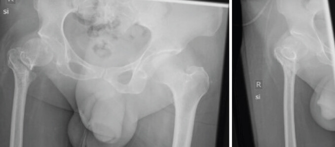
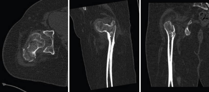
Plain radiographs revealed a pertrochanteric femur fracture (AO 31-A1.2) and a dysplastic proximal femur with coxa valga and severe narrowing of the canal (Fig. 1 and 2). Because of the trochanteric deformity and stenosis, an intramedullary device such as a Gamma 3 nail could not be introduced safely.
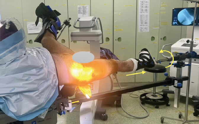
Under spinal anesthesia, closed reduction was achieved on a traction table; 35° internal rotation was required to obtain anatomical alignment in both anteroposterior and axial planes (Fig. 3). Fixation was performed with a Synthes Pediatric Hip Plate (120°, four-hole, 5.0 mm) through a lateral trans-muscular approach. Intraoperative fluoroscopy confirmed optimal reduction and implant position. The procedure was uncomplicated.
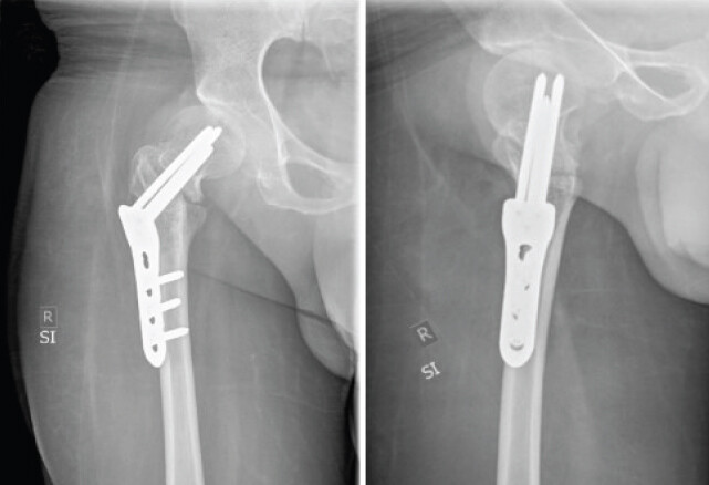
Postoperatively, the patient was mobilized with toe-touch partial weight bearing using his orthosis. The wound healed uneventfully, and radiographs obtained on postoperative day 2 confirmed anatomical reduction and stable implant positioning (Fig. 4).
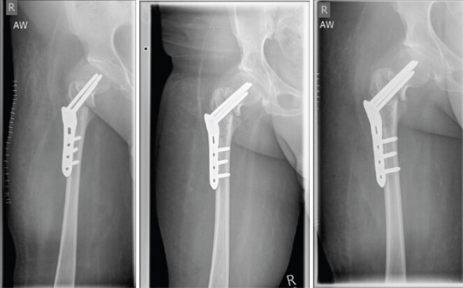
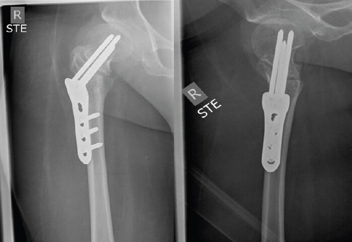
At 6 weeks, the patient reported minimal pain, with maintained alignment on radiographic evaluation (Fig. 5). By 12 weeks, radiographs demonstrated incomplete bony consolidation; however, alignment remained stable, and the position of the surgical implants was unchanged (Fig. 6). The patient regained his pre-injury level of orthosis-assisted mobility.
Discussion
Managing proximal femoral fractures in patients with post-polio residual paralysis (PPRP) remains complex. Decades of muscle imbalance lead to abnormal biomechanics, cortical thinning, and coxa valga deformity, while osteopenia further compromises implant fixation [1,2,3]. Standard cephalomedullary nailing requires a reproducible trochanteric entry point and sufficient canal width, conditions typically absent in the dysplastic PPRP femur [1,2,3,8].
The use of a pediatric LCP hip plate in adult patients represents an off-label application but was deemed anatomically and biomechanically justified in the presence of severe deformity and medullary canal stenosis.
In contrast, prospective and randomized studies in the general population have demonstrated that intramedullary fixation provides superior biomechanical stability, lower rates of implant failure, and earlier mobilization compared with extramedullary systems, particularly in unstable fractures [5]. These data, however, presuppose normal proximal femoral anatomy. When severe deformity and medullary canal narrowing are present, intramedullary nailing carries a substantial risk of malreduction or cortical perforation.
Several case series involving post-polio patients emphasize the importance of individualized surgical planning and adapting fixation strategies to altered anatomy. Gupta et al. reported that narrow medullary canals and abnormal femoral geometry frequently preclude standard nailing, necessitating modified plate constructs [1,3]. Although locking plate osteosynthesis represents a valid alternative in view of the altered anatomy in these patients, the risk of implant failure must be considered, as asymmetric muscle paralysis results in abnormal loading and altered biomechanics. Castellanos-Alonso et al. similarly concluded that fractures in post-polio patients require tailored fixation strategies, as both bone fragility and deformity limit the applicability of conventional approaches [2].
Among the therapeutic options described for patients with extremely narrow medullary canals, the use of elastic intramedullary devices such as the titanium elastic nailing system has been proposed. However, these implants do not reliably provide rotational stability and are generally better suited for diaphyseal fractures. External fixation has also been described, although it is more commonly reserved for shaft fractures rather than proximal femoral injuries [3,4]. Mingo-Robinet et al. suggested sliding hip screw constructs and discussed pediatric plates as a potential alternative in altered femoral anatomy, although no clinical cases using such implants were reported [9].
The use of extramedullary plating as a valid alternative in patients with preexisting femoral deformities has been described by several authors. El-Sayed Khalil et al. reported a series of 13 post-polio patients treated with LCP, including a single AO/OTA 31 fracture, and concluded that these implants yielded favorable clinical outcomes, with only one case of nonunion at 24 months of follow-up [10]. Similarly, Fan et al. published a retrospective study of 37 patients with pertrochanteric femoral fractures treated with LCP fixation, nine of whom had post-polio syndrome. The authors reported bone healing at a mean of 5.1 months, with excellent functional outcomes and only one case of nonunion [8].
In this patient population, comprehensive preoperative radiographic evaluation is essential and should include assessment of greater trochanter anatomy, femoral neck and shaft morphology, as well as measurement of the femoral canal diameter [1]. Gupta et al. proposed that intramedullary nailing should be considered only in patients with a canal diameter >9 mm. In our case, the maximum canal diameter was 7 mm.
In the present case, the pediatric LCP hip plate provided a small, low-profile construct capable of achieving angular-stable fixation in a dysplastic proximal femur. Originally validated in pediatric and cerebral palsy populations, this implant offers reliable screw purchase in osteopenic bone and allows restoration of alignment without extensive surgical exposure [6,7]. Rutz and Brunner reported stable fixation using this plate in osteoporotic pediatric femora, and Joeris et al. confirmed its favorable biomechanical characteristics [6,7]. Interestingly, as described in previous reports, excessive femoral anteversion, typical in post-polio patients, may require substantial internal rotation to achieve optimal closed reduction [3,11]. In our case, 35° of internal rotation on the traction table was necessary to obtain an anatomical reduction.
Among the potential complications reported in these patients are malunion, nonunion, and peri-implant fractures.
The incomplete bony union observed at the 12-week postoperative follow-up in our patient is likely related to the underlying poor bone quality as well as reduced mechanical loading of the affected limb due to orthosis-assisted ambulation with crutches. As proposed by Gupta et al., the primary goal of treatment in these patients is not necessarily osseous union, but rather the facilitation of early rehabilitation and mobility.
In line with this, our patient regained his pre-injury level of mobility and remains pain-free.
This case supports the off-label yet rational use of a pediatric LCP hip plate for fracture fixation in adult patients with post-polio deformity, balancing mechanical stability with a minimally invasive approach.
The main limitations of this report include the short-term follow-up and the inherent constraints of a single-case design. Nevertheless, this case demonstrates a feasible and reproducible surgical option for a particularly challenging clinical scenario.
Conclusion
In adult patients with post-polio femoral deformity and canal stenosis, pediatric LCP hip plates can provide stable, anatomically conforming fixation when intramedullary nailing is not feasible. This approach achieves reliable reduction, maintained alignment, and return to pre-injury function with minimal soft-tissue trauma.
Clinical Message
In pertrochanteric fractures with severe post-polio femoral deformity and medullary canal stenosis, intramedullary nailing may be anatomically unsafe; in such cases, a pediatric LCP hip plate represents a viable angular-stable alternative for achieving anatomical reduction and reliable fixation.
Conflict of Interest:
Nil
Source of Support:
Nil
Consent:
The authors confirm that informed consent was obtained from the patient for publication of this article
How to Cite this Article
Dietz B, Ariza ME, Schneider M. Pertrochanteric Femur Fracture in a Post-Polio Patient treated with a Pediatric Hip Plate: A Case Report. Journal of Orthopaedic Case Reports 2026 October;16(10):40-44.
References
- Gupta A, Saurabh S, Trikha T, Karpe A, Mittal S. Femoral shaft fracture in post-polio syndrome patients: Case series from a Level-I trauma center and review of literature. Indian J Orthop 2022;56:1339-46. [Google Scholar] | [PubMed]
- Castellanos-Alonso S, Tomás-Hernández J, Teixidor-Serra J, Selga-Marsà J, Piedra-Calle CA, Andrés-Peiró JV. Non-prosthetic implant-related femur fractures in post-polio patients-surgical management. Trauma Case Rep 2023;46:100843. [Google Scholar] | [PubMed]
- Gupta M, Jain VK, Upadhyaya GK, Arya RK. Comprehensive review of challenges associated with management of lower limb fractures in poliomyelitis patients. J Clin Orthop Trauma 2016;7:276-81. Clinical Message. [Google Scholar] | [PubMed]
- Raju S, Singhi PK, Thangamani V, Muthu C. A study of surgical intervention in fractures of post-polio residual paralytic lower limb, challenges encountered, and outcome analysis. J Orthop Jt Surg 2020;2:10-6. [Google Scholar] | [PubMed]
- Yu X, Wang H, Duan X, Liu M, Xiang Z. Intramedullary versus extramedullary internal fixation for unstable intertrochanteric fracture: A meta-analysis. Acta Orthop Traumatol Turc 2018;52:299-307. [Google Scholar] | [PubMed]
- Rutz E, Brunner R. The pediatric LCP hip plate for fixation of proximal femoral osteotomy in cerebral palsy and severe osteoporosis. J Pediatr Orthop 2010;30:726-31. [Google Scholar] | [PubMed]
- Joeris A, Audigé L, Ziebarth K, Slongo T. The locking compression paediatric hip plate: Technical guide and critical analysis. Int Orthop 2012;36:2299-306. [Google Scholar] | [PubMed]
- Fan S, Yin M, Xu Y, Ren C, Ma T, Lu Y. Locking compression plate fixation of femoral intertrochanteric fractures in patients with preexisting proximal femoral deformity: A retrospective study. J Orthop Surg Res 2021;16:285. [Google Scholar] | [PubMed]
- Mingo-Robinet J, Alonso JA, Moreno-Barrero M, González-García L, Garcia-Virto V, Aguado HJ. Technical aspects and complications in the surgical treatment of poliomyelitis-affected lower limb fractures. Rev Esp Cir Ortop Traumatol (Engl Ed) 2018;62:257-66. [Google Scholar] | [PubMed]
- El-Sayed Khalil A. Locked plating for femoral fractures in polio patients. Arch Orthop Trauma Surg 2010;130:1299-304. [Google Scholar] | [PubMed]
- Lai MC, Ng SH, Premchand AX. The surgical challenges of intertrochanteric hip fracture in a patient with polio dysplastic hip and previous distal femur fracture: A case report. Malays Orthop J 2022;16:130-3. [Google Scholar] | [PubMed]
© 2026 Journal of Orthopaedic Case Reports - Published by Indian Orthopaedic Research Group
About the Authors
-

Benjamin Dietz
-

Marco Edgar Ariza
-

Michael Schneider
Related Articles in Journal of Orthopaedic Case Reports
 August 1, 2026 The “Terrible Tetrad” of the Elbow: A Case Series of Two Patients with a Rare Quadruple-Component Injury and a Proposed New Classification Term
 August 1, 2026 Augmentation Plating with Intramedullary Nailing for Aseptic Long Bone Non-union: A Prospective Observational Study of Clinical, Radiological, and Functional Outcomes
 August 1, 2026 Predictors of Delayed Union Following Locking Compression Plate Fixation of Tibial Shaft Fractures: A Prospective Observational Study
 June 1, 2026 Clinical Implications of ChatGPT-assisted Multimodal Pre-operative Assessment in Elderly Pertrochanteric Fracture Patients: An Exploratory Study