

Unilateral Bi-portal endoscopy (UBE) offers a safe, effective, and minimally invasive alternative for treating lumbar disk herniation and canal stenosis, with faster recovery, minimal complications, and a shorter learning curve for spine surgeons.
Dr. Shashank Nahar, Department of Neurosurgery, All India Institute of Medical Sciences, Bhopal, Madhya Pradesh, India. E-mail: naharshashank@gmail.com
Abstract
Introduction: Lumbar degenerative pathologies such as lumbar disk herniation (LDH) and lumbar canal stenosis (LCS) are among the most common causes of chronic low back and radicular pain and require surgery in patients with failed conservative management. The Unilateral Bi-portal Endoscopy (UBE) technique has emerged as a minimally invasive alternative to surgically treat these pathologies, offering reduced morbidity and faster recovery. This study evaluates the clinical outcomes and safety of UBE in patients with LDH and LCS based on a fellowship-based experience.
Materials and Methods: A case series of eight patients with LCS and LDH operated on using the UBE technique during our spine endoscopy fellowship training at a high-volume spine center in the central part of India in October 2025. Intraoperative parameters (duration, complications) and postoperative outcomes (visual analog scale [VAS] for back and leg pain, hospital stay, complications) were recorded. Data were statistically summarized using mean and standard deviation.
Results: The mean operative time for a single level was 41.6 ± 7.8 min. No intraoperative or postoperative complications occurred. Mean pre-operative VAS for back pain (BP) was 5.75 ± 1.17, and for lower limb pain (LL) was 7.00 ± 1.31. On post-operative day 1, mean BP reduced to 2.87 ± 0.60 and LL to 3.37 ± 0.92; by post-operative day 7, BP and LL further decreased to 1.37 ± 0.50 and 2.12 ± 1.13, respectively. All patients were discharged within 3 days and resumed daily activities within a week.
Conclusion: Based on our fellowship experience, we believe that UBE is a safe, effective, and minimally invasive surgical option for managing LDH and canal stenosis. It offers significant pain relief, reduced blood loss, faster recovery, and excellent patient satisfaction. Furthermore, this minimally invasive spine technique has a less steep learning curve, and its use of low-cost instruments provides a valuable learning opportunity for spine surgeons who are already performing conventional open surgical approaches.
Keywords: Unilateral biportal endoscopy, lumbar disk herniation, lumbar canal stenosis, minimally invasive spine surgery, visual analog scale, minimally invasive lumbar discectomy.

Lumbar degenerative diseases, including lumbar disk herniation (LDH) and lumbar canal stenosis, are among the leading causes of chronic low back pain (BP) and sciatica [1]. These conditions commonly arise due to degenerative changes such as intervertebral disk degeneration, hypertrophy of the facet joints, and thickening of the ligamentum flavum, which together contribute to neural compression and canal narrowing. The global annual incidence of lumbar degenerative disease is estimated to affect approximately 266 million individuals [2]. Clinically, patients may present with a spectrum of symptoms ranging from localized low BP and radiculopathy to paresthesia, motor weakness, and neurogenic claudication. In advanced cases, severe neural compression may lead to bowel or bladder dysfunction [3]. Surgical intervention is typically reserved for cases where conservative management fails after 6 weeks–2 months, as indicated by many studies. This approach can lead to significant improvements in pain in patients requiring surgery [4].
Various surgical techniques for LDH have been described in the literature. The conventional method of open lumbar laminectomy and discectomy has been considered the standard surgical treatment for LDH since 1929 and is widely practiced worldwide by spine surgeons [5]. However, conventional methods raise several concerns, including iatrogenic paraspinal muscle injury, postoperative pain, increased blood loss, prolonged hospital stay, and delayed recovery. To address these limitations, there has been a gradual shift toward tissue-sparing approaches, leading to the development of minimally invasive surgical techniques. “Minimally invasive” lumbar discectomy encompasses a spectrum – from tubular microdiscectomy and microendoscopic discectomy to percutaneous endoscopic lumbar discectomy (PELD) – united by smaller exposures and muscle-splitting or serial dilatation approaches [6,7].
PELD has gained widespread recognition among both surgeons and patients in recent years, as the preferred surgical approach for LDH [8]. However, PELD has the drawbacks of a small surgical field, low surgical efficiency, a long learning curve, and difficulty in mastering [9]. Hence, to overcome these limitations of PELD, the Bi-portal endoscopy procedure is now widely gaining popularity in clinical practice due to its short learning curve, low-cost instruments, and provides a wide surgical field.
To evaluate the clinical effectiveness and safety of the bi-portal endoscopic procedure in patients with LDH and canal stenosis, we present a case series of 8 patients who underwent the procedure during a fellowship training program at a high-volume center in Pune.
What is Bi-portal spinal endoscopy?
Unilateral Bi-portal Endoscopy (UBE) is an endoscope-assisted spinal surgery technique that involves creating two unilateral portals – a working portal and an endoscopic portal – and utilizing a saline-based irrigation system to perform disk surgery.
Surgical indications of the UBE technique in Lumbar degenerative diseases
In lumbar degenerative disease, the UBE technique has been proven to be feasible for various pathologies, including lumbar canal stenosis (both central and lateral recess), all types of disk herniation based on location (central, paracentral, lateral, foraminal, and extraforaminal), recurrent disk herniation, calcified disk herniation, and spondylolisthesis [10,11].
We included a case series of 8 patients with lumbar canal stenosis and LDH who were admitted and operated on using the UBE technique at a central part of India – a fellowship training center for bi-portal endoscopy – during October 2025. Our inclusion and exclusion criteria were as follows:
Inclusion criteria
- Radiologically confirmed diagnosis of LDH and lumbar canal stenosis, including central, lateral recess, and foraminal stenosis
- Presence of typical symptoms of neurogenic intermittent claudication or lower limb (LL) radicular pain with or without associated low backache, with inadequate relief following at least 6 weeks of conservative therapy
- Age between 18 and 80 years
- Ability to provide informed consent, either independently or with assistance from family members, and to read, comprehend, and respond to follow-up assessments
- Willingness to participate in the study and adhere to all aspects of the clinical protocol, including scheduled follow-up visits and required evaluations.
Exclusion criteria
- Lumbar canal stenosis or disk herniation associated with segmental instability or spondylolisthesis, or an infective cause
- Patients <18 years or >80 years
- Patients who gave negative consent for surgery and lost follow-up.
The clinical effectiveness and safety of the bi-portal endoscopic technique were evaluated based on the type and location of disk herniation, using intraoperative parameters such as duration of surgery and intraoperative complications, as well as postoperative parameters including pain scores (visual analog scale [VAS] scale) for low back and LL pain, duration of hospital stay, return to routine activities, and postoperative complications.
Case 1
A 45-year-old male patient was admitted with primary complaints of low BP. He also reported pain radiating to both lower limbs, with more discomfort on the right side than the left. These symptoms had been present for 5 months and were associated with tingling and numbness along the L5 dermatome. The patient did not exhibit any motor deficits, and no involvement of the bladder or bowel.
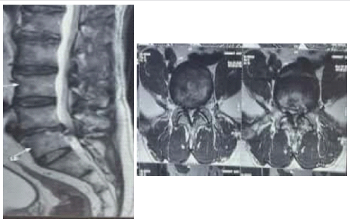
Figure 1: Magnetic resonance imaging (T2) Sagittal and axial cut shows central canal stenosis with bilateral lateral recess stenosis, with right lateral prolapse intervertebral disk.
A magnetic resonance imaging (MRI) (Fig. 1) showed L4-L5 central canal stenosis with bilateral foraminal stenosis, along with a right lateral protrusion of the intervertebral disk compressing both traversing L5 nerve roots. The patient underwent surgery using the UBE technique. The docking of the endoscopic and working portal was performed from a left-sided approach. Unilateral left-sided foraminal ligament and thickened ligamentum flavum causing central canal decompression were excised along with the undercutting of the hypertrophied superior articular process (SAP). The “over the top technique” or “contralateral sub-laminar approach” was utilized by thinning or removing the base of the spinous process from inside, and the contralateral sub-laminar surface was approached, hypertrophied ligamentum flavum was excised, followed by L4-L5 discectomy and excision of the contralateral foraminal ligament. The contralateral hypertrophied SAP was also punched out.
Intraoperatively, effective spinal and bilateral nerve root decompression was achieved. Cord pulsations were observed, and the nerve roots changed from pale white to pink due to the restoration of microvascular blood flow over the nerve roots. Finally, a Romovac drain was inserted into the epidural space to prevent post-operative epidural hematoma formation, and the skin was closed by subcuticular sutures.
Preoperatively, the VAS score for low BP was 7, and it was also 8 for LL pain. On postoperative day 1, the VAS score decreased to 3 for both low back and LL pain. After 1 week of follow-up, the VAS score further improved to 1. The patient was discharged on the 2nd postoperative day without any complications.
Case 2
Patient Manisha Pawar 45-year-old female, came with chief complaints of BP with pain radiating to bilateral lower limbs for 5 months, associated with tingling and numbness involving the left L4 dermatome and right L5 dermatome along bilateral lower limbs. MRI shows (Fig. 2) right lateral recess stenosis with left foraminal disk protrusion at the L4-L5 level. Pt was operated on from a left-sided approach by the UBE technique, and left L4-L5 foraminal discectomy was done with contralateral (“over the top approach”) to decompress the right lateral recess by undercutting the
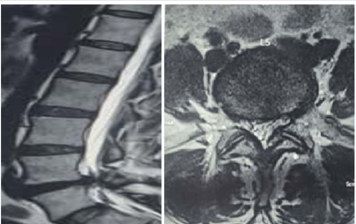
Figure 2: Magnetic resonance imaging shows right lateral recess stenosis with left L4-L5 foraminal disk prolapse in sagittal and axial cuts.
hypertrophied right SAP. Pt was self-ambulatory on 1st post-operative day and was discharged on 2nd post-operative day with intraoperative complications. VAS score in the preoperatively and postoperatively periods was noted as shown in Table 1.
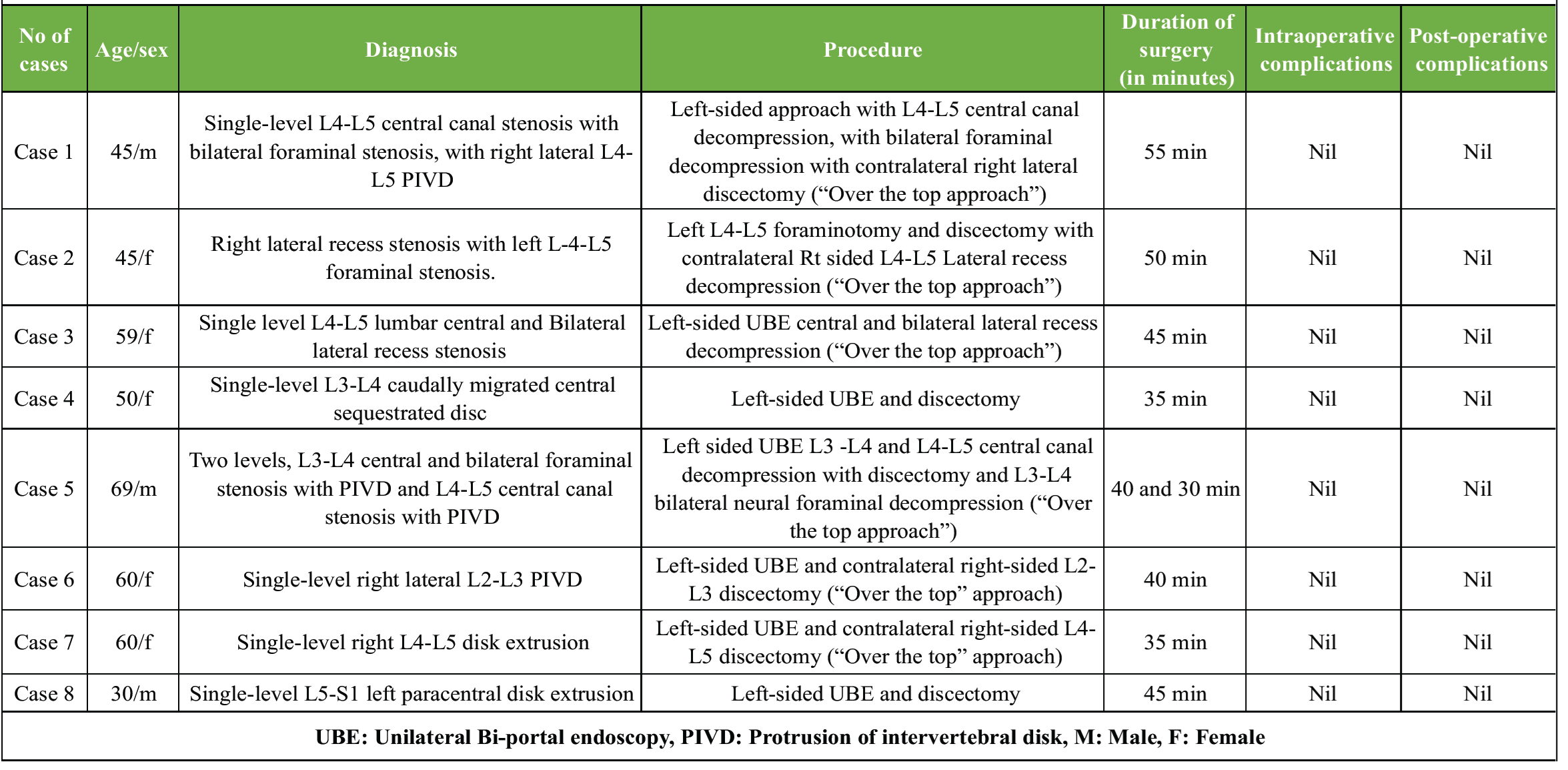
Table 1: The duration of surgery and intraoperative and post-operative complications in all the patients who underwent UBE surgery
Surgical technique for creation of UBE portals
Creation of portal
In all cases, being right-handed surgeons, we approach from the left side. Hence, we make the portal incisions on the left side.
- First, place a metal marker horizontally at the L4-L5 disk level under anteroposterior (AP) fluoroscopy, as shown in 3a, and draw a horizontal reference line (black marker line in Fig. 3b).
- Next, draw a vertical midline corresponding to the spinous process, as illustrated in 3b.
- Then, mark the instrumental (working) portal approximately 5 cm caudal to the horizontal reference line, ensuring it remains medial to the pedicle under AP fluoroscopic guidance. (As shown in orange color in Fig. 3b).
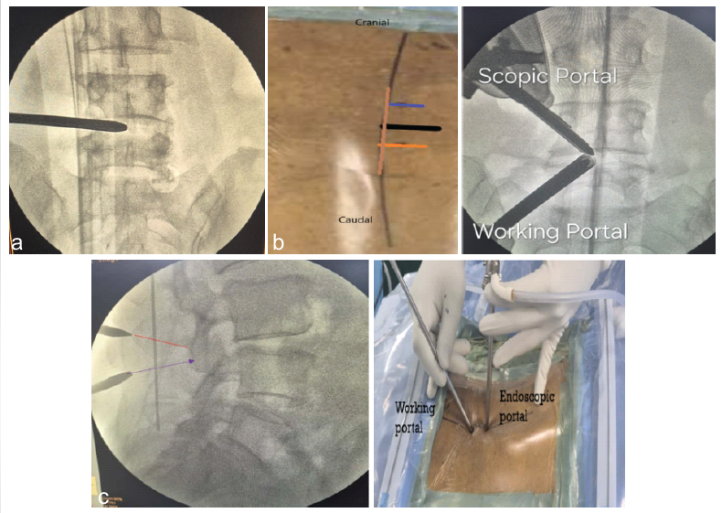
Figure 3: The initial marking step for making the endoscopic portal cranially and the working portal site caudally. (a) Firstly keeping the metal marker horizontally placed at the L4-L5 disk level at anteroposterior fluoroscopy. (b) The vertical brown line passing over the midline spinous process at the L4-L5 level. The dark black horizontal reference line corresponds to the L4-L5 disk level as shown in anteroposterior (AP) fluoroscopy. The second horizontal orange line is marked 1.5 cm caudally from the horizontal reference black line for the instrument portal. The third horizontal blue line is marked 1.5 cm cranially from the reference horizontal black line for the scopic portal. The second AP fluoroscopy image shows the insertion and direction of the scopic and instrumental portal at the L4-L5 level. (c) That the endoscopic cranial portal should be directed toward the L4 lower lamina, and the instrumental caudal portal should also be directed towards the L4 lamina, forming triangulation as shown in lateral fluoroscopy.
Similarly, mark the endoscopic (scopic) portal 1.5 cm cranial (as shown in blue color) from the horizontal reference line (marked in black color), also staying medial to the pedicle. The distance between the endoscopic portal and working instrumental portal should be at least 3 cm apart.
- After confirming the markings on AP fluoroscopy, make a transverse skin incision deep to the fascia with a knife using 11 no blade at each portal site. After this, adequate muscle dissection with a serial dilator is necessary to split the muscle.
- On fluoroscopic imaging, both the cranial (endoscopic) and caudal (instrumental) portals should be directed along the spino-laminar junction.
- Finally, take the lateral fluoroscopy ( 3c) image to confirm that the endoscope should correspond and directed to the L4 lower lamina, and the working portal should also be directed to the L4 lower lamina, forming a triangulation felt by both portal ends touching each other inside.
- Finally, by the interlaminar approach, we do an L4 lower laminectomy and L4-L5 discectomy using normal saline (0.9%) irrigation, and by the “over the top technique” to address contralateral pathology on the right side (Fig. 4).
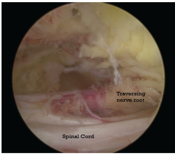
Figure 4: Intraoperative unilateral Bi-portal endoscopy image showing the lax right L4 traversing nerve root with a reddish-pink appearance following right lateral L4-L5 discectomy, excision of thickened ligamentum flavum, and undercutting of the hypertrophied superior articular process in a patient with right lateral L4-L5 protrusion of intervertebral disk and lateral recess stenosis. The change in nerve root color from pale white to reddish-pink indicates restoration of blood flow in the vessels overlying the nerve root after effective decompression.
UBE spine surgery is gaining popularity nowadays as a minimally invasive endoscopic spinal procedure. With its short learning curve and wide viewing angle compared to other endoscopic spine procedures, it is gaining popularity among practicing spine surgeons. It has the advantages of reduced postoperative pain, minimal muscle trauma, and faster recovery [12]. UBE has the potential to replace conventional open approaches for managing lumbar canal stenosis and LDH in the near future.
Our case series demonstrated that the UBE interlaminar technique is effective for a wide range of LDH s, irrespective of disk morphology – whether bulge, protrusion, extrusion, or sequestration – and for all disk locations, including central, paracentral, lateral, and foraminal herniations. Although studies have shown that UBE can also be used successfully for extraforaminal disk herniations through a paraspinal approach but such cases were not encountered in our series [13].
In terms of disk migration, we observed that caudally migrated discs were effectively decompressed using the UBE interlaminar approach after removing the upper one-third lamina of the lower vertebrae, along with the removal of the lower lamina of the above vertebrae. Previous reports have also supported the efficacy of UBE in both caudally and cranially migrated herniated lumbar disk [14,15]. Notably, even a hard calcified disk in one of our patients was successfully removed using this technique, demonstrating its versatility.
For lumbar canal stenosis – including central, lateral recess, and foraminal stenosis – UBE provided effective decompression in all our cases. Regarding the approach, as right-handed surgeons, we generally operated from the left side, maintaining the endoscopic (scopic) portal cranially and the working portal caudally. This setup allowed us to address both ipsilateral and contralateral pathologies efficiently. For right-sided lumbar pathology, we used the “over-the-top” contralateral approach. This method involves undercutting the base of the spinous process to access the contralateral sublaminar area, facilitating decompression of the contralateral ligamentum flavum, foraminal ligament, SAP, and even contralateral disk material. In our experience, this technique proved to be safe and effective.
The average operative time for single-level lumbar disk pathology was approximately 41.66 min. No intraoperative or postoperative complications were encountered in our series. However, literature reports possible complications associated with UBE, such as dural tears, epidural hematoma, transient nerve root dysesthesia, nerve root injury, and infection [16]. The use of small incisions (0.8–1.0 cm for the endoscopic portal and 1.0–1.5 cm for the working portal), along with a muscle-splitting rather than a muscle-cutting approach, significantly minimizes tissue trauma, postoperative muscular pain, and atrophy. Small incisions and muscle-splitting also decrease dead space, which prevents cerebrospinal fluid leak from very small dural tears [17]. Continuous normal saline irrigation during surgery provides effective hemostasis by reducing the risk of postoperative epidural hematoma formation, and sometimes it acts as a tamponade due to its external pressure, which seals off the small, non-visible dural tears [18]. Based on our experience, we recommend placing a drain in all cases to prevent epidural hematoma from unnoticed small bleeders, which can otherwise lead to postoperative low BP.
When comparing pre- and post-operative pain outcomes, the mean pre-operative VAS score for BP was 5.75 ± 1.17, and for LL radicular pain was 7.00 ± 1.31. On postoperative day 1, these values reduced to 2.87 ± 0.60 and 3.37 ± 0.92, respectively. At 1-week follow-up, the scores further improved to 1.37 ± 0.50 for BP and 2.12 ± 1.13 for LL pain (Table 2). This demonstrates that UBE effectively reduces both back and radicular pain in the immediate post-operative period and during early follow-up.
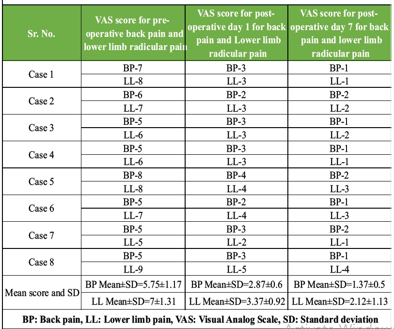
Table 2: Compares the mean VAS score pre-operatively and postoperatively in all cases
All patients were discharged on post-operative day 2. Patients were advised to avoid prolonged sitting, forward bending exercises, and heavy lifting (>10 kg) for at least 6 months. Most patients returned to routine daily activities within 1 week. The mean postoperative satisfaction score (0–10 scale) was 8.7 ± 0.7, indicating a high level of patient satisfaction.
Our findings align with previous studies demonstrating that UBE is an effective and safe alternative to open decompression with its superior cosmetic and recovery benefits as discussed above.
Based on our fellowship-based experience, we conclude that UBE is a safe, effective, and minimally invasive surgical technique for managing a wide spectrum of lumbar spine pathologies, including various types of disk herniations and lumbar canal stenosis. The procedure offers several advantages, such as reduced post-operative back and leg pain, minimal muscle trauma, improved hemostasis, shorter hospital stays, and faster return to daily activities.
Furthermore, the bi-portal endoscopic technique utilizes many of the working instruments employed in conventional open spine surgery, making it a cost-effective option with a relatively shorter learning curve compared to full-endoscopic procedures. Thus, surgeons proficient in conventional open spine surgery may find the transition to the UBE technique smoother, allowing for an easier and more confident shift towards minimally invasive spine surgery.
Unilateral Bi-portal endoscopy (UBE) is a safe, effective, and minimally invasive technique for managing LDH and canal stenosis. It provides excellent visualization, reduced tissue trauma, faster recovery, and high patient satisfaction. With a relatively shorter learning curve and the use of standard spine instruments, UBE offers an accessible and cost-effective alternative for surgeons transitioning from conventional open to minimally invasive spine surgery.
References
- 1. Benzakour T, Igoumenou V, Mavrogenis AF, Benzakour A. Current concepts for lumbar disc herniation. Int Orthop 2019;43:841-51. [Google Scholar] [PubMed]
- 2. Ravindra VM, Senglaub SS, Rattani A, Dewan MC, Härtl R, Bisson E, et al. Degenerative lumbar spine disease: Estimating global incidence and worldwide volume. Global Spine J 2018;8:784-94. [Google Scholar] [PubMed]
- 3. Wong AP, Smith ZA, Stadler JA 3rd, Hu XY, Yan JZ, Li XF, et al. Minimally invasive transforaminal lumbar interbody fusion (MI-TLIF): Surgical technique, long-term 4-year prospective outcomes, and complications compared with an open TLIF cohort. Neurosurg Clin N Am 2014;25:279-304. [Google Scholar] [PubMed]
- 4. Kögl N, Petr O, Löscher W, Liljenqvist U, Thomé C. Lumbar disc herniation-the significance of symptom duration for the indication for surgery. Dtsch Arztebl Int 2024;121:440-8. [Google Scholar] [PubMed]
- 5. Alvi MA, Kerezoudis P, Wahood W, Goyal A, Bydon M. Operative approaches for lumbar disc herniation: A systematic review and multiple treatment meta-analysis of conventional and minimally invasive surgeries. World Neurosurg 2018;114:391-407.e2. [Google Scholar] [PubMed]
- 6. Choi JY, Park SM, Kim HJ, Yeom JS. Recent updates on minimally invasive spine surgery: Techniques, technologies, and indications. Asian Spine J 2022;16:1013-21. [Google Scholar] [PubMed]
- 7. Skovrlj B, Gilligan J, Cutler HS, Qureshi SA. Minimally invasive procedures on the lumbar spine. World J Clin Cases 2015;3:1-9. [Google Scholar] [PubMed]
- 8. Gadjradj PS, Harhangi BS, Amelink J, Van Susante J, Kamper S, Vleggeert-Lankamp CL, et al. Percutaneous transforaminal endoscopic discectomy versus open microdiscectomy for lumbar disc herniation: A systematic review and meta-analysis. Spine (Phila Pa 1976) 2021;46:538-49. [Google Scholar] [PubMed]
- 9. Hsu HT, Chang SJ, Yang SS, Chai CL. Learning curve of full-endoscopic lumbar discectomy. Eur Spine J 2013;22:727-33. [Google Scholar] [PubMed]
- 10. Hwa Eum J, Hwa Heo D, Son SK, Park CK. Percutaneous biportal endoscopic decompression for lumbar spinal stenosis: A technical note and preliminary clinical results. J Neurosurg Spine 2016;24:602-7. [Google Scholar] [PubMed]
- 11. Choi DJ, Jung JT, Lee SJ, Kim YS, Jang HJ, Yoo B. Biportal endoscopic spinal surgery for recurrent lumbar disc herniations. Clin Orthop Surg 2016;8:325-9. [Google Scholar] [PubMed]
- 12. Januário G. Unilateral biportal endoscopy (UBE): Recommendations for the interlaminar approach. J Neurosci Neurol Surg 2025;17:1-8. [Google Scholar] [PubMed]
- 13. Reis JP, Pinto EM, Teixeira A, Frada R, Rodrigues D, Cunha R, et al. Unilateral biportal endoscopy: Review and detailed surgical approach to extraforaminal approach. EFORT Open Rev 2025;10:151-5. [Google Scholar] [PubMed]
- 14. Chen WC, Wang WT, Pao JL. Unilateral biportal endoscopic discectomy via translaminar approach for highly upward-migrated lumbar disc herniation: A technical note and preliminary treatment outcomes. BMC Musculoskelet Disord 2024;25:722. [Google Scholar] [PubMed]
- 15. Cheng W, Gao W, Zhu C, Shao R, Wang D, Pan H, et al. Contralateral translaminar endoscopic approach for highly down-migrated lumbar disc herniation using percutaneous biportal endoscopic surgery: Original research. BMC Surg 2024;24:58. [Google Scholar] [PubMed]
- 16. Yang L, Yu T, Jiao J, Hou T, Wang Y, Zhao B, et al. Comprehensive analysis of UBE-related complications: Prevention and management strategies from 4685 patients. Med Sci Monit 2024;30:e944018. [Google Scholar] [PubMed]
- 17. Tosun B, Ilbay K, Kim MS, Selek O. Management of persistent cerebrospinal fluid leakage following thoraco-lumbar surgery. Asian Spine J 2012;6:157-62. [Google Scholar] [PubMed]
- 18. Soliman HM. Irrigation endoscopic discectomy: A novel percutaneous approach for lumbar disc prolapse. Eur Spine J 2013;22:1037-44. [Google Scholar] [PubMed]




Related Articles in Journal of Orthopaedic Case Reports
 March 10, 2024 Unilateral Biportal Endoscopy for L5-S1 Extraforaminal Stenosis (Far Out Syndrome) – Technical note With Literature Review
 October 1, 2026 Comparative Study of Effect of Periarticular Fentanyl and Dexmedetomidine Injection for Pain Management in Total Knee Arthroplasty a Randomized Control Trial
 October 1, 2026 Prevalence and Clinical Impact of Lumbosacral Transitional Vertebrae in Chronic Low Back Pain: A Cross-Sectional Study from Central India
 October 1, 2026 Efficacy of Transforaminal Nerve Root Block for Lumbar Disc Herniation